0869
Attention-Boosted CNN for Improving the Classification of IDH and TERTp Mutation Status in Gliomas Based on Dynamic Susceptibility Contrast MRI1Institute of Biomedical Imaging, Bogazici University, Istanbul, Turkey, 2Department of Medical Pathology, Acibadem University, Istanbul, Turkey, 3Brain Tumor Research Group, Acibadem University, Istanbul, Turkey, 4Department of Molecular Biology and Genetics, Acibadem University, Istanbul, Turkey, 5Department of Neurosurgery, Acibadem University, Istanbul, Turkey, 6Department of Radiology, Acibadem University, Istanbul, Turkey
Synopsis
Keywords: Diagnosis/Prediction, Data Processing
Motivation: Molecular markers, such as IDH and TERTp, have been reported as significant prognostic factors in gliomas.
Goal(s): The aim of this study is to predict IDH and TERTp mutational subtypes in gliomas non-invasively using deep-learning applied to rCBV images derived from DSC-MRI.
Approach: We proposed a deep-learning approach with attention gates to classify IDH- and TERTp-mutation subgroups of gliomas using rCBV images along with anatomical-MRI. Additionally, Grad-CAM approach was employed to provide an explanation of which image sections played a role in decision-making.
Results: Attention-boosted deep learning-based classification model yielded high accuracy rates. GradCAM approach also highlighted the significance of different tumor components.
Impact: The proposed attention-boosted deep learning based method might have the potential to assist clinicians in the noninvasive identification of IDH and TERTp mutations at the pre-surgery point and potentially enhance treatment strategies and patient outcomes.
Introduction
Molecular markers, such as isocitrate dehydrogenase (IDH) and telomerase reverse transcriptase gene promoter (TERTp), have been reported as significant prognostic factors in gliomas.1 IDH mutations have been associated with lower angiogenesis, and IDH mutant (IDH-mut) gliomas have been reported to have a better prognosis and longer overall survival than IDH wildtype (IDH-wt) gliomas.2 On the other hand, IDH-wt gliomas with the TERTp mutation (TERTp-only) have been reported to have the worst overall survival rates.3 Recent studies have shown that the relative cerebral blood volume (rCBV) images derived from dynamic susceptibility contrast MRI (DSC-MRI) can be utilized to identify these genetic subtypes in gliomas.4 Additionally, several studies have proposed convolutional neural network (CNN) architectures to classify different subtypes of gliomas using MRI.5, 6 Moreover, the attention mechanism enables CNN models to automatically learn and focus on specific target structures within the images and to boost the performance of classification. Therefore, this study aimed to develop a deep-learning approach with attention-gates to classify IDH and TERTp mutational subgroups of gliomas using rCBV images along with anatomical-MRI while providing an explanation of which tumor parts played a role in decision-making during the classification process using gradient-weighted class activation mapping (Grad-CAM).Methods
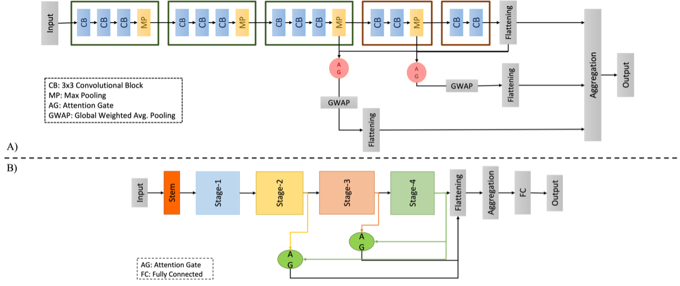
The patient cohort contained 162 gliomas (100 males/62 females, mean age: 46.8±14.7 years, range: 20-84 years; 88 glioblastoma-IDH wildtype, 74 IDH-mut, 64 TERTp-wt, and 74 TERTp-mut patients). The brain tumor MRI protocol included pre- and post-contrast T1-weighted TSE (TR=532 ms, TE=9.2 ms, voxel size=0.69 mm x 0.69 mm x 3.6 mm), T2-weighted TSE (TR=4250 ms, TE=99 ms, voxel size =0.22 mm x 0.22 mm x 3.6 mm), and T2*-weighted gradient-echo EPI DSC-MRI (TR=1610 ms, TE=30 ms voxel size=1.80 mm x 1.80 mm x 6.5 mm ). The rCBV maps were generated on the scanner console from the DSC-MRI (Siemens Healthcare, Erlangen, Germany).The whole tumor volumes were segmented on T2-weighted MRI, and the necrotic cores were segmented on post-contrast T1-weighted MRI using 3D slicer (http://slicer.org/). Each subject's tumor and necrotic areas were registered to the rCBV images using ANTs.7 First, multimodal images were created by placing the post-contrast T1-weighted, T2-weighted, and rCBV slices with the largest tumor area at the three RGB channels. All images were automatically cropped to a rectangular bounding box, including the tumor area, and resized to 128 × 128. The cohort was then split into training, validation, and held-out test sets with 3:1:1 ratios. Afterwards, various pre-trained network models, including ResNet50 and VGG16, were used to determine the best-performing network architecture for the classification tasks. Next, two attention gates were added to the same networks to assess the results (Figure 1). Since the dataset is relatively small, data augmentation techniques were also incorporated to minimize overfitting. After training and validation, the best-performing models were tested on the held-out test set to report accuracy, specificity, and sensitivity for the classification tasks. Grad-CAM was also employed in this study to investigate the effect of signal intensities at different tumor parts on identifying genetic subtypes.
Results
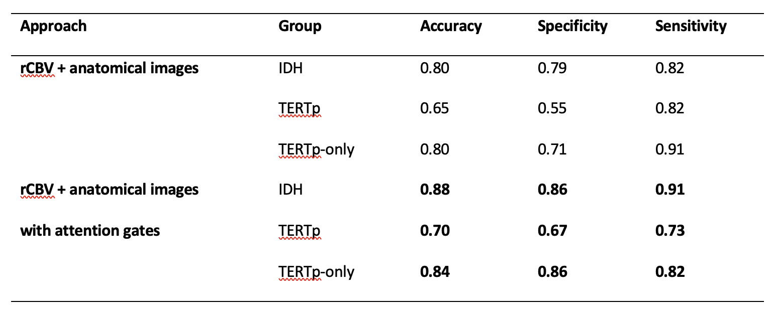
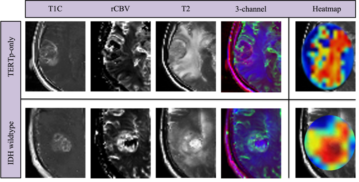
The classification accuracies without attention gates reached up to 80% (79% specificity and 82% sensitivity) for the IDH subgroup, 65% (82% specificity and 55% sensitivity) for the TERTp subgroup, and 80% (71% specificity and 91% sensitivity) for the TERTp-only versus the others (Table 1). After the addition of attention gates, the model accuracies were 88% (86% specificity and 91% sensitivity) for the classification of the IDH subgroup, 70% (67% specificity and 73% sensitivity) for the TERTp subgroup, and 84% (86% specificity and 82% sensitivity) for the TERTp-only versus others classifications. The heatmaps obtained from the Grad-CAM analysis along with anatomical-MRI and rCBV images and the RGB image created from these three modalities at the corresponding slice for an IDH-wt and a TERTp-only patient examples are shown in Figure 2. The heatmaps represented the salient features, where the red regions were the most important pixels for the CNN model. For the TERTp-only mutation example, the most important regions were located in the edema part, which the CNN model primarily focused on. Conversely, the model predominantly concentrated on the tumor core and necrotic regions in the case of the IDH-mut example.Conclusion and Discussion
This study demonstrated that incorporating rCBV images obtained from DSC-MRI into a deep learning approach provided high-performance differentiation of glioma subgroups based on IDH and TERTp mutations. Our results also indicated that an attention-boosted structure could improve the performance of the classification. The proposed methods might have the potential to assist clinicians in identifying molecular subtypes of gliomas at the pre-surgery point for improving treatment planning and patient outcomes.Acknowledgements
This study was supported by the Scientific and Technological Research Council of Turkey (TUBITAK) grant 216S432.References
1. Eckel-Passow JE, Lachance DH, Molinaro AM, Walsh KM, Decker PA, Sicotte H, et al. Glioma groups based on 1p/19q, IDH, and TERT promoter mutations in tumors. New England Journal of Medicine 2015;372(26):2499-508.
2. Yan H, Parsons DW, Jin G, McLendon R, Rasheed BA, Yuan W, et al. IDH1 and IDH2 mutations in gliomas. New England Journal of Medicine 2009;360(8):765-73.
3. Ozturk-Isik E, Cengiz S, Ozcan A, Yakicier C, Ersen Danyeli A, Pamir MN, et al. Identification of IDH and TERTp mutation status using (1) H-MRS in 112 hemispheric diffuse gliomas. Journal of Magnetic Resonance Imaging 2020;51(6):1799-809.
4. Kickingereder P, Sahm F, Radbruch A, Wick W, Heiland S, Deimling Av, et al. IDH mutation status is associated with a distinct hypoxia/angiogenesis transcriptome signature which is non-invasively predictable with rCBV imaging in human glioma. Scientific Reports 2015;5(1):16238.
5. Bangalore Yogananda CG, Shah BR, Vejdani-Jahromi M, Nalawade SS, Murugesan GK, Yu FF, et al. A novel fully automated MRI-based deep-learning method for classification of IDH mutation status in brain gliomas. Neuro-Oncology 2019;22(3):402-11.
6. Cluceru J, Interian Y, Phillips JJ, Molinaro AM, Luks TL, Alcaide-Leon P, et al. Improving the noninvasive classification of glioma genetic subtype with deep learning and diffusion-weighted imaging. Neuro-Oncology 2022;24(4):639-52.
7. Avants BB, Tustison NJ, Song G, Cook PA, Klein A, Gee JC. A reproducible evaluation of ANTs similarity metric performance in brain image registration. Neuroimage 2011;54(3):2033-44.
Figures