0866
MRI-Based Habitat, Radiomics, and Deep Learning for Assessing Response of Platinum-Based Chemotherapy in HGSOC Patients1the First People’s Hospital of Yunnan Province, Kunming, China, 2Jinshan Hospital, Fudan University, Shanghai, China, 3MR Research Collaboration Team, Siemens Healthineers Ltd., Shanghai, China
Synopsis
Keywords: Diagnosis/Prediction, Pelvis
Motivation: High-grade serous ovarian carcinoma (HGSOC) poses a significant challenge due to platinum resistance and the inherent difficulty in its prediction.
Goal(s): We aimed to explore MRI-based habitat model for predicting response of platinum-based chemotherapy in HGSOC patients, and compared with radiomics and deep learning models.
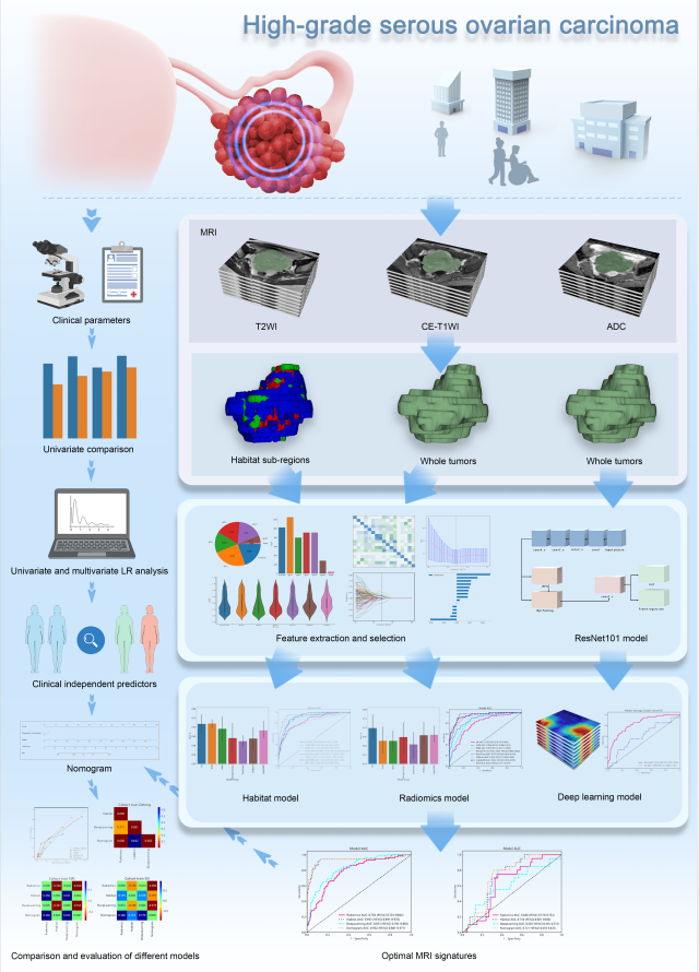
Approach: We leveraged the K-means algorithm for clustering on multiparameter MRI data. Then the radiomics, habitat, and deep learning models were constructed.
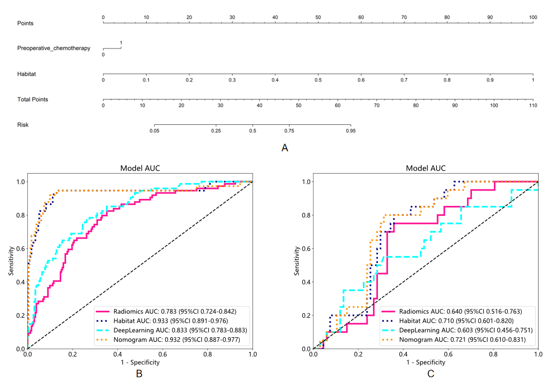
Results: Habitat model had the potential to predict platinum resistence, with a superior performance to radiomics and deep learning models. The nomogram integrating habitat with neoadjuvant chemotherapy yielded a better performance compared to others.
Impact: This study holds substantial clinical significance as it establishes a foundational framework for the customization of treatment strategies for patients afflicted with HGSOC.
Introduction
High-grade serous ovarian carcinoma (HGSOC) is the most common and aggressive subtype of ovarian cancer [1]. Up to 70% of HGSOC patients experience relapse, with approximately 15% of these cases attributed to platinum resistance [2].MRI-based radiomics and deep learning to predict platinum resistance in ovarian carcinoma patients [3,4]. However, these methods fail to account for spatial heterogeneity and intricate biological details within tumors. Habitat imaging enables a non-invasive evaluation of intratumoral heterogeneity [5].
This study aims to explore the feasibility of MRI-based habitat model for predicting platinum resistance in HGSOC patients and compare it with radiomics and deep learning models. Additionally, we construct a nomogram by integrating these findings with independent clinical predictors for a comprehensive evaluation of platinum-based chemotherapy response.
Methods
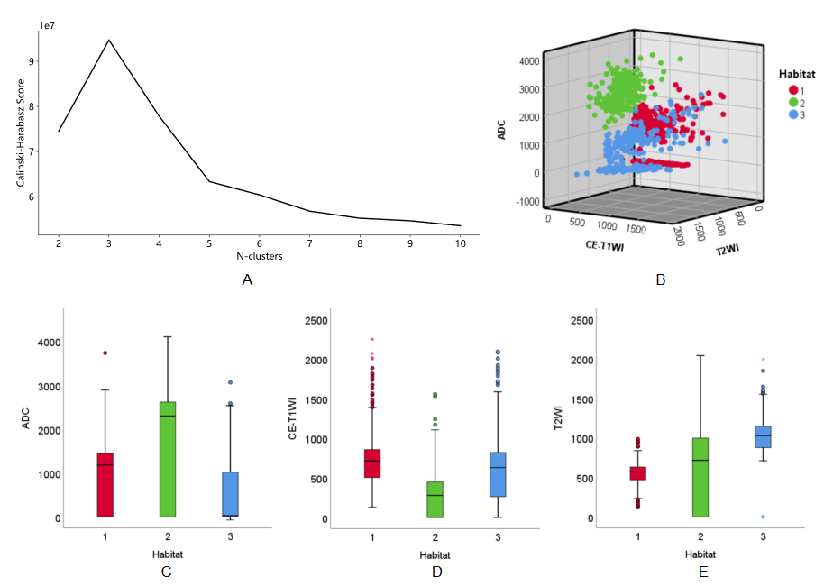
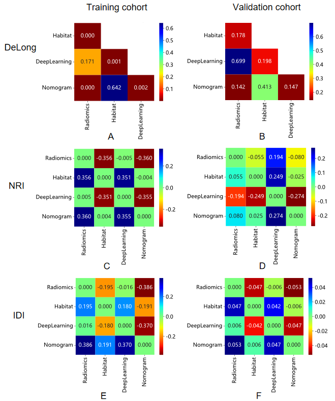
A retrospective study involving HGSOC patients from three hospitals was conducted. Pelvic MRI was performed using 1.5T or 3T scanners (Siemens Magnetom Prisma, Siemens Magnetom Vida, Philips Ingenia, GE Signa Pioneer, and GE Signa HDXt). MRI habitat clustering was performed on T2-weighted images (T2WI), contrast-enhanced T1-weighted images (CE-T1WI), and apparent diffusion coefficient (ADC) maps. After image preprocessing and space registration, region of interest of the lesion was manually delineated layer by layer to form volume of interest by two radiologists on T2WI.The individual voxels in each cluster were grouped based on their similarities using the K-means algorithm based on cohort with squared Euclidean distances between voxel intensities as the similarity metric. All voxels were assigned to one of the clusters and visualized as spatial habitats in the original image space. We used the Calinski-Harabasz score to determine the optimal number of clusters, with a cluster range from two to ten. After feature extraction and selection, radiomics and habitat models were constructed to identify platinum-resistant and platinum-sensitive patients. A deep learning model was trained using the ResNet101 framework. Subsequently, a nomogram was developed by combining the most effective model with clinical independent predictors. Model performance and benefit was assessed using the area under the receiver operating characteristic curve (AUC), net reclassification index (NRI), and integrated discrimination improvement (IDI).
Results
The study included 394 eligible patients, revealing the existence of three distinctive habitats. A significant difference in habitat 2 (weak enhancement, high ADC values, and moderate T2WI signal intensity) was found between the platinum-resistant and platinum-sensitive groups.The habitat model exhibited superior performance with an AUC of 0.710 compared to the radiomics model (AUC=0.640) and the deep learning model (AUC=0.603). The nomogram, incorporating habitat signature with a clinical independent predictor (neoadjuvant chemotherapy), yielded the highest AUC of 0.721, along with positive NRI and IDI.
Discussion
Habitat 2 belonged to a low-vascularity low-cellularity (LV-LC), which indicated reduced sensitivity to cytotoxic therapy like chemotherapy [6]. Syed et al. [7] discovered the LV-LC habitat may represent regions of necrosis. Hence, the necrotic regions may be associated with platinum resistance of HGSOC, which supports our findings.Habitat model integrated the advantages of conventional radiomics and intratumoural spatial heterogeneity, achieving success in the evaluation of cancers. Wang et al. [8] found habitat radiomics could accurately predict the Ki-67 expression and outperformed conventional radiomics model in HGSOC patients, which was consistent with our findings.
In addition, a nomogram combining with clinical predictor and habitat signature demonstrated the best predictive performance for platinum resistence in patients with HGSOC, with positive model gains. The reason for the improvement may be that the nomogram is essentially an integrated model, which can explore tumour heterogeneity information from diverse dimensions [9].
Conclusion
In conclusion, MRI-based habitat model holds significant potential in predicting response of platinum-based chemotherapy in HGSOC patients. The nomogram, enriched with habitat signature, represents the most robust model, offering substantial improvements in identifying platinum-resistant patients.Acknowledgements
No acknowledgement found.References
1. Barnes BM, Nelson L, Tighe A, et al. Distinct transcriptional programs stratify ovarian cancer cell lines into the five major histological subtypes. Genome Med. 2021; 13: 140.
2. Le Saux O, Ray-Coquard I, Labidi-Galy SI. Challenges for immunotherapy for the treatment of platinum resistant ovarian cancer. Semin Cancer Biol. 2021; 77: 127-43.
3. Li H, Cai S, Deng L, et al. Prediction of platinum resistance for advanced high-grade serous ovarian carcinoma using MRI-based radiomics nomogram. Eur Radiol. 2023; 33: 5298-5308.
4. Lei R, Yu Y, Li Q, et al. Deep learning magnetic resonance imaging predicts platinum sensitivity in patients with epithelial ovarian cancer. Front Oncol. 2022; 12: 895177.
5. Napel S, Mu W, Jardim-Perassi BV, Aerts H, Gillies RJ. Quantitative imaging of cancer in the postgenomic era: Radio(geno)mics, deep learning, and habitats. Cancer-Am Cancer Soc. 2018; 124: 4633-49.
6. Kazerouni AS, Hormuth DN, Davis T, et al. Quantifying tumor heterogeneity via MRI habitats to characterize microenvironmental alterations in HER2+ breast cancer. Cancers. 2022; 14:1837.
7. Syed AK, Whisenant JG, Barnes SL, Sorace AG, Yankeelov TE. Multiparametric analysis of longitudinal quantitative mri data to identify distinct tumor habitats in preclinical models of breast cancer. Cancers. 2020; 12: 1682.
8. Wang X, Xu C, Grzegorzek M, Sun H. Habitat radiomics analysis of PET/CT imaging in high-grade serous ovarian cancer: application to Ki-67 status and progression-free survival. Front Physiol. 2022; 13: 948767.
9. Bi Q, Wang Y, Deng Y, et al. Different multiparametric MRI-based radiomics models for differentiating stage IA endometrial cancer from benign endometrial lesions: A multicenter study. Front Oncol. 2022; 12: 939930.
Figures