0853
Investigation of Metabolic Heterogeneity in von Hippel Lindau (VHL) Patient Derived Renal Tumor cells using NMR Stable Isotope Tracing1Urologic Oncology Branch, NCI, National Institutes of Health, Bethesda, MD, United States
Synopsis
Keywords: Cancer, Cancer, Clear Cell Renal Cell Carcinoma
Motivation: To develop targeted therapies for Clear Cell Renal Cell Carcinoma (ccRCC).
Goal(s): Investigation of metabolic activity in von Hippel-Lindau (VHL) patient derived renal tumor cells using NMR based stable isotope tracing to develop targeted tumor therapies.
Approach: Cultured VHL patient derived renal tumor cells (UOK270 and UOK 366) were harvested and extracted upon 24 h of [U-13C] Glucose tracing and analyzed using NMR based stable isotope Resolved Metabolomics (SIRM).
Results: After 24 h of [U-13C] Glucose tracing, we determined the relative levels of 13C incorporation in Glycolysis and (Tri Carboxylic Acid) TCA cycle intermediates in UOK 270 and UOK366 using NMR based SIRM.
Impact: This is the first-time investigation of metabolic activity in VHL patient derived renal tumor cells generated in house (UOK270 and UOK366) impacting on ccRCC treatment development.
Introduction
VHL disease is a rare autosomal dominant hereditary disorder caused by germline mutations in the tumor suppressor gene VHL, and VHL patients are at risk of developing bilateral multifocal clear cell renal tumors (ccRCC) throughout life [1]. Developing targeted therapies based on metabolic abnormalities specific to ccRCC is a strategy for developing effective treatments. NMR based stable isotope Resolved Metabolomics (SIRM) can facilitate an improved understanding of metabolic processes by direct analysis of metabolic intermediates. We aim to investigate the metabolic pathways in familial VHL patient derived tumor cells. We utilized [U-13C]-glucose tracing to analyze glycolysis and tricarboxylic cycle (TCA) intermediates by NMR.Methods and Material
UOK270 and UOK266 tumor cells were derived from VHL patients who underwent surgery at the NIH Clinical Center. Tracer experiments were carried out with [U-13C]-glucose. Following 24h incubation, the cells were quenched and extracted in acetonitrile:water:chloroform (2:1.5:1) [2]. An aliquot of the polar fraction was dissolved in D2O for NMR analyses, which were performed at 293 K on 700 MHz, and Bruker cryoprobe at 293K. 1D proton, 1D 1H{13C}-HSQC, and 2D {1H, 1H} TOCSY spectra were acquired. TOCSY spectra were recorded with acquisition times of 0.14 s in t2, 0.02 s in t1 with a MLEV17, spin lock of 80 ms mixing time, 32 scans, spectral width was set at 11 ppm in both dimensions and a B1 field strength of 6.5 kHz. 1D 1H{13C}-HSQC spectra were recorded with 13C adiabatic decoupling during the acquisition time of 0.21 s for 1024 scans. The spectral width was set at 12 ppm and recycle delay was set to 1.75 s. A total of 3500 data points were collected and zero filled to 16k points with a 5 Hz line-broadening exponential. Fractional enrichments were calculated from 2D TOCSY spectra, and peak areas were measured from 1D 1H{13C}-HSQC spectra.Results
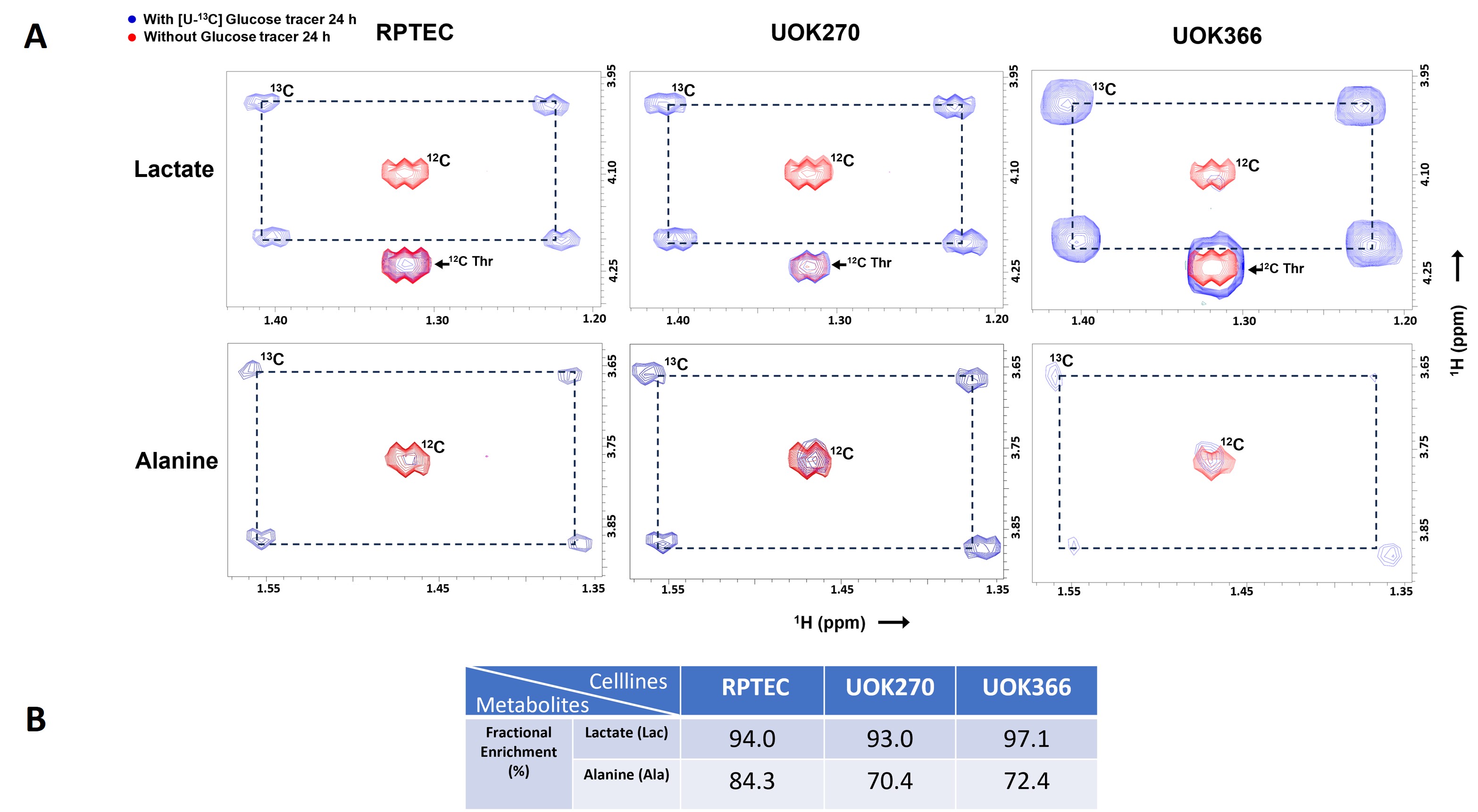
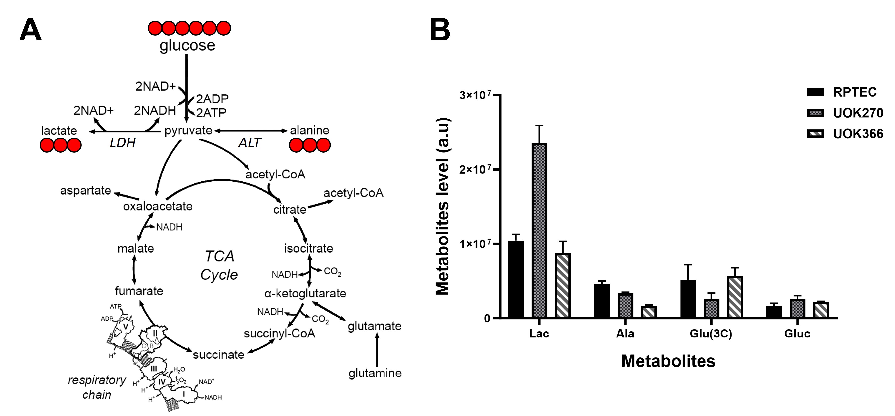
We determined the 13C fractional enrichment of lactate and alanine from [U-13C]-glucose and observed above 90 % fractional enrichments for lactate in all three cells whereas the alanine fractional enrichment was different, showing 84.3 %, 70.4 % and 72.4 % in RPTEC, UOK270 and UOK366 respectively (Figure 1). Relative levels of [13-3C]-Lactate, [13-3C]-Alanine, [13-3C]-Glutamate and 13C-glucose were determined from the 1D 1H{13C}-HSQC spectra (Figure 2B). Substantial differences in [13-3C]-Alanine abundance were observed between the two VHL-deficient ccRCC tumor cell lines, suggesting metabolic heterogeneity. Notably, significant labeling of [13-3C]-Glutamate was observed, demonstrating entry of glucose into the TCA cycle in these cells.Discussion
Our data indicate a significant metabolic heterogeneity in two ccRCC tumor cell lines derived from hereditary VHL patients. Differing amounts and fractional enrichments of 13C-lactate and 13C-alanine from glucose suggest that these two tumor cell lines divert different amounts of glucose-derived carbon to the lactate dehydrogenase and alanine aminotransferase reactions. Further work including mass spectrometry as well as NMR analysis of lipid biosynthesis in these cell lines will provide a more complete picture of the metabolic underpinnings of ccRCC caused by loss of function of the VHL gene.Acknowledgements
This work was supported by the Intramural Research Program of the National Cancer InstituteReferences
1. Linehan, W.M., et al., The Metabolic Basis of Kidney Cancer. Cancer Discov, 2019. 9(8): p. 1006-1021.
2. Crooks, D.R., T.W. Fan, and W.M. Linehan, Metabolic Labeling of Cultured Mammalian Cells for Stable Isotope-Resolved Metabolomics: Practical Aspects of Tissue Culture and Sample Extraction. Methods Mol Biol, 2019. 1928: p. 1-27.
Figures