0843
The feasibility of APT and IVIM in renal malignancies: A Preliminary Study1Department of Radiology, Xi'an GaoXin Hospital, Xi'an, China, 2Philips Healthcare, Beijing, China
Synopsis
Keywords: Kidney, Kidney, Amide proton transfer; renal malignancies; Intravoxel incoherent motion
Motivation: Amide proton transfer (APT) imaging studies are gradually expanding to the body, with few applications in renal diseases and a lack of tumor studies.
Goal(s): In the present study, we aimed to investigate the ability of APT and voxel-wise intravoxel incoherent motion (IVIM) imaging to detect renal malignancies.
Approach: We preliminarily analyzed the ability of APT imaging to discriminate malignant tumors from normal renal tissues, combined with each parameter of IVIM imaging, which has high diagnostic efficacy.
Results: The results showed that APT imaging was feasible and superior to IVIM imaging in discriminating renal malignant tumors from normal renal tissues.
Impact: APT imaging has potential clinical applications in diagnosing of renal malignancies and provides a reference for subsequent accurate studies of APT imaging of renal tumors.
Introduction
Amide proton transfer (APT) imaging can noninvasively reflect the distribution of proteins and peptides within a lesion and provide more detailed information for the diagnosis and treatment of diseases [1]. Currently, APT imaging has been applied to glioma, prostate cancer, rectal cancer and other fields [2-4]. However, APT imaging has been rarely used in the kidney and lacks studies in oncology. Intravoxel incoherent motion (IVIM) has been widely used in renal tumor diagnosis as a special diffusion-weighted imaging. In this study, we investigated the ability of APT and IVIM imaging to detect renal malignancies.Methods
This retrospective study included nineteen patients with surgically confirmed renal malignancies, encompassing 17 cases of clear cell renal cell carcinoma (ccRCC), one papillary renal cell carcinoma, and one cystic renal cell carcinoma. All patients underwent conventional T2WI, APT and IVIM imaging scans. APT scanning was performed with breath-holding mode to observe the APT signal characteristics of the solid mass component. The APT values, diffusion coefficient(D), perfusion-related diffusion coefficient fraction(D*) and perfusion fraction (f) values of the solid component of the mass and the normal tissues on the APT and IVIM images were evaluated. An independent sample t-test was utilized to ascertain the statistical differences between tumor and normal tissues' parameter values. The diagnostic performance of APT and D* values was appraised using Receiver Operating Characteristic (ROC) curve analysis. The DeLong test was employed to evaluate the statistical differences between the areas under the ROC curves (AUC).Results
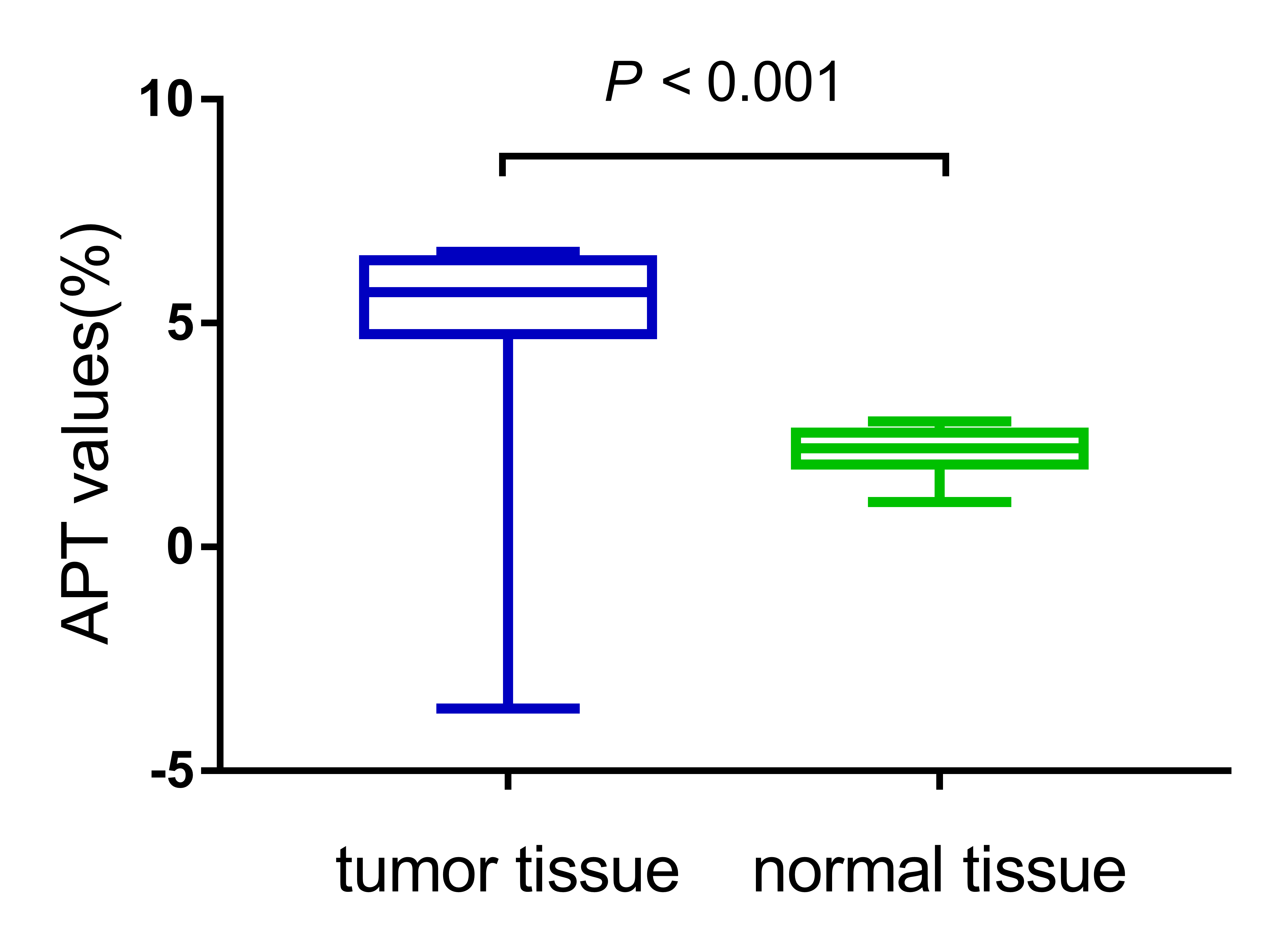
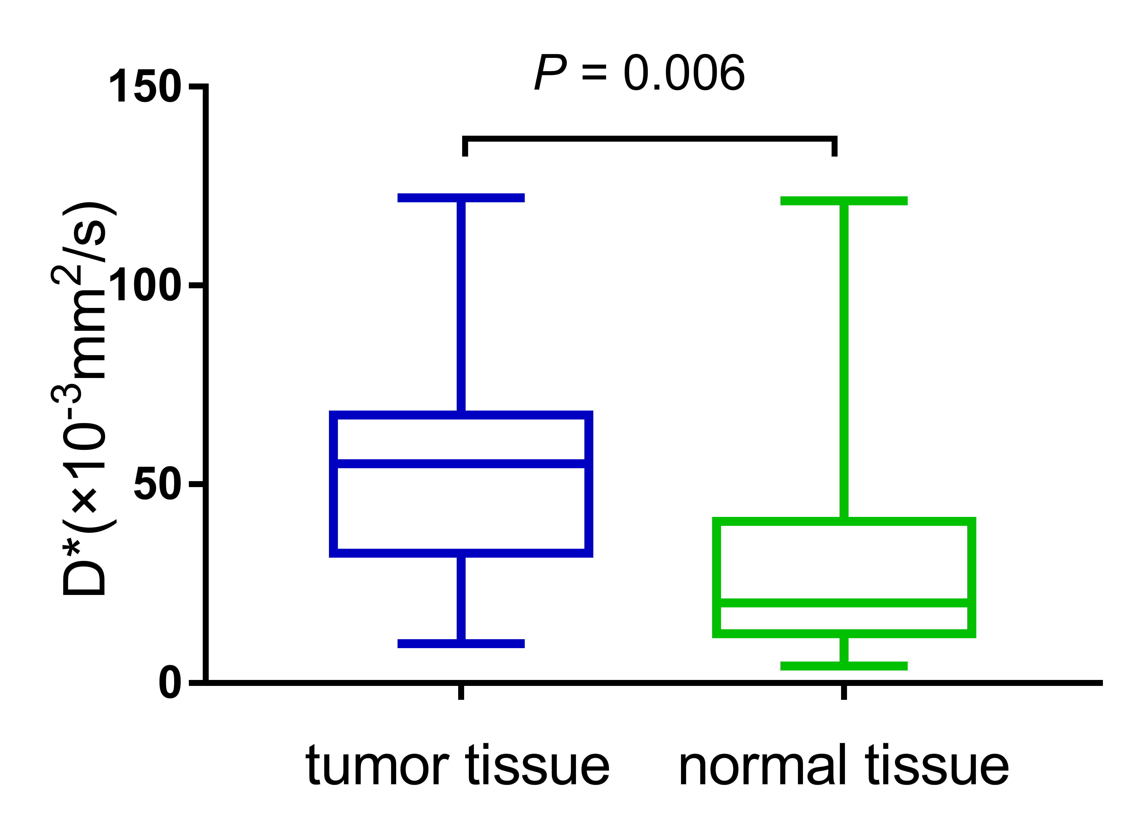
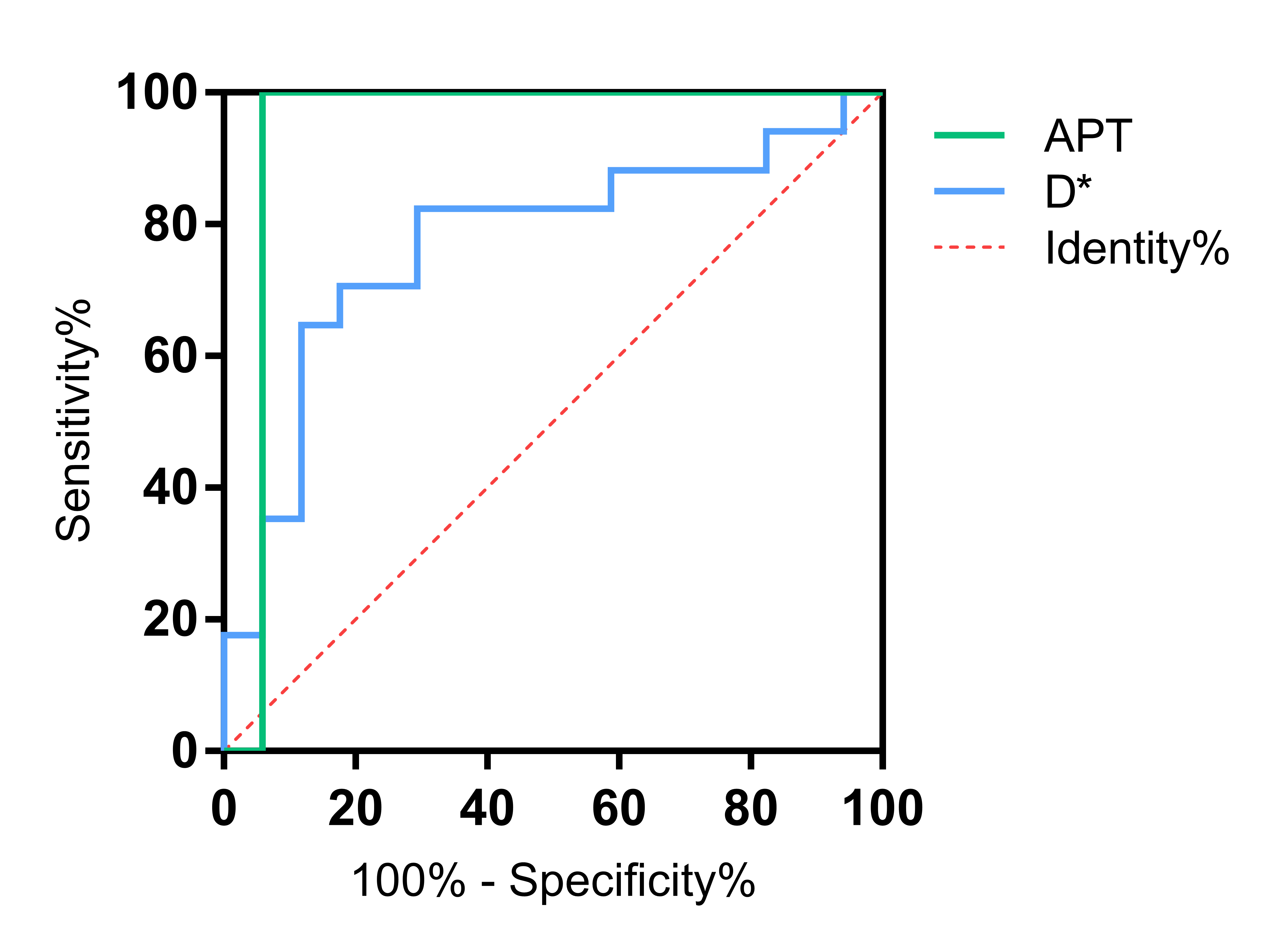
APT values of tumor tissues (4.93% ± 2.41%) were significantly higher than those of normal tissues (2.15% ± 0.47%) (P < 0.001) (Figure 1); D* values of tumor tissues (53.27 ± 23.44×10⁻³mm2/s) were higher than those of normal tissues (29.91 ± 30.05×10⁻³mm2/s) (P < 0.05) (Figure 2). ROC curve analysis showed that APT values had the highest efficacy in discriminating renal malignant and normal tissues (AUC = 0.941), superior to D* (AUC = 0.772) (Figure 3). However, Delong's test showed no statistically significant difference (P = 0.118).Discussion
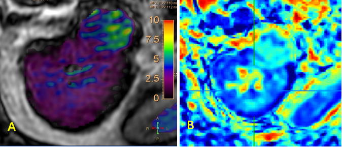
The APT value of tumor tissues in this study was significantly higher than that of normal tissues(Figure 4), indicating that the content of free proteins and peptides in malignant tumor tissues is significantly higher than that in normal tissues, and peptide content of malignant tumor tissues is significantly higher than that of normal tissues, and their cells are metabolically active and produce a large number of proteins. Previous studies have also shown that the APT values of tumor tissues of prostate cancer patients were significantly higher than those of normal tissues [2]. In addition, this result may also be due to tumor angiogenesis, which produces proteins, peptides, and other substances, and is presumed to be the reason why the D* values of tumor tissues are higher than those of normal tissues, which can be used to reflect the degree of tissue vascular proliferation noninvasively.Conclusion
APT and IVIM imaging techniques are feasible for detecting renal malignant tumors, and the diagnostic efficacy of APT imaging is higher compared with IVIM, which can provide crucial clinical reference value.Acknowledgements
NoReferences
1. He YL, Li Y, Lin CY, et al. Three-dimensional turbo-spin-echo amide proton transfer-weighted mri for cervical cancer: A preliminary study[J]. J Magn Reson Imaging, 2019, 50(4): 1318-1325. DOI:10.1002/jmri.26710.
2. Jia G, Abaza R, Williams JD, et al. Amide proton transfer MR imaging of prostate cancer: a preliminary study[J]. J Magn Reson Imaging, 2011, 33(3): 647-654. DOI:10.1002/jmri.22480.
3. Xu Z, Ke C, Liu J, et al. Diagnostic performance between MR amide proton transfer (APT) and diffusion kurtosis imaging (DKI) in glioma grading and IDH mutation status prediction at 3 T[J]. Eur J Radiol, 2021, 134: 109466. DOI:10.1016/j.ejrad.2020.109466.
4. Chen W, Li L, Yan Z, et al. Three-dimension amide proton transfer MRI of rectal adenocarcinoma: correlation with pathologic prognostic factors and comparison with diffusion kurtosis imaging[J]. Eur Radiol, 2021, 31(5): 3286-3296. DOI:10.1007/s00330-020-07397-1.
Figures