0832
Common coordinate framework of neonate macaque brain based on ultra-high-resolution diffusion MRI1Department of Radiology, Children's Hospital of Philadelphia, Philadelphia, PA, United States, 2Department of Bioengineering, University of Pennsylvania, Philadelphia, PA, United States, 3Department of Anatomy and Neurobiology, Shandong University, Jinan, China, 4Department of Radiology, University of Pennsylvania, Philadelphia, PA, United States, 5Department of Neuroscience, University of Wisconsin-Madison, Madison, WI, United States, 6Department of Neurology, University of California San Francisco, San Francisco, CA, United States
Synopsis
Keywords: Normal Development, Normal development, large animals-nonhuman primates, ultra-high resolution, diffusion MRI, common coordinate framework
Motivation: Macaque brain structures change dramatically from birth to adulthood. However, there is no neonate macaque brain common coordinate framework (CCF) serving as neuroanatomical reference for mapping genetic, cellular, and molecular information.
Goal(s): To establish an ultra-high-resolution CCF for macaque brain at birth.
Approach: We acquired ultra-high resolution diffusion MRI (dMRI) of neonate macaque brain and annotated fine neuroanatomical structures and investigated white matter tract development from neonate to adult macaque through dMRI-based tractography.
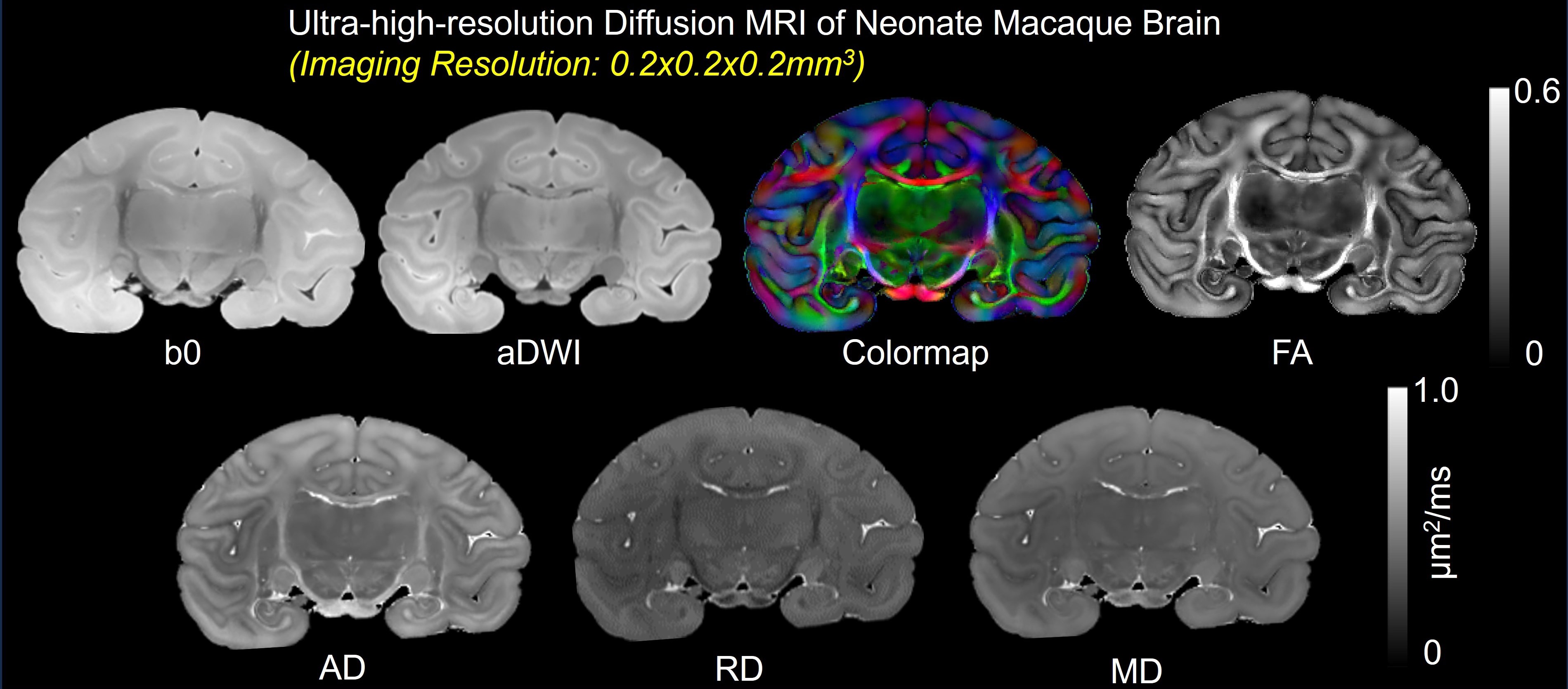
Results: The established neonate macaque brain CCF is featured with 0.2mm isotropic ultra-high diffusion imaging resolution, comprehensive gray and white matter labels, and a coordinate framework.
Impact: This first neonate macaque brain CCF with 0.2mm isotropic ultra-high diffusion imaging resolution serves as neuroanatomical reference, enables mapping genetic, cellular, and molecular information, and provides image templates, laying the foundation for the brain development and evolution discoveries.
Introduction
Macaque brain structures change dramatically from birth to adulthood. However, there is no neonate macaque brain common coordinate framework (CCF) serving as neuroanatomical reference for mapping genetic, cellular, and molecular information. A common coordinate framework (CCF) is critical for accurate anatomical atlas mapping, facilitating the integration of microstructural, cellular, genetic, functional, and neurophysiological data. However, there has not been reported ultra-high-resolution developmental CCF for macaque brain. We hypothesized that significant and selective white matter development occurs from neonate to adult macaque brain. In this study, we aimed to create CCF and compare the 3D pathways of corresponding white matter tracts between neonate and adult macaque brains using DTI tractography. The comparison was conducted in all six categorized functional tract groups.Methods
Acquisition of ultra-high-resolution dMRI data of neonate postmortem macaque brainA Bruker 9.4T scanner was used for ultra-high-resolution dMRI data acquisition. A 2D spin echo diffusion sequence was used. DMRI parameters were b = 1500 s/mm2, 30 unique gradient directions, TE = 35ms, TR = 7.1s, FOV = 54 × 68 mm2, imaging matrix = 266 × 340 ×172 for an imaging resolution of 0.2×0.2×0.2 mm3. Four repetitions were performed with a total imaging time of 48 hours.
Acquisition of dMRI data of young adult postmortem macaque brain
A Bruker 4.7T scanner was used in 8 linearly independent directions with b value 1000 s/mm2 was used. DWI parameters were TE = 32.5ms, TR = 0.7s, FOV = 78 x 56 mm2, imaging matrix = 200 × 108 × 108 for a nominal resolution of 0.39×0.52×0.54mm3. Two repetitions were performed with a total imaging time of 45 hours.
Diffusion tensor imaging (DTI) processing
Marcenko-Pastur principal component analysis (PCA) denoising1 was performed on the raw dMRI data with patch radius=2. Automated image registration (AIR)2 was conducted on raw DWIs. The tensor fitting was conducted with DtiStudio3.
Creation of CCF
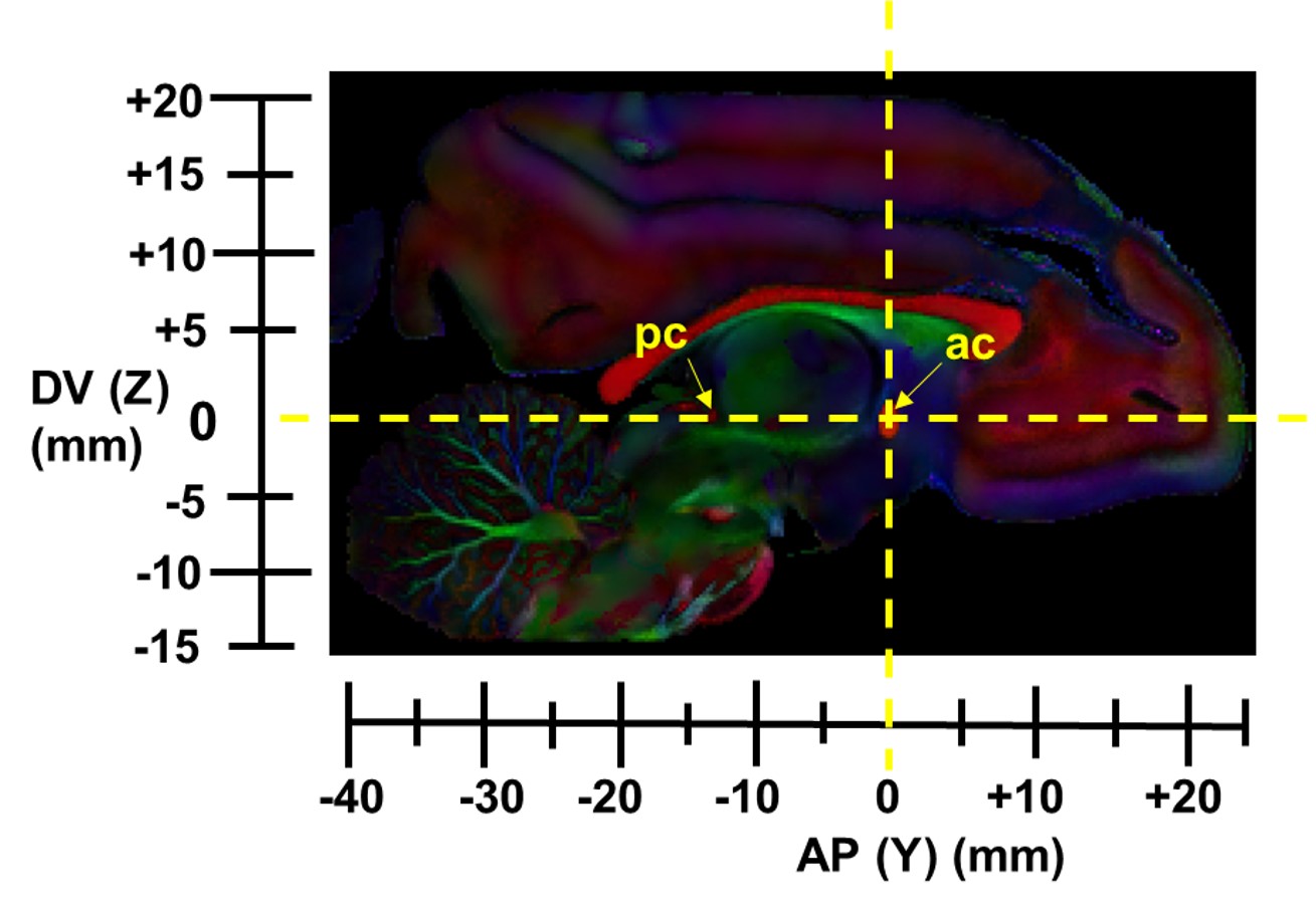
The origin of the CCF is chosen at the mid-sagittal plane of the anterior commissure (ac). A line connecting the posterior commissure and ac in the medial sagittal plane is defined as the anterior posterior (AP), and the dorsal-ventral (DV) is perpendicular to the AP axis in the mid-sagittal plane (Fig. 2). Manual delineation of brain regions was performed by an experienced neuroanatomist based on DTI-derived contrasts and a high resolution dMRI based adult macaque atlas4.
DTI tractography in six tract groups of macaque white matter tracts
Streamline tractography5 was used for DTI fiber tracking of all ex-vivo neonate and young adult macaque brain DTI data. The tractography protocol for tracing six categories of white matter (WM) tracts in human brain6 was also used to trace tracts in the macaque brains.
Results
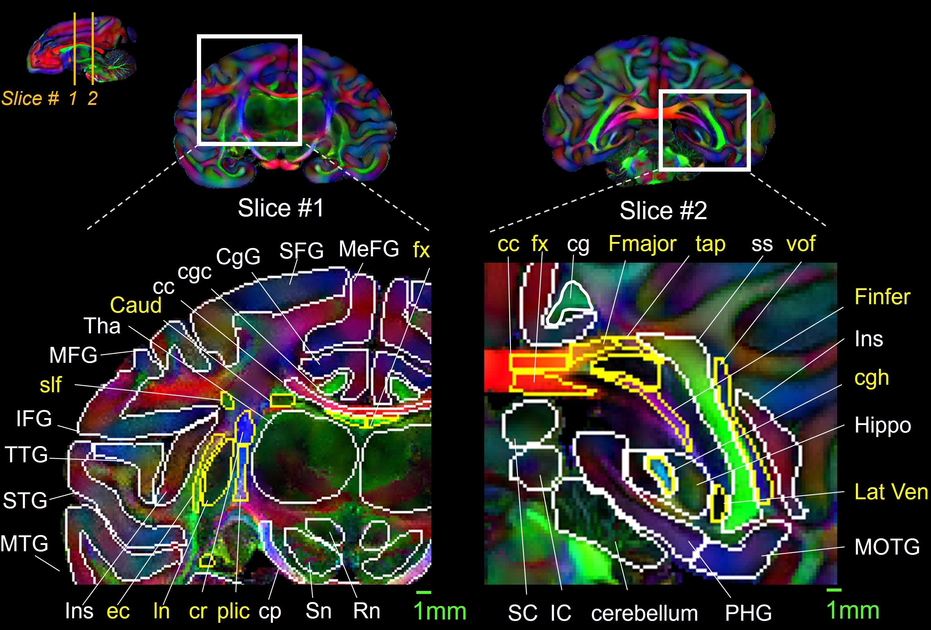
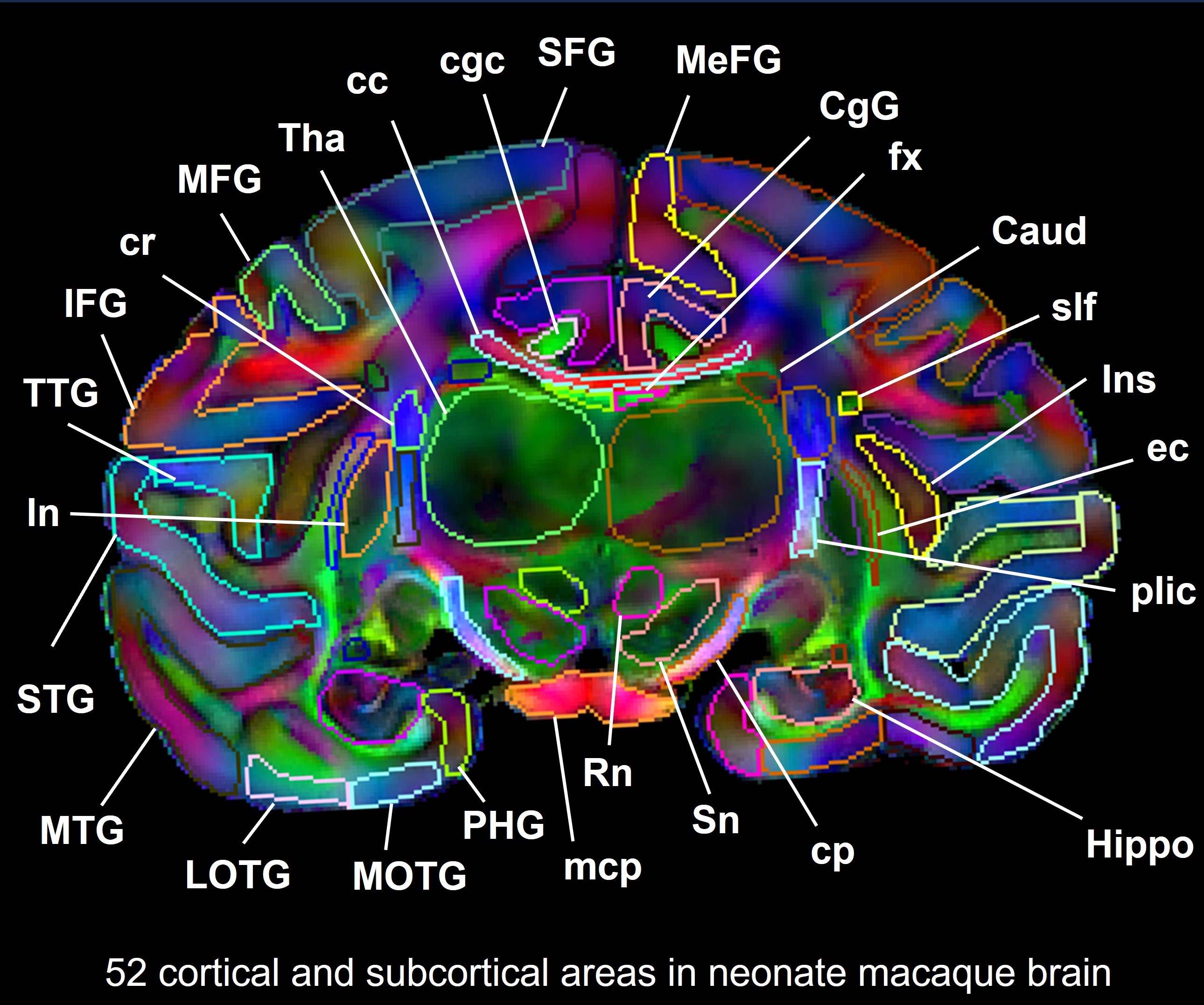
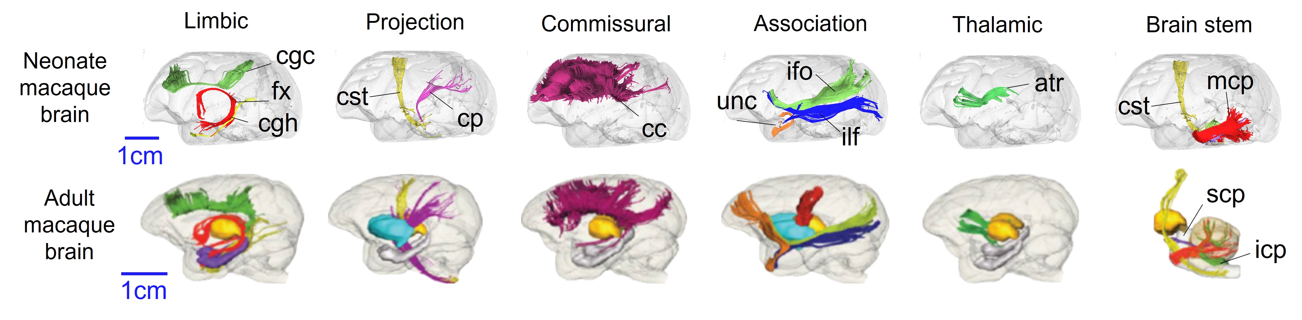
DTI derived maps from the first ultra-high resolution (0.20x0.20x0.20 mm3) dMRI dataset for a neonate macaque is shown in Fig 1. In this dataset, we defined the first developmental macaque CCF (Fig. 2) in which the brain is conveniently oriented for the registration of 2D histological, cellular, and genetic data. Representative slices from our developmental atlas are shown in Fig. 3 to delineate fine adjacent WM structures such as plic (posterior limb of internal capsule), and cr (corona radiation) (left panel). We annotated 52 cortical and subcortical areas in neonate macaque brain in Fig. 4. DTI tractography results show distinctive WM tract development from neonate to adult macaque brain in the association, limbic, and projections tract groups, whereas commissural, thalamic, and brain stem tract groups are consistent (Fig. 5). The uncinate fasciculus (unc), crucial for processing novel information7, does not extend far into the prefrontal lobe in the neonate macaque brain compared to adults. Similarly, in the neonate macaque brain, the cingulum in the cingulate gyrus (cgc), a component of limbic system communication, does not reach into the superior parietal lobe, but it is clearly delineated in adults. Moreover, the cerebral peduncle (cp), which plays a role in coordinating bodily functions, exhibits a reduced quantity in the neonate compared to adults.Discussion and conclusion
We have built the first neonate macaque brain CCF based on ultra-high-resolution diffusion MRI. The labels of fine neuroanatomical structures revealed detailed information of cortical gyri, subcortical nuclei, and WM tracts in neonate macaque brain. The tracing of WM tracts from neonate and adult macaque brain demonstrates distinctive changes of WM tracts in limbic, commissural, thalamic, brain stem, projection, and association tract groups. This first neonate macaque brain CCF with 0.2mm isotropic ultra-high diffusion imaging resolution serves as neuroanatomical reference, enables mapping genetic, cellular, and molecular information, and provides image templates, laying the foundation for the brain development and evolution discoveries.Acknowledgements
This study is funded by NIH R01MH092535, R01MH125333, R01EB031284, R01MH129981, R21EB009545, R21MH123930, UM1MH130991 and P50HD105354.References
1. Veraart J, Fieremans E, Novikov DS. Diffusion MRI noise mapping using random matrix theory. Magn Reson Med. 2016;76(5):1582-93.
2. Woods RP, Grafton ST, Holmes CJ, Cherry SR, Mazziotta JC. Automated image registration: I. General methods and intrasubject, intramodality validation. J Comput Assist Tomogr. 1998;22(1):139-52.
3. Jiang H, van Zijl PC, Kim J, Pearlson GD, Mori S. DtiStudio: resource program for diffusion tensor computation and fiber bundle tracking. Comput Methods Programs Biomed. 2006;81(2):106-16.
4. Feng L, Jeon T, Yu Q, Ouyang M, Peng Q, Mishra V, et al. Population-averaged macaque brain atlas with high-resolution ex vivo DTI integrated into in vivo space. Brain Struct Funct. 2017;222(9):4131-47.
5. Mori S, Crain BJ, Chacko VP, van Zijl PC. Three-dimensional tracking of axonal projections in the brain by magnetic resonance imaging. Ann Neurol. 1999;45(2):265-9.
6. Wakana S, Jiang H, Nagae-Poetscher LM, van Zijl PC, Mori S. Fiber tract-based atlas of human white matter anatomy. Radiology. 2004;230(1):77-87.
7. Olson IR, Von Der Heide RJ, Alm KH, Vyas G. Development of the uncinate fasciculus: Implications for theory and developmental disorders. Dev Cogn Neurosci. 2015;14:50-61.
Figures