0830
Inhomogeneous Magnetization Transfer Imaging in Extremely Preterm Neonates at 7T.1Department of Neonatology, University Medical Center Utrecht, Utrecht, Netherlands, 2Department of Radiology, University Medical Center Utrecht, Utrecht, Netherlands, 3Department of Radiology, Leiden University Medical Center, Leiden, Netherlands, 4Philips Healthcare, Best, Netherlands
Synopsis
Keywords: Neonatal, Neonatal, Neuro; Pediatrics; High-Field-MRI
Motivation: Extremely preterm neonates often show myelination delay in the brain, which is associated with long-term neurodevelopmental outcome impairments. Traditional imaging lacks myelin specificity, therefore, we implemented inhomogeneous magnetization transfer (ihMT).
Goal(s): This study explores the use of ihMT at 7 Tesla for myelin assessment in extremely preterm neonates.
Approach: The ihMT data was acquired from a phantom, demonstrating its specificity for myelin content. Six neonates and five adults were scanned with ihMT, and an ROI-based analysis was performed.
Results: Phantom and human data confirm ihMT's potential for myelin evaluation. As expected, neonates exhibit lower ihMTR values in key brain regions compared to adults.
Impact: This study is an important first step in discovering myelin development in the extremely preterm neonatal brain. Differences in myelin development across the extremely preterm population could be used to predict long-term neurodevelopmental outcome in the future.
Introduction
Extremely preterm (EPT) neonates are born before 28 weeks of gestation. From around 28 weeks of gestation, myelin is formed by the maturation of pre-oligodendrocytes (pre-OLs) to myelinating oligodendrocytes (OLs)1,2. The development of myelination ascends from the lower brain regions through the subcortical structures towards the cortex1. EPT neonates are born when pre-OLs are at maximum vulnerability. Additionally, EPT neonates often become subject to many medical complications. This is causing pre-OL death and dysmaturation, leading to a delay in myelination2, which is associated with a decrease in cognitive performance later in life3,4.Early myelination evaluation in EPT neonates is typically conducted using T1-weighted images or DTI measures, serving as proxies for brain development, but lacking specificity for myelin content. In contrast, inhomogeneous magnetization transfer (ihMT) imaging offers a myelin-specific approach by capitalizing on non-motion averaged dipolar couplings in the myelin phospholipid bilayer, resulting in a long dipolar order relaxation time (T1d)5. The ihMT approach has been used to characterize demyelinating conditions like multiple sclerosis in adults6.
Previously, we showed that it is safe and feasible to scan neonates at 7T7,8. In this study, we take advantage of the ultrahigh field and show the feasibility of ihMT in neonates at 7 Tesla (T).
Methods
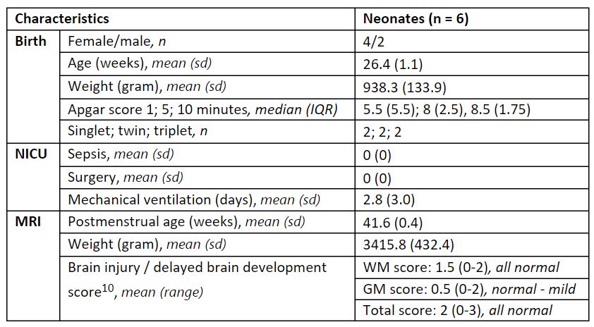
We acquired ihMT data in a phantom, six EPT neonates (Table 1), and five healthy adults (3/2 male/female; 22.5-29.3 years, all born at term age) on a 7T MRI scanner (Philips Healthcare, Best, The Netherlands), using a 2Tx/32Rx head coil (Nova Medical, Wilmington, MA).The phantom contained eight boxes filled with three solutions: NaCl, hair conditioner (TRESemmé®) and NaCl + Gadolinium (Gd) (Figure 1A). Hair conditioner was chosen as a myelin proxy due to its similar molecular and lamellar structure9. EPT neonates were scanned at term-equivalent age, at the 7T MRI directly after their clinical 3T MRI scan, without the use of sedation. If necessary, feeding was given to promote natural sleep. The presence of brain injury or growth delay was determined based on their 3T MRI scan10.
Data Acquisition
A pulsed ihMT-TFE sequence was implemented using 100 Hyperbolic-Secant pulses with an inter-pulse-gap of 0.45 ms and a B1 of 14µT. Scan parameters were: off-resonance frequency (ΔkHz): 10 kHz; FOV: 255x255x35 mm; voxel size: 2.5x2.5x5 mm; flip angle (FA): 7°. Additionally, a T1 map (MP2RAGE) was acquired.
Data Processing
The five ihMT images (S0, S+, S-, 2xS±), were spatially normalized using affine edge registration (Elastix toolbox11,12). Thereafter, the ihMT-ratio (ihMTR) was calculated as:
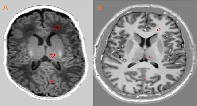
The mean ihMTR was calculated in three regions of interest (ROIs) per subject: the thalamus region of the deep gray matter (DGM), the frontal white matter (WM), and the occipital cortical gray matter (CGM) (Figure 2).
Results
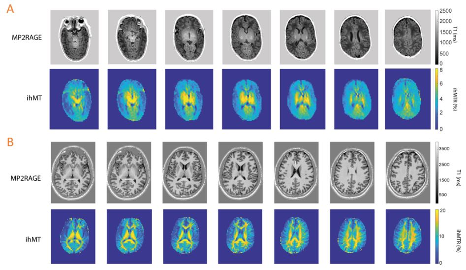
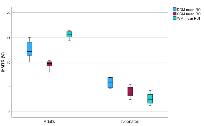
Data from the phantom shows that ihMTR is specific for hair conditioner (i.e., a proxy for myelin) and not dominated by T1 relaxation, since the addition of Gd did not change the contrast (Figure 1).Figure 3 shows an example of the ihMTR in a neonate and adult. The ihMTR values in adults were highest in the WM (15.5±0.8%), followed by the DGM (12.5±2.0%) and the CGM (9.5±0.9%). The ihMTR values in neonates were highest in the DGM (5.9±1.0%), followed by the CGM (3.9±1.4%) and the WM (2.6±1.1%). When comparing ihMTR values between adults and neonates, the ihMTR value in adults was 52.8% higher in the DGM, 58.4% higher in the CGM and 83.2% higher in the WM (Figure 4).
Discussion
It is feasible to perform ihMT sequence scans on neonates at 7T, and the ihMTR contrast is not dominated by T1-differences. As expected, ihMTR values are higher in adults than in neonates, as myelination is far from complete in neonates1. In adults, the ihMTR values are highest in the WM, while in neonates the ihMTR values are highest in the DGM. This observation is consistent with the ascending pattern of myelination in the neonatal brain, whereby subcortical structures, such as the thalamus, tend to undergo myelination earlier than WM regions1.7T MRI often exhibits non-uniform B1+ patterns, which were not corrected for in our study. However, due to the use of adiabatic saturation pulses, we anticipated a greater impact on regions with lower B1+ strengths, like the outer brain regions, with less effect on central areas where the adiabatic condition is met.
We plan to expand our study by including EPT neonates with brain damage and a growth delay. This broader sample will help us better understand how myelin development correlates with neurodevelopmental outcomes, possibly serving a predictive marker in the future.
Acknowledgements
We would like to thank all neonates and their parents, and volunteers who participated in this study. Also, we gratefully acknowledge funding from the Wilhemina Children's Hospital fund 2021 Wiegers for this study.References
- Yakovlev, P. I., Lecours, A. R., Minkowski, A., & Davis, F. A. (1967). Regional development of the brain in early life.
- Volpe, J. J. (2019). Dysmaturation of premature brain: importance, cellular mechanisms, and potential interventions. Pediatric neurology, 95, 42-66.
- Vandewouw, M. M., Young, J. M., Shroff, M. M., Taylor, M. J., & Sled, J. G. (2019). Altered myelin maturation in four year old children born very preterm. NeuroImage: Clinical, 21, 101635.
- Corrigan, N. M., Yarnykh, V. L., Huber, E., Zhao, T. C., & Kuhl, P. K. (2022). Brain myelination at 7 months of age predicts later language development. Neuroimage, 263, 119641.
- Alsop, D. C., Ercan, E., Girard, O. M., Mackay, A. L., Michal, C. A., Varma, G., & Duhamel, G. (2023). Inhomogeneous magnetization transfer imaging: Concepts and directions for further development. NMR in Biomedicine, 36(6), e4808.
- Van Obberghen, E., Mchinda, S., Le Troter, A., Prevost, V. H., Viout, P., Guye, M., & Duhamel, G. (2018). Evaluation of the sensitivity of inhomogeneous magnetization transfer (ihMT) MRI for multiple sclerosis. American Journal of Neuroradiology, 39(4), 634-641.
- Annink, K. V., Van Der Aa, N. E., Dudink, J., Alderliesten, T., Groenendaal, F., Lequin, M., & Benders, M. J. N. L. (2020). Introduction of ultra-high-field MR imaging in infants: preparations and feasibility. American Journal of Neuroradiology, 41(8), 1532-1537.
- Van Ooijen, I. M., Annink, K. V., Benders, M. J. N. L., Dudink, J., Alderliesten, T., Groenendaal, F., & van der Aa, N. E. (2023). Introduction of ultra-high-field MR brain imaging in infants: vital parameters, temperature and comfort. Neuroimage: Reports, 3(2), 100175.
- Malyarenko, D. I., Zimmermann, E. M., Adler, J., & Swanson, S. D. (2014). Magnetization transfer in lamellar liquid crystals. Magnetic Resonance in Medicine, 72(5), 1427-1434.
- Kidokoro, H., Neil, J. J., & Inder, T. E. (2013). New MR imaging assessment tool to define brain abnormalities in very preterm infants at term. American Journal of Neuroradiology, 34(11), 2208-2214.
- Klein, S., Staring, M., Murphy, K., Viergever, M. A., & Pluim, J. P. (2009). Elastix: a toolbox for intensity-based medical image registration. IEEE transactions on medical imaging, 29(1), 196-205.
- Shamonin, D. P., Bron, E. E., Lelieveldt, B. P., Smits, M., Klein, S., Staring, M., & Alzheimer's Disease Neuroimaging Initiative. (2014). Fast parallel image registration on CPU and GPU for diagnostic classification of Alzheimer's disease. Frontiers in neuroinformatics, 7, 50.
Figures
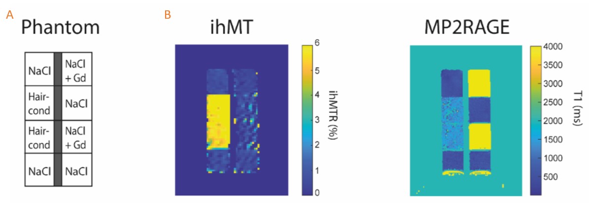
Gd: gadolinium; NaCl: natrium chloride; ihMTR: inhomogeneous magnetization transfer ratio; MP2RAGE: magnetization prepared 2 rapid gradient echoes.