0828
High-resolution Susceptibility Weighted Fetal Brain using ZOOMit EPI and Slice to Volume reconstruction1Computational Radiology Laboratory, Boston Children's Hospital, and Harvard Medical School, Boston, MA, United States, 2Massachusetts General Hospital and Harvard Medical School, Boston, MA, United States
Synopsis
Keywords: Fetal, Fetus
Motivation: Susceptibility weighted imaging is of great interest in fetal MRI. Conventional single-shot EPI often suffers from field distortion and motion artifacts due to long readout.
Goal(s): To develop a susceptibility-weighted isotropic fetal brain imaging technique with reduced artifacts.
Approach: ZOOMit EPI was used for data acquisition. A slice-to-volume reconstruction was further employed to correct the motion between slices and reconstruct an isotropic high-resolution volume of the fetal brain.
Results: Zoomit EPI produces images with reduced artifacts due to reduced acquisition time. The slice-to-volume reconstruction further corrects the motion between slices and reconstruct an isotropic high-resolution SWI-weighted volume of the fetal brain.
Impact: Zoomit EPI offers faster scan times and produces images with reduced artifacts. With Zoomit EPI, high-resolution SWI-weighted isotropic fetal brain imaging has been achieved using slice-to-volume reconstruction.
Introduction
Strokes affect neurovascular structures, especially the different brain vessels, and lead to brain injury. Fetuses suffering from stroke in utero are at a higher risk of neurological and motor disorders, and severe cognitive impairment. Accurate delineation of the neurovascular structures using MRI, especially susceptibility-weighted imaging (SWI) has the potential to enable early diagnosis and prediction of disease progression [1-4]. SWI is often achieved using an EPI-based Gradient-echo readout. While commonly used, the long readout makes EPI sensitive to motion and field distortion artifacts, especially for fetal scans. ZOOMit is an emerging technique that enables imaging of objects at high spatial resolution under reduced field of view (FOV) [5-7]. This results in faster scan times and thus could produce images with reduced artifacts. For fetal imaging, the resolution of the acquired data may still not be sufficient for accurately delineating the structure of the vessels. To address this issue, we further employ a slice-to-volume reconstruction pipeline to correct the motion between slices and reconstruct an isotropic high-resolution volume of the fetal brain.Methods
The fetal EPI data was acquired on Siemens 3T Trio/Prisma scanners by imaging pregnant women after obtaining their informed consent. Data was acquired using a 46-channel phased-array cardiac coil without maternal breath-hold. Six datasets were used in the study. The scan protocol consisted of multiple acquisitions in the fetal axial, coronal, and sagittal planes using a ZOOMit MR sequence with the following scan parameters: TR values ranging from 3.02 to 13.44 s, TE values ranging from 49 to 105 ms, FOV between 154x52 and 220x96 mm2 with matrix size between 256x48 and 256x104, and slice thickness between 2 and 3.5 mm. For comparison, conventional EPI was performed on the same subject with typical parameters: TR=10 s, TE=81 ms, slice thickness 2 mm, FoV = 256x256 mm2 with a matrix size of 250x250. Slice-to-volume reconstruction (SVR)The stack of 2D slices corresponding to every acquisition was passed through the SVR pipeline [8] to correct for motion between slices and reconstruct a super-resolved volume with isotropic resolution.Results
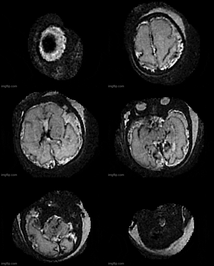
Figure 1 shows a comparison of conventional EPI (left) and ZOOMit EPI (right) for susceptibility-weighted imaging of two fetal brains (top: axial plane, bottom: coronal plane). With a reduced acquisition time, ZOOMit EPI generates images with visually less distortion artifacts than conventional EPI. Figure 2 presents all three image orientations (axial, sagittal, coronal) of the SVR reconstructions for conventional EPI and ZOOMit EPI, respectively. In line with Figure 1, ZOOMit EPI-based SVR reconstruction shows a visually much better image quality than conventional EPI. Figure 3 demonstrates an axial view of slice-to-volume reconstructed high-resolution isotropic images for the subject presented in Figure 2.Discussion and Conclusion
Initial results demonstrate high-resolution isotropic fetal SWI imaging can be achieved via ZOOMit EPI and slice-to-volume reconstruction. Although visually ZOOMit EPI shows a better image quality than conventional EPI, more quantitative evaluations are needed to confirm the above findings.Acknowledgements
No acknowledgement found.References
1. Gholipour, A., Estroff, J. A., Barnewolt, C. E., Robertson, R. L., Grant, P. E., Gagoski, B., Warfield, S. K.,Afacan, O., Connolly, S. A. & Neil, J. J. Fetal MRI: a technical update with educationalaspirations. Concepts in Magnetic Resonance Part A, 2014, 43, 237-266.
2. Kang, D.-W., Jeong, H.-G., Yang, W. & Lee, S.-H. Prediction of Stroke Subtype and RecanalizationUsing Susceptibility Vessel Sign on Susceptibility-Weighted Magnetic Resonance Imaging. Stroke, 2017, 48,1554-1559.
3. Khalil, A. A., Ostwaldt, A.-C., Nierhaus, T., Ganeshan, R., Audebert, H. J., Villringer, K., Villringer, A. &Fiebach, J. B. Relationship Between Changes in the Temporal Dynamics of the Blood-Oxygen-Level-Dependent Signal and Hypoperfusion in Acute Ischemic Stroke. Stroke, 2017, 48, 925-9314.
4. Haacke, E. M., Mittal, S., Wu, Z., Neelavalli, J. & Cheng, Y.-C. Susceptibility-weighted imaging: technicalaspects and clinical applications, part 1. American Journal of Neuroradiology, 2009, 30, 19-30.
5. Feinberg DA, Hoenninger JC, Crooks LE, et al. Inner volume MR imaging: technical concepts and their application. Radiology. 1985;156:743–747.
6. Frahm J, Merboldt K-D, Hänicke W. Localized proton spectroscopy using stimulated echoes. J Magn Reson 1987; 72:502-508.
7. Riffel P, Michaely HJ, Morelli JN, et al. Zoomed EPI-DWI of the pancreas using two-dimensional spatially-selective radiofrequency excitation pulses. PLoS One. 2014; 9:e89468.
8. Uus, A., Zhang, T., Jackson, L., Roberts, T., Rutherford, M., Hajnal, J.V., Deprez, M. Deformable Slice-to-Volume Registration for Motion Correction in Fetal Body MRI and Placenta. IEEE Transactions on Medical Imaging, 2020, 39(9), 2750-2759.
Figures
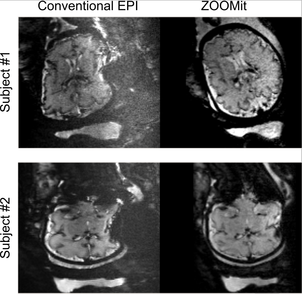
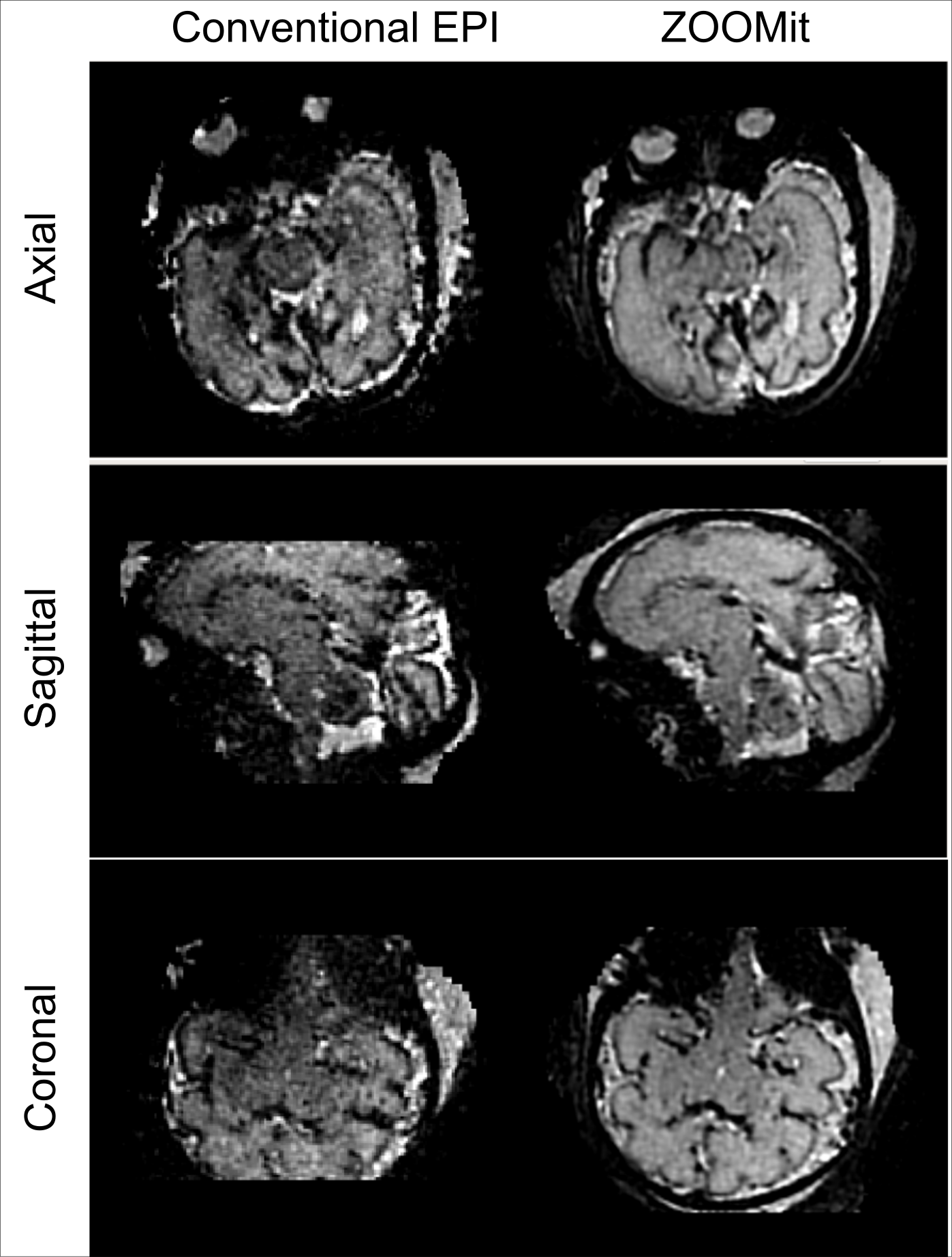
Figure 2. Comparison of slice-to-volume reconstructions based on conventional EPI (left) and ZOOMit EPI (right) for an example subject. All three orientations are presented (from top to bottom: axial, sagittal and coronal). ZOOMit-EPI-based slice-to-volume reconstruction demonstrates a much better image quality than conventional EPI.