0827
MRI-Based Quantitative Analysis of Placenta and Fetal Brain in SGA Pregnancies: Feasibility Insights1Radiology, International Peace Maternity and Child Health Hospital, Shanghai, China, 2MR Research Collaboration, Siemens Healthineers, Shanghai, China, 3School of Biomedical Engineering, Shanghai Jiao Tong University, Shanghai, China, 4The National Engineering Research Center of Advanced Magnetic Resonance Technologies for Diagnosis and Therapy (NERC-AMRT), Shanghai Jiao Tong University, Shanghai, China, 5the School of Information Science and Technology, ShanghaiTech University, Shanghai, China
Synopsis
Keywords: Prenatal, Prenatal
Motivation: Understanding placental dysfunction's role in small-for-gestational-age (SGA) fetal neurodevelopment is crucial; this study seeks to fill that gap, enhancing prenatal care.
Goal(s): The primary goal is to assess whether MRI can effectively quantify the relationship between placental function and fetal brain development in SGA pregnancies.
Approach: The study used IVIM, and T2* mapping to evaluate placental and fetal brain development, applying Pearson correlation and t tests for comparative analysis.
Results: Significant differences in placental perfusion and cortical properties between control and SGA groups were reported, demonstrating the feasibility of using MRI for in-utero assessment.
Impact: This study’s MRI approach could change prenatal care, allowing earlier detection of small-for-gestational age–related brain development issues, prompting interventions, and guiding research into neurodevelopmental support for affected neonates, with potential long-term cognitive benefits.
Introduction
Intravoxel incoherent motion (IVIM) imaging and T2* mapping have garnered increasing attention as tools for quantifying placental perfusion and oxygen content, facilitating the identification of placental dysfunction (1,2). This dysfunction is a crucial contributing factor for small-for-gestational-age (SGA) fetuses, which can subsequently impact neurodevelopmental processes in the fetal brain (3).Currently, the evaluation of brain development of SGA fetuses typically occurs postnatally, with limited research focusing on the correlation between placental function and fetal brain development during the middle and third trimester of pregnancy.Current fetal brain research predominantly relies on volumetric measurements (4). Nevertheless, recent studies have suggested that assessing the geometric properties of cortical development in the fetal brain can provide valuable insights (5).Therefore, this study aimed to incorporate the geometric properties of cortical development to quantitatively analyze the relationship between impaired placental function and fetal brain development in pregnancies with SGA fetuses.Methods
The study included 43 pregnant women with appropriate gestational age, having a mean gestational age of 27.7 weeks (range: 23-38.6 weeks), and 27 pregnant women with SGA pregnancies, having a mean gestational age of 29.3 weeks (range: 23.3-36.3 weeks). After excluding 25 cases with normal fetal brain data but considerable motor artifacts, 29 fetal brains from the normal group (mean age: 27.4 weeks, range: 23-38.6 weeks) and 16 fetal brains from the SGA group (mean age: 28.1 weeks, range: 23.3-33.7 weeks) were included in this study. Examinations were conducted using a 1.5 T MRI system (MAGNETOM Aera; Siemens Healthineers AG, Erlangen, Germany) with an 18-channel body coil. The main scanning sequence and corresponding parameters are listed in Table 1. To assess fetal brain development, various cortical measurements, such as convexity, curvature, sulcal depth, and cortical thickness, were conducted for each participant. SGA was defined as ultrasonically estimated fetal weight below the 10th percentile. In both the control and SGA groups, Pearson correlation analysis was conducted to examine the correlation between cortical surface properties of various brain regions and gestational age, as well as placental function parameters. For continuous variables, the Shapiro-Wilk test for normality and Levene F test for equal variance were performed. If the data were normally distributed and had equal variance, the Student t test was used. Otherwise, the Mann-Whitney U test was performed. A P value <.05 was statistically significant.Results
The FP and T2* values significantly decreased in SGA pregnancies compared with the control group (P <.05), indicating impaired placental growth. In both the control and SGA groups, the FP and T2* values exhibited a negative correlation with gestational age (r = –0.352, P =.021 and r = –0.589, P <.01 in the control group; r = –0.382, P =.0496 and r = –0.63, P <.01 in the SGA group). We selected 7 representative cortical regions, as detailed in Table 2. These brain regions demonstrated a strong correlation with gestational age. In the control group, many indicators demonstrated strong correlations with DP and T2* values, but these correlations were absent in the SGA group. Furthermore, 8 representative cortical surface properties exhibited significant differences (P <.01) between the normal and the SGA groups (Table 3).Discussion/Conclusions
Both T2* and FP values could serve as noninvasive measures for assessing placental dysfunction in vivo, and they played a considerable role in distinguishing the normal group from the SGA group, which was consistent with previous research (1,6). Especially in the middle and superior frontal and temporal gyrus. We identified a correlation between fetal brain development and DP and T2* values in the normal group by comparing geometric properties of cortical development with placental functional indicators. However, this correlation was not observed in the SGA group. This suggested that growth-restricted, chronically hypoxic fetuses tend to redistribute cardiac output preferentially to support brain growth, leading to asymmetric fetal growth or so-called "brain sparing", and the middle and superior frontal and temporal gyrus have high priority for oxygen supply; however, this does not guarantee normal neurodevelopment (3).Moreover, when comparing fetal brains between the normal and SGA groups, we identified substantial differences in specific cortical characteristics, particularly in the orbital part of the middle and superior frontal gyrus and temporal pole (superior temporal gyrus), the brain surface convexity and curvature had greater absolute values in the SGA group compared to the control group. These structural changes may correspond to the development of short- and long-term motor skills, cognitive abilities, memory, and neuropsychological dysfunction observed in school-aged children with fetal growth restriction.This feasibility study demonstrated the potential of MRI in quantitatively evaluating placental function and fetal brain development in SGA fetuses.Acknowledgements
This study received funding from the Shanghai Jiao Tong University Medical Engineering Cross-Fund (YG2023QNB29).References
(1) He J, Chen Z, Chen C, Liu P. Comparative study of placental T2* and intravoxel incoherent motion in the prediction of fetal growth restriction. Placenta. 2021 Aug;111:47-53.(2) He J, Chen Z, Wen T, Xu L, Chen C, Liu P. Utility of placental diffusion-weighted magnetic resonance imaging in prenatal diagnosis of small for gestational age infants and pregnancy outcome prediction. Placenta. 2022 Apr;121:91-98.
(3) Miller SL, Huppi PS, Mallard C. The consequences of fetal growth restriction on brain structure and neurodevelopmental outcome. J Physiol. 2016 Feb 15;594(4):807-23.
(4) Grinberg A, Katorza E, Hoffman D, Ber R, Mayer A, Lipitz S. Volumetric MRI Study of the Brain in Fetuses with Intrauterine Cytomegalovirus Infection and Its Correlation to Neurodevelopmental Outcome. AJNR Am J Neuroradiol. 2019 Feb;40(2):353-358.
(5) Wu J, Sun T, Yu B, Li Z, Wu Q, Wang Y, Qian Z, Zhang Y, Jiang L, Wei H. Age-specific structural fetal brain atlases construction and cortical development quantification for chinese population. Neuroimage. 2021 Nov 1;241:118412.
(6) He J, Chen Z, Chen C, Liu P. Differences in placental oxygenation and perfusion status between fetal growth-restricted and small-for-gestational-age pregnancies: a functional magnetic resonance imaging study. Eur Radiol. 2023 Mar;33(3):1729-1736.
Figures
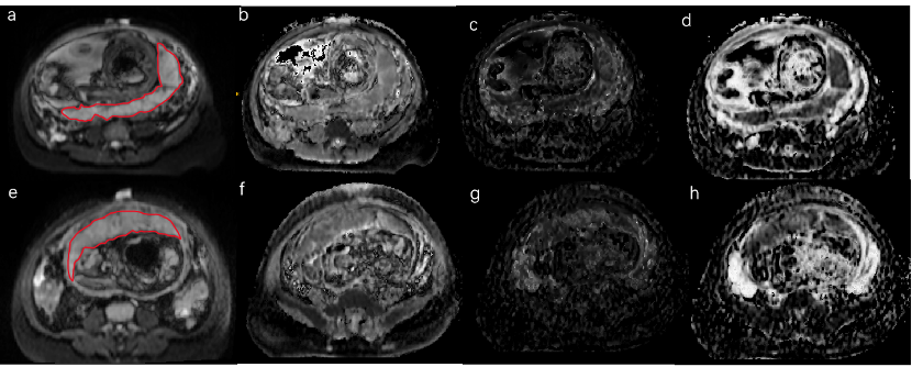
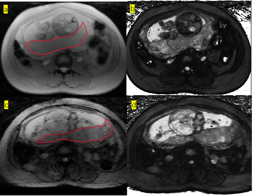
c-d Representative patient in the SGA group(scanned at 27+4 gestation week).Example of ROI on the T2* images,and the measurement of T2* value was 73.4 ms.

Table 1. Main scanning sequences and corresponding parameters
Abbreviations: IVIM=Intravoxel Incoherent Motion; TR = repetition time; TE = echo time; FA = flip angle; FOV = the field of view; T2w SSFSE:T2-weighted half-Fourier acquisition single-shot turbo/fast spin-echo.
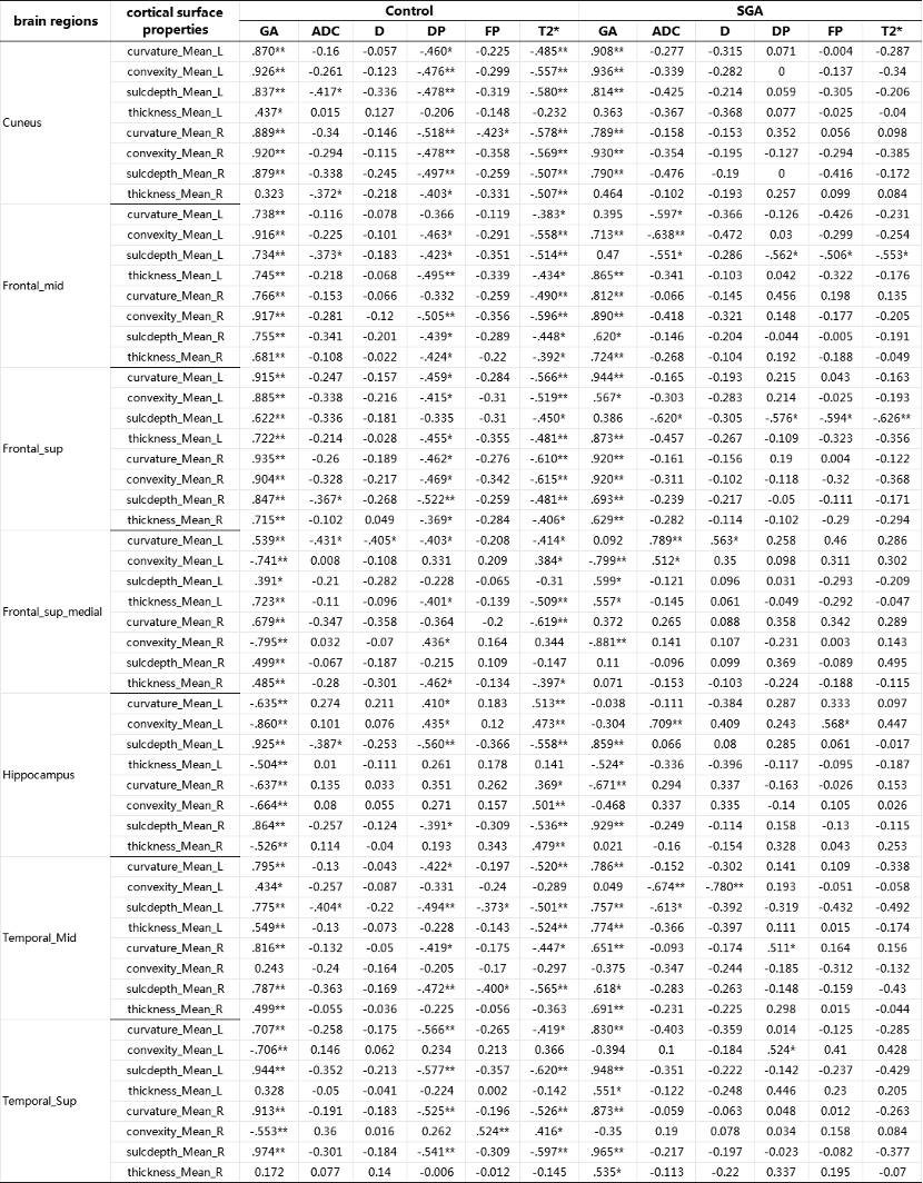
Table 2. Pearson correlation coefficients between cortical surface properties of various brain regions and gestational age, as well as placental function parameters both in the control and SGA groups
Abbreviations: SGA, Small-for-gestational age; D, diffusion coefficient; DP, pseudo diffusion coefficient; FP, perfusion fraction; Frontal-mid, middle frontal gyrus; Frontal-sup, superior frontal gyrus(dorsolateral); Frontal-sup-medial, superior frontal gyrus(medial); Temporal_Mid, middle temporal gyrus; Temporal-Sup, superior temporal gyrus
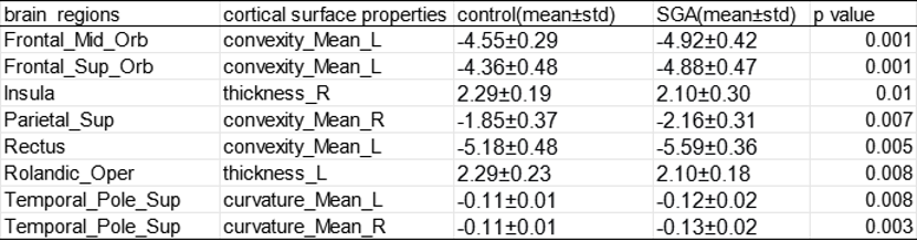
Table 3. Comparison of the average values of surface properties in 6 representative cortical regions between the control and SGA groups
Abbreviations: SGA, Small-for-gestational age; Frontal_Mid_Orb,Middle frontal gyrus(orbital part); Frontal_Sup_Orb,superior frontal gyrus(orbital part); Parietal_Sup,superior parietal gyrus; Rectus,gyrus rectus; Rolandic_Oper,rolandic operculum; Temporal_Pole_Sup,temporal pole(superior temporal gyrus)