0826
Machine learning and the prediction of enlarged lateral ventricular postnatal development trend in fetuses with isolated ventriculomegaly1Department of Radiology, the Affiliated Suzhou Hospital of Nanjing Medical University, Suzhou Municipal Hospital, Suzhou, China, 2Department of Radiology, the First Affiliated Hospital of Soochow University, Suzhou, China, 3Philips Healthcare, Shanghai, China
Synopsis
Keywords: Fetal, Machine Learning/Artificial Intelligence
Motivation: To evaluate the intracranial structures and distinct components (grew matter [GM] and white matter [WM]) adjacent to the occipital horn of the lateral ventricle T2WI radiomics features in healthy fetuses and fetuses with ventriculomegaly (FVs),
Goal(s): and to predict postnatal changes in the size of the enlarged lateral ventricle in FVs.
Approach: Utilizing WM-radiomics on the affected sides of FVs, the SVM algorithm effectively predicted the changes in ventricle size,
Results: as evidenced by the highest area under the curve (AUC) values of 0.771 and 0.738 in both the training and validation sets based on DeLong’s test (all P < 0.05).
Impact: An MRI-based occipital WM-radiomics model holds the potential to predict trends in changing ventriculomegaly.The image-based predictive model exhibits applicability in prenatal care. Leveraging image analysis and machine learning techniques may provide further insight into the pathophysiologic features of ventriculomegaly.
Introduction
Fetal ventriculomegaly stands as the most prevalent central nervous system (CNS) abnormality during pregnancy, signifying the expansion of the cerebral ventricles1 with a reported incidence ranging from 0.3% to 2%2-3. Isolated ventriculomegaly refers to cases without any other associated anatomical or genetic abnormalities, with 6% to 10% of cases potentially leading to significant neurodevelopmental impairment4. Currently, there are no prenatal markers that can reliably indicate the postnatal development trend of dilated lateral ventricles. The occipital horn of the lateral ventricle demonstrates the initial dilation, while the atrium dilates to a greater extent than the frontal horns in fetuses with ventriculomegaly (FVs)5. The dysregulation of neural stem cells in the ventricular neuroepithelium leads to impaired morphological development of the cerebral cortex, which is associated with ventriculomegaly6. The production and circulation of cerebrospinal fluid (CSF) in the ventricles generate a constant positive pressure that must be counteracted by the surrounding brain tissue or parenchyma to maintain a consistent ventricular size. A close relationship exists between lateral ventricle morphology and the surrounding brain structure6-9. Our objective was to determine whether an appropriate machine learning algorithm, based on the morphological features of bilateral ventricles and brain parenchymal radiomics features around the enlarged occipital horn, could be applied to predict postnatal changes in ventriculomegaly.Methods
This retrospective investigation involved 141 normal fetuses (NFs) and 101 FVs, who underwent brain MRI scans from January 2014 to July 2023 (Figure 1). FV-stable was defined as an enlargement of less than 2 mm postnatally, while FV-resolved referred to a decrease of 2 mm or more. Fetal MRI was conducted on a 1.5-T MRI system (Philips Achieva; Philips Medical Systems) using a 5-channel cardiac coil, without the use of sedation. T2-weighted balanced steady-state free precession (BSSFP) sequences were acquired in the coronal, sagittal, and axial planes of the fetal brain. Manual segmentation of the bilateral ventricles, grey matter (GM), and white matter (WM) surrounding the bilateral occipital horn in NFs, as well as the GM and WM around the enlarged occipital horn of FVs, was performed using the ITK-SNAP software (version 3.4.0; http://www.itksnap.org) (Figure 2). PyRadiomics (version 3.0.1; http://github.com/Radiomics/pyradiomics) was employed to extract the morphology of the ventricles and brain parenchyma radiomics features. Independent predictors without high collinearity were used to establish five distinct models and predictive models, respectively, employing the support vector machine (SVM) algorithm: bilateral ventricles morphology, GM-radiomics, WM-radiomics, (GM+WM) combined-radiomics, and (bilateral ventricles morphology + GM + WM) mixed models. AUC (area under the curve) with 95% confidence interval (CI), specificity, sensitivity, accuracy, positive predictive value (PPV), and negative predictive value (NPV) were calculated and compared using DeLong’s test.Results
In terms of baseline clinical characteristics, the three groups demonstrated a well-matched distribution. Significant differences in intracranial structures were observed between the NFs and FVs groups, whereas no significant differences were found between the FV-resolved and FV-stable groups. The AUC values for morphology, GM-radiomics, WM-radiomics, combined-radiomics, and mixed-models in distinguishing NFs from FVs were all found to be higher than 0.93 in the validation sets (Table 1 and Figure 3A). The AUC values for the five models in predicting the postnatal development trend of enlarged lateral ventricles were 0.462, 0.662, 0.738, 0.691, and 0.665 in the validation dataset, respectively (Table 1 and Figure 3B). DeLong’s test showed that the WM-radiomics model achieved the highest AUC values in both the training and validation sets (all P < 0.05).Discussion
Predicting postnatal alterations in the lateral ventricles of a fetus with ventriculomegaly could serve as a valuable guide to facilitate the timely completion of prenatal examinations10. The WM-radiomics model based on the abnormal side(s) may help predict the lateral ventricle changes (e.g., remaining the same or becoming smaller) of FVs after birth. One of the earliest pathogenic effects of ventricular dilatation is periventricular axon destruction11. Subsequent ventricle enlargement disrupts the ependyma and distorts and compresses the microvasculature. The severity of ventriculomegaly determines the degree of ependymal injury. Axonal and secondary myelin damage occurs through a combination of ischemic and mechanical effects. Compression of the extracellular space as a result of lateral ventricular dilatation in the cortex may hinder waste product elimination, leading to increased damage to the WM11. This result was constant in another study, which showed the displaced inferiorly and appeared thinner/inconsistency with the enlarged ventricles, especially the inferior longitudinal fasciculus12.Conclusion
Our findings suggest that the microstructure of WM around the occipital horn of lateral ventricle may be associated with the progression of ventriculomegaly.Acknowledgements
None.References
1 Cardoza JD, Goldstein RB, Filly RA (1988) Exclusion of fetal ventriculomegaly with a single measurement: the width of the lateral ventricular atrium. Radiology 169:711-714. Doi:10.1148/radiology.169.3.30550342
2 Pilu G, Hobbins JC (2002) Sonography of fetal cerebrospinal anomalies. Prenat Diagn 22:321-330. Doi:10.1002/pd.3103
3 Salomon LJ, Garel C (2007) Magnetic resonance imaging examination of the fetal brain. Ultrasound Obstet Gynecol 30:1019-1032. Doi:10.1002/uog.51764
4 Tarui T, Madan N, Graham G et al (2023) Comprehensive quantitative analyses of fetal magnetic resonance imaging in isolated cerebral ventriculomegaly. Neuroimage Clin 37:103357. Doi:10.1016/j.nicl.2023.1033575
5 Pisapia JM, Rozycki M, Akbari H et al (2017) Correlations of atrial diameter and frontooccipital horn ratio with ventricle size in fetal ventriculomegaly. Journal of Neurosurgery-Pediatrics 19:300-306. Doi:10.3171/2016.9.Peds1621066
6 Duy PQ, Rakic P, Alper SL et al (2022) Brain ventricles as windows into brain development and disease. Neuron 110:12-15. Doi:10.1016/j.neuron.2021.12.0097
7 Svancer P, Spaniel F (2021) Brain ventricular volume changes in schizophrenia. A narrative review. Neurosci Lett 759:136065. Doi:10.1016/j.neulet.2021.1360658
8 Pang D, Altschuler E (1994) Low-pressure hydrocephalic state and viscoelastic alterations in the brain. Neurosurgery 35:643-655; discussion 655-646. Doi:10.1227/00006123-199410000-000109
9 Peña A, Harris NG, Bolton MD, Czosnyka M, Pickard JD (2002) Communicating hydrocephalus: the biomechanics of progressive ventricular enlargement revisited. Acta Neurochir Suppl 81:59-63. Doi:10.1007/978-3-7091-6738-0_1510
10 McKechnie L, Vasudevan C, Levene M (2012) Neonatal outcome of congenital ventriculomegaly. Seminars in Fetal & Neonatal Medicine 17:301-307. Doi:10.1016/j.siny.2012.06.00111
11 Varela MF, Miyabe MM, Oria M (2020) Fetal brain damage in congenital hydrocephalus. Childs Nerv Syst 36:1661-1668. Doi:10.1007/s00381-020-04657-912
12 Horgos B, Mecea M, Boer A et al (2023) White matter changes in fetal brains with ventriculomegaly. Front Neuroanat 17:1160742. Doi:10.3389/fnana.2023.1160742
Figures
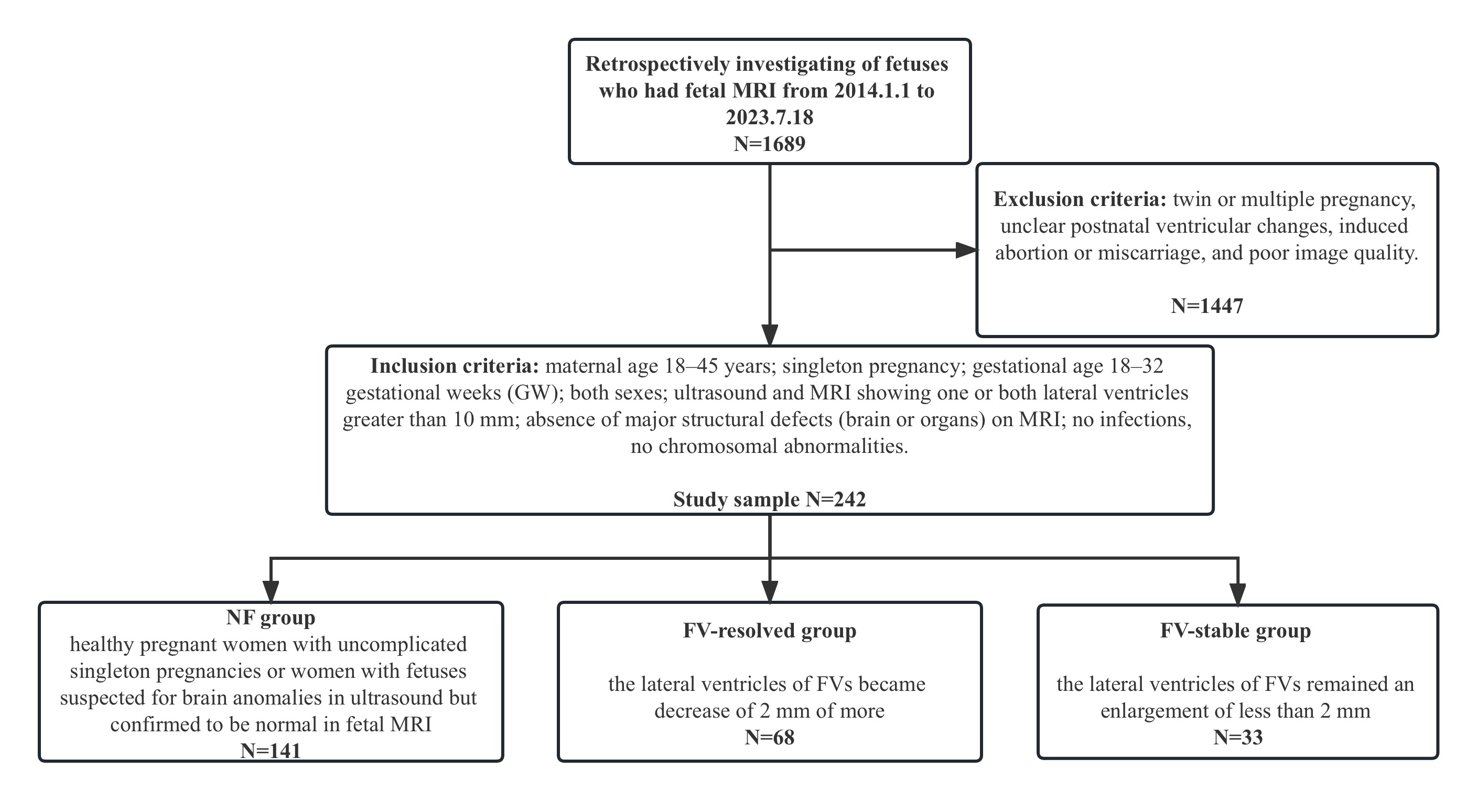
Fig. 1 Recruitment flowchart for including study samples in the study.
Abbreviations: NFs, normal fetuses; FVs, fetuses with ventriculomegaly.
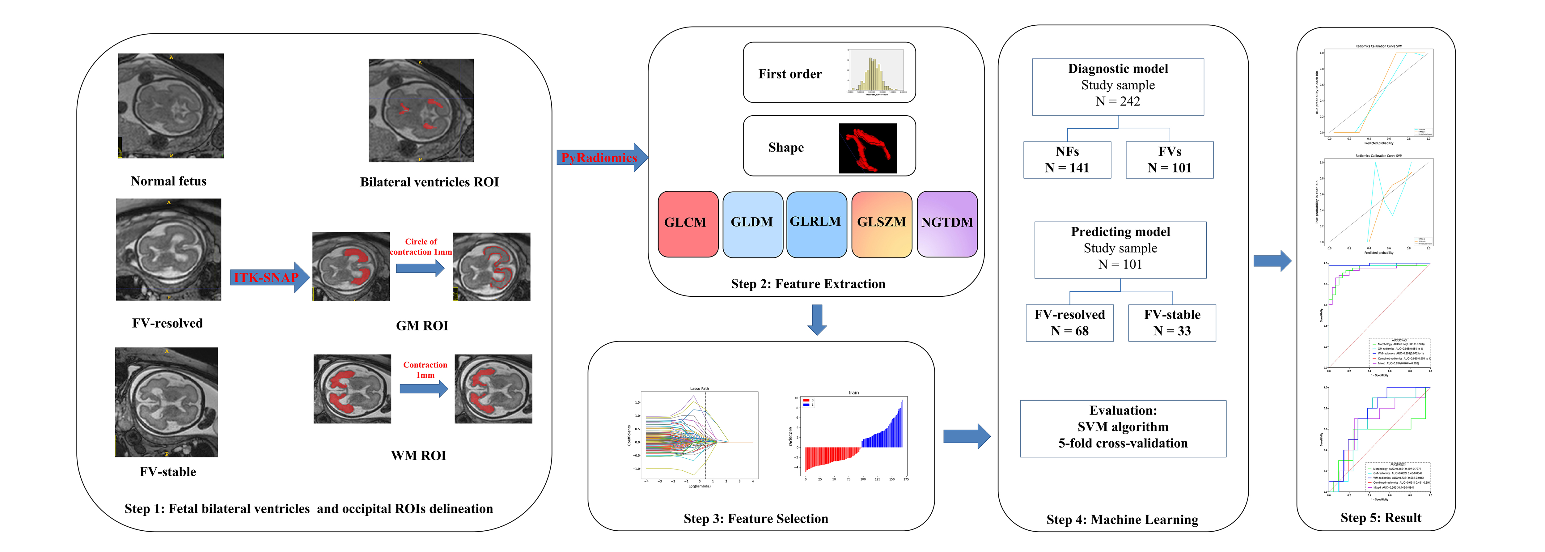
Fig. 2 Radiomics signature workflow.
Abbreviations: GM, grew matter; WM, white matter; SVM, support vector machine.
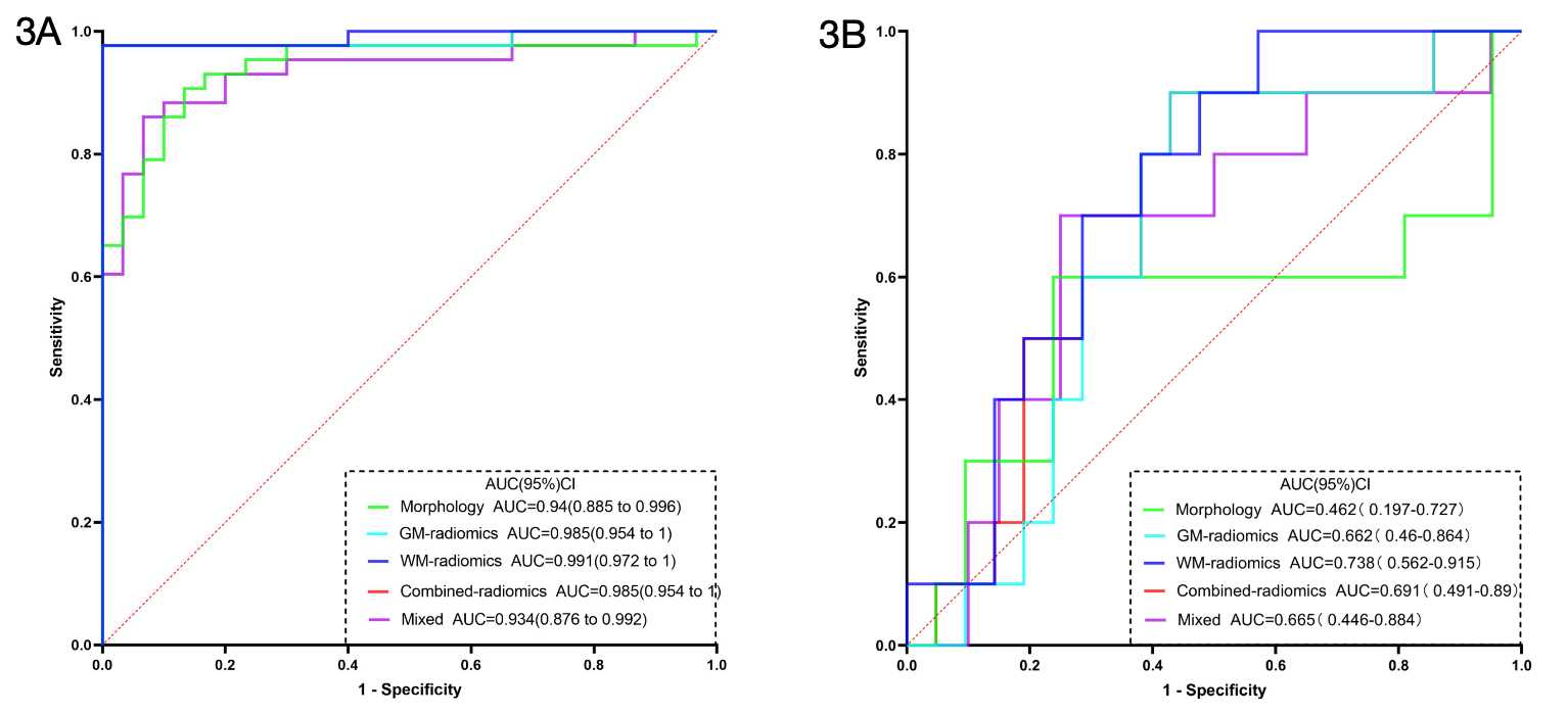
Fig. 3 The receiver operating characteristic curves (ROC) of the diagnostic and predictive models based on SVM algorithm.
(3A) The ROC curves of five diagnostic models in the validation sets. (3B) The ROC curves of five predictive models in the validation sets.
Abbreviations: ROC, receiver operating characteristic curves; CI, confidence interval; AUC, area under the curve; SVM, support vector machine.
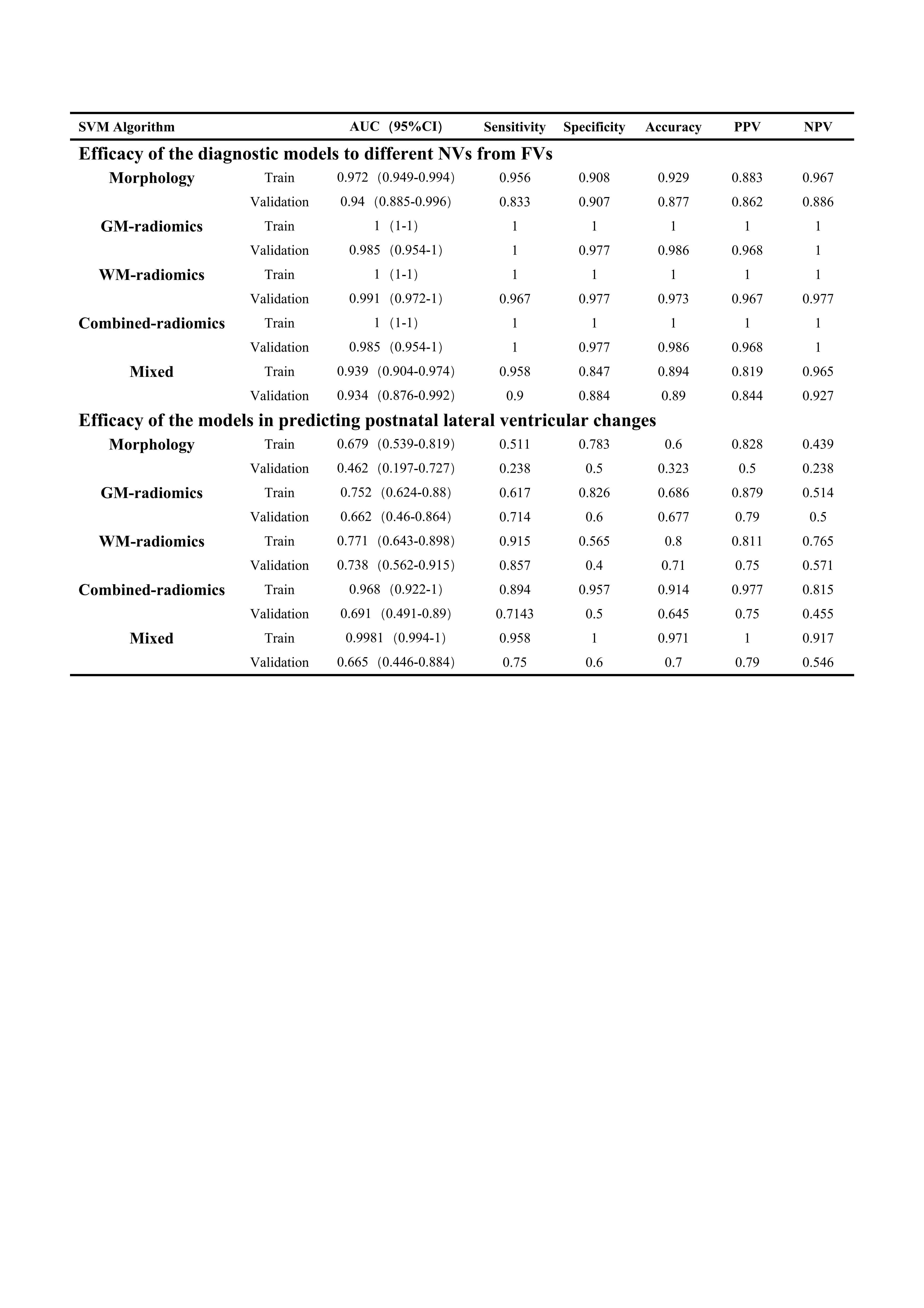
Table 1. The efficacy of diagnostic and predictive models based on SVM algorithm.
Abbreviations: CI, confidence interval; AUC, area under the curve; PPV, positive predictive value; NPV, negative predictive value.