0825
Normal subcortical nucleiand and cortex Growth ,and Lateral Asymmetries at Fetal Brain MRI1Lanzhou University Second Hospital, Lanzhou, China, 2School of information Science& Engineering,Lanzhou University, Lanzhou, China
Synopsis
Keywords: Fetal, Fetus
Motivation: The subcortical nuclei have important brain connectivity, which are rarely studied in fetal brain development.
Goal(s): The aim of this study is to analyze the developmental characteristics of fetal subcortical nuclei and their relationship with gestational age and cortical development.
Approach: The subcortical nuclei and cortical volumes were manually segmented from the three-dimensional (3D) volume parameters of tomosynthesis to volume reconstruction (SVR) images
Results: We found a good linear relationship between subcortical nuclei and gestational age, with agreement on the left and right sides. In addition, we found good coupling between the subcortical nuclei and right cerebral cortex development.
Impact: 3D Volumetric MR to assess the developmental characteristics of fetal subcortical nuclei and their relationship with gestational age. The normative values of fetal intracranial structures across a range of gestations could e used as a reference tool in prenatal counseling.
Introduction
Subcortical nucleus is a functional whole composed of a series of neural nuclei in the deep brain. It is nvolved in higher cognitive functions such as memory, emotion and reward learning[1]. With the advent of rapid magnetic resonance imaging (MRI) technology and the development of motion correction algorithms, it is possible to obtain high-quality 3D images of the fetal brain[2]. Most of the previous studies focused on cerebral cortex and cortical folds, while few studies on subcortical nuclei. In this study, we aimed to evaluate the development of fetal subcortical nuclei by 3D volumetric MRI, and to analyze its relationship with gestational age and cortical development.Methods
172 healthy fetuses between 22 and 36 weeks of gestation were recruited and underwent scanning on a 3T MR scanner (Philips Healthcare, the Netherlands). The following parameters were used for the single-shot fast spin-echo (SSFSE) sequence: TR/TE: 12,000/80 ms, matrix: 236 × 220, flip angle: 90 degrees, field of view: 260–355 mm2, and slice thickness: 2 mm with 0-mm spacing. The scan time of SSFSE sequence was 15–45 s. A single 3D motion-corrected high-resolution brain volume was reconstructed from the 2D SSTSE imaging stacks using a slice-to-volume reconstruction (SVR) method. First, we used an atlas-based method to extract a mask of the brain by defining a region of interest (ROI) from surrounding fetal and maternal tissue in each of the 3 principal planes, namely, sagittal, coronal, and axial. After that, the high-resolution isotropic reconstructed 3D volumetric images with the resolution of 0.5 × 0.5 × 0.5 mm were reconstructed from the registered low resolution and motion-corrupted 2D slices by using the Gauss–Seidel and super-resolution reconstruction method. The volume of subcortical nuclei was measured by manual segmentation by editing using ITK-SNAP software (version3.8, http://www.itksnap.org/). Scatter plots were drawn according to the segmented volumes against GA [3]. The specific process is shown in Figure 1Results
After excluding 125 normal fetal brain data with significant motion artifacts and reconstruction failures, a total of 47 normal fetal brains between 22 and 36 GA were analyzed. As shown in FIG. 2... The volume measurements of each nucleus showed a significant positive correlation with GA, with good agreement between the left and right sides. The quadratic line of the CI for each GA was determined from the best regression fit for each structure as shown in FIG. 3 below. One point of note is that we found an inverted "U" shape in the fitting curve of amygdala development to gestational age, peaking at about 32 to 33 weeks. In addition, we found some coupling between subcortical nuclei and the development of the cerebral cortex by Pearson analysis, and the left and right nuclei were strongly correlated with the development of the right cerebral cortex (P<0.05), as shown in Figure 4.Discussion
This study showed normative data of the subcortical nucleus of normal health fetuses between 22 and 36 GA, and all volumetric measurements had significant positive correlations with GA. Quantitative image analysis of the human brain in utero plays an important role in clinical decision-making and neuroscience investigation. With the advent of image post-processing technology and motion correction algorithms to obtain high-quality 3D images it is now possible to improve the accuracy of manual segmentation of the fetal brain in the early and middle trimesters[4]. The maturation process of gray matter nuclei includes the increase of basal dendrites, the proliferation of cell membranes and organelles and the change of cell density, resulting in an increase in nuclear volume. Cai et al showed that the increase in brain volume is linear from 20 to 28 GW[5]. Our findings are also consistent with overall fetal development. In addition, we found that there was a certain coupling between the subcortical nuclei and the development of the cerebral cortex, but the left and right nuclei were strongly correlated with the development of the right cerebral cortex. This finding has not been mentioned in other studies. This may be due to that during the early stages of brain development, the right side of the brain develops better, or asymmetry or lateralization during brain development. It may also be because our sample size is small, and there are certain errors in the reconstruction algorithm or manual sketch.Conclusion
The development of fetal subcortical nuclei is one of the important indexes to evaluate the central nervous system. Volumetric growth of the fetal brain follows a complex trajectory that is dependent on structure, GA etal. High-resolution 3D volumetric MRI can be used to assess the development of fetal brain structure.Acknowledgements
This work was supported by Gansu Province Clinical Research Center for Functional and Molecular Imaging (No. 21JR7RA438) , Gansu Provincial Science and Technology Program Projects (Key Research and Development Program) (No. 22JR5RA997) and Lanzhou talent innovation and entrepreneurship project(2022-RC-74).References
[1] Ruffle JK, Coen SJ, Giampietro V, Williams SCR, Apkarian AV, Farmer AD, Aziz Q. Morphology of subcortical brain nuclei is associated with autonomic function in healthy humans. Hum Brain Mapp. 2018 Jan;39(1):381-392. doi: 10.1002/hbm.23850. Epub 2017 Oct 28. PMID: 29080228; PMCID: PMC6866383.
[2] Benkarim OM, Sanroma G, Zimmer VA, Muñoz-Moreno E, Hahner N, Eixarch E, Camara O, González Ballester MA, Piella G. Toward the automatic quantification of in utero brain development in 3D structural MRI: A review. Hum Brain Mapp. 2017 May;38(5):2772-2787. doi: 10.1002/hbm.23536. Epub 2017 Feb 14. PMID: 28195417; PMCID: PMC6867074.
[3] Ren JY, Zhu M, Wang G, Gui Y, Jiang F, Dong SZ. Quantification of Intracranial Structures Volume in Fetuses Using 3-D Volumetric MRI: Normal Values at 19 to 37 Weeks' Gestation. Front Neurosci. 2022 May 12;16:886083. doi: 10.3389/fnins.2022.886083. PMID: 35645723; PMCID: PMC9133784.
[4] Machado-Rivas F, Gandhi J, Choi JJ, Velasco-Annis C, Afacan O, Warfield SK, Gholipour A, Jaimes C. Normal Growth, Sexual Dimorphism, and Lateral Asymmetries at Fetal Brain MRI. Radiology. 2022 Apr;303(1):162-170. doi: 10.1148/radiol.211222. Epub 2021 Dec 21. PMID: 34931857; PMCID: PMC8962825.
[5] Cai S, Zhang G, Zhang H, Wang J. Normative linear and volumetric biometric measurements of fetal brain development in magnetic resonance imaging. Childs Nerv Syst. 2020 Dec;36(12):2997-3005. doi: 10.1007/s00381-020-04633-3. Epub 2020 May 28. PMID: 32468242.
Figures
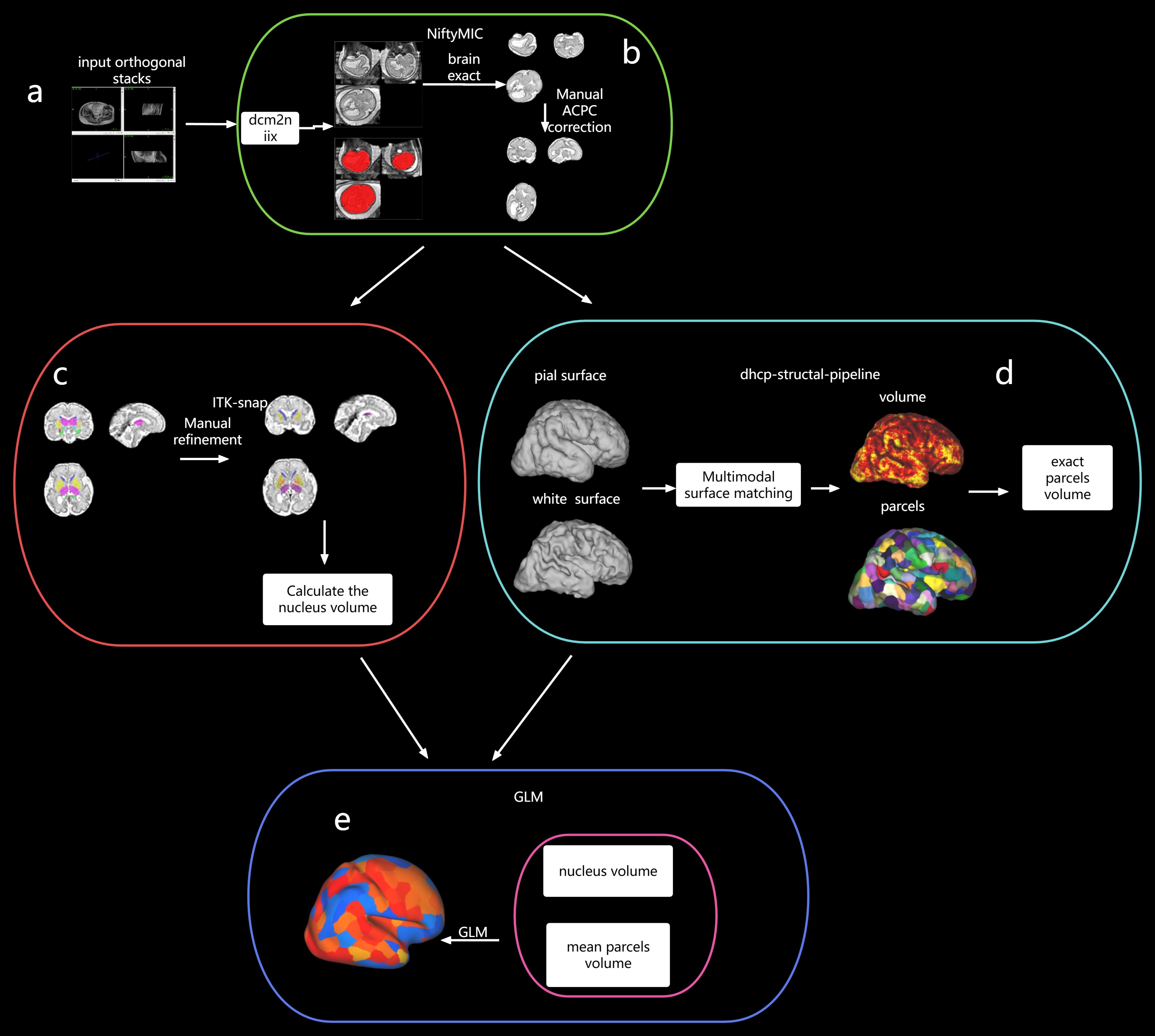
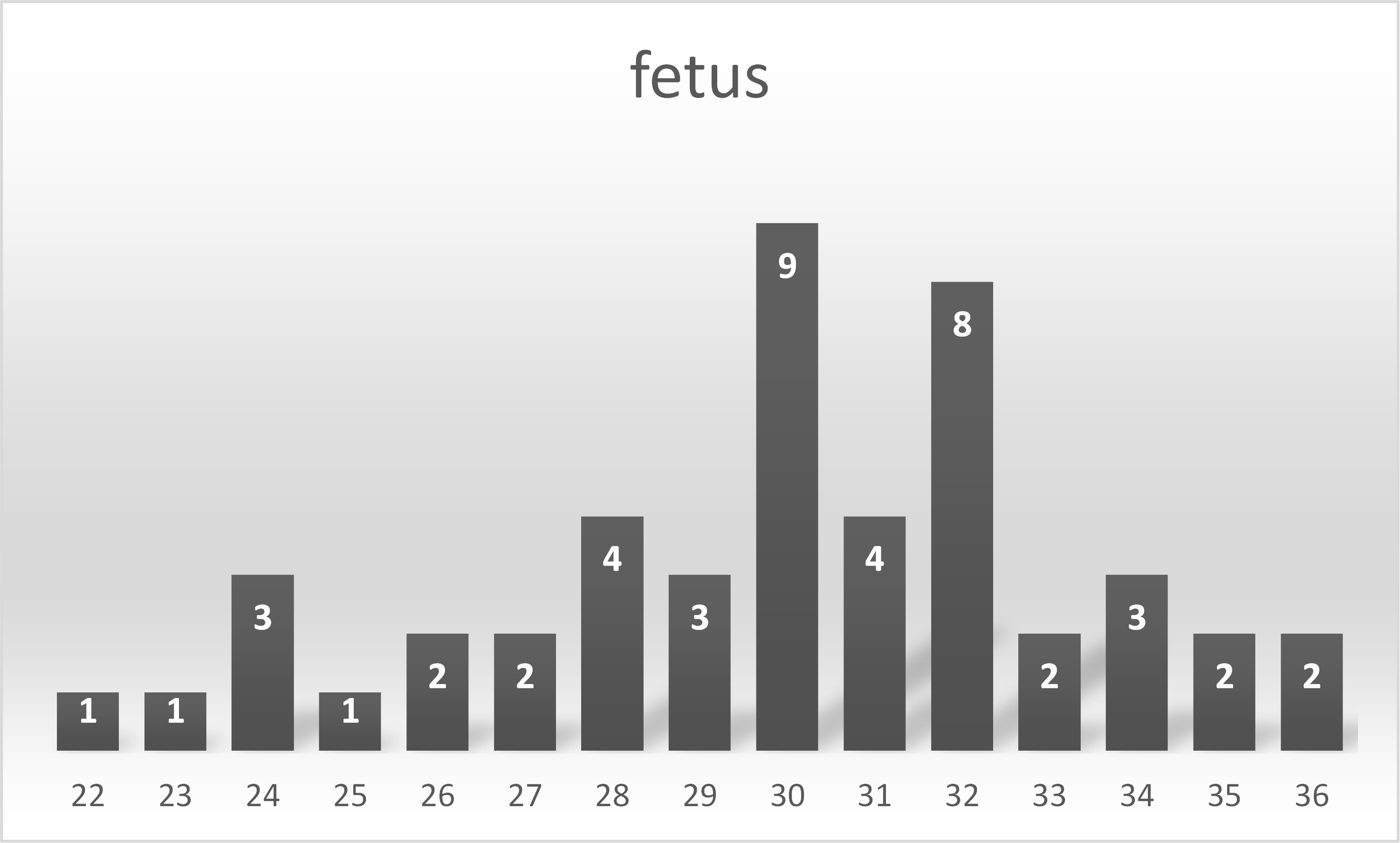
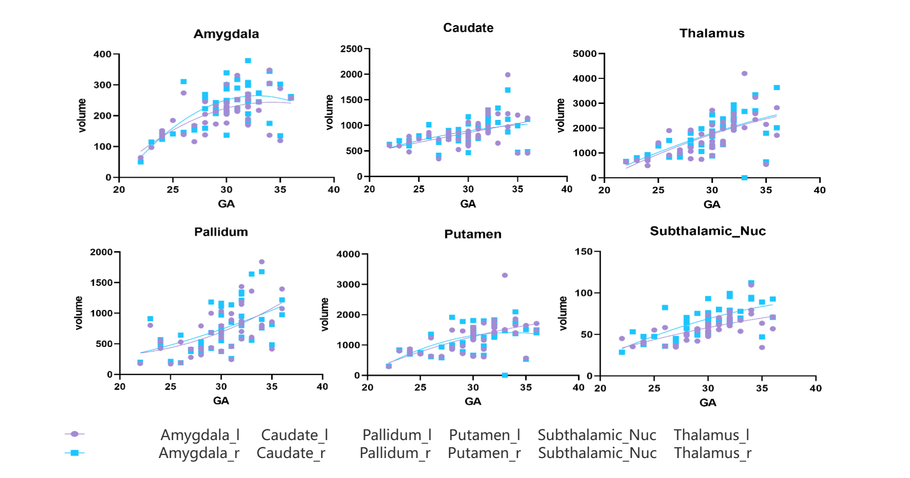
Figure 3 Correlation between subcortical nuclei and GA
The volume measurements of each nucleus showed a significant positive correlation with GA, with good agreement between the left and right sides. One point of note is that we found an inverted "U" shape in the fitting curve of amygdala development to gestational age, peaking at about 32 to 33 weeks.
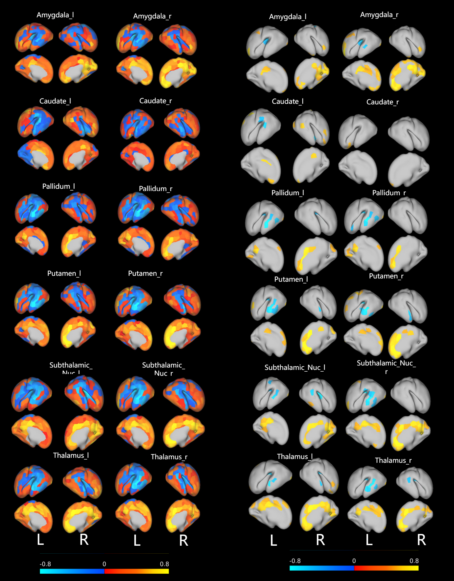
Figure 4 Correlation of subcortical nuclei with cortical development
The left side of the map shows the correlation values of the nucleus and each brain region. Each brain region has different colors corresponding to different correlation values, and the color bar at the bottom of the map shows the corresponding values of different colors. Right is also a nucleus and different brain regions, but due to the left side of the figure of the effect is not very significant, so only show the area of p value is less than 0.05.