0824
A framework for abnormality detection in developing white matter in utero, applied to fetuses with Congenital Heart Disease1Centre for the Developing Brain, King's College London, London, United Kingdom, 2KU Leuven, Leuven, Belgium
Synopsis
Keywords: Fetal, Fetus
Motivation: In utero neurodevelopment is complex and not well understood, particularly in fetuses with Congenital Heart Disease.
Goal(s): Normatively model microstructural maturation in transient fetal compartments
Approach: Diffusion MRI was acquired in a healthy control cohort of 235 fetuses (22–37 weeks gestation). White matter bundles were estimated and divided into cross-sections. Gaussian Process Regression models were fit to diffusion metrics in each cross-section, and Z-scores calculated along the tract for 26 fetuses with CHD.
Results: We observe gradients of change, highlighting abnormal regions along the white matter unique to each subject. We did not find consistent patterns or associations with a specific diagnosis.
Impact: We establish normative trajectories in diffusion MR signal at the level of individual fetal brain compartments that reflect developing microstructure, improving understanding of dynamic fetal brain development and allowing us to predict deviations from the norm.
Introduction
White matter injury is common in infants with congenital heart disease (CHD) (Morton et al., 2015). The volumes of transient fetal compartments underlying white matter are also notably reduced in fetuses with CHD, suggesting impaired white matter development in utero (Rollins et al., 2021). However, microstructural WM development in CHD has not been investigated. We present an approach to identify individual deviations from the normal trajectory of diffusion contrast change, with high spatiotemporal specificity, within specific white matter fibre bundles as they emerge from transient fetal compartments. We aimed to explore whether the volumetric differences reported by previous work (Rollins et al., 2021, Cromb et al., 2023, Limperopoulous et al., 2010) are accompanied by changes at the microstructural level, using high angular resolution, multi-shell diffusion imaging (HARDI), comparing healthy control fetuses to a cohort with various forms of CHD.Methods
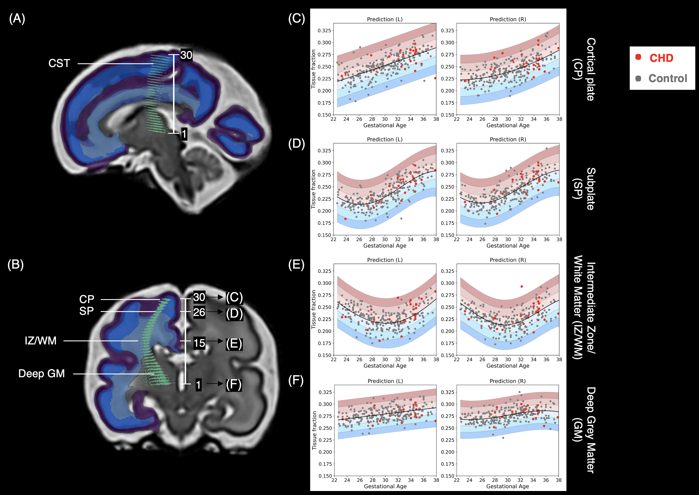
The study population included 235 healthy controls (22 – 37 weeks GA, 129 male, 106 female) and 26 fetuses with CHD (23 – 38 weeks GA, 19 male, 7 female). Informed, written consent was acquired prior to MRI (Ethical approval CHD: 21/WA/0075; dHCP: 14/LO/1169). T2 and HARDI volumes were acquired with the Developing Human Connectome Project acquisition protocol (Price et al., 2019) on a Philips Achieva 3T system, with a 32-channel cardiac coil. HARDI data was collected with a combined spin echo and field echo (SAFE) sequence at 2 mm isotropic resolution, using a multi-shell diffusion encoding that consists of 15 volumes at b=0 s/mm2, 46 volumes at b=400 s/mm2, and 80 volumes at b=1000 s/mm2 (Christiaens et al., 2019). HARDI datasets were reconstructed to 0.8 mm, using a data driven representation of the spherical harmonics and radial decomposition (SHARD). The SHARD pipeline caters to the motion corrupted fetal data, using dynamic distortion correction and a slice-to-volume motion correction framework (Cordero-Grande et al., 2019, Christiaens et al., 2021). All subsequent diffusion processing and tractography to estimate thalamocortical pathways was performed using MRtrix3 (Tournier et al., 2019, Wilson et al., 2023). We used multi-shell multi-tissue constrained spherical deconvolution (MSMT-CSD) to deconvolve the diffusion signal, and quantified the anisotropic component based on the mature white matter response function, to obtain the 'tissue fraction' (Jeurissen et al., 2013, Wilson et al., 2021, 2023). 30 cross-sections were taken along the tracts (Figure 1A,B), and values of underlying tissue fraction were sampled and averaged within each slice. We used Gaussian process regression (GPR) implemented in GPy, to predict and characterise the normative range of tissue fraction values within each cross-section (Figure 1C-F). We quantified the deviation from normal using a z-score, computing the difference between the predicted and the observed value, normalised by the uncertainty of the prediction (Marquand et al. 2016).Results
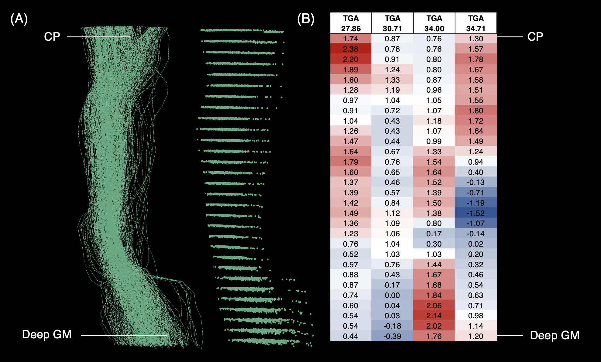
We identified unique maturational trends within different fetal tissue types across the second to third trimester (Figure 1). We observe different rates of change in tissue fraction maturation between compartments, and in the intermediate zone/white matter compartment, tissue fraction maturation follows a parabolic curve. We also observe fluctuations in the level of variability between individuals along the tract. When examining the z-scores of fetuses with CHD, all fetuses showed a high proportion of normal z-scores along the white matter, with isolated regions of deviations from the normal mean. We observe gradients of change from normal to abnormal in regions along the white matter in some subjects. The regions of abnormality appeared to be specific to each subject, and we did not find consistent patterns across the cohort or for subjects with a specific diagnosis.Discussion
The analysis framework highlights unique maturational trends for different fetal tissue types across the second to third trimester. In fetuses with CHD we observed normal z scores along a large proportion of the white matter, but identified specific regions of abnormality. The abnormal regions were unique to individuals, reflecting the highly dynamic development of the fetal brain and the heterogeneity of CHD subtypes within this cohort. Further post-hoc testing and clustering approaches will be required to investigate this further. Although this approach is tailored to detect differences within individuals, we were statistically underpowered to draw conclusions about CHD at the group level.Acknowledgements
We would like to thank the families who participated in this study, along with the research radiologists, radiographers, and scanning team at the Centre for the Developing Brain at King's College London. We also thank the staff from the St Thomas’ Hospital Neonatal Intensive Care Unit and the Evelina London Children’s Hospital. This research was funded by the Medical Research Council UK (MR/ L011530/1; MR/V002465/1), the British Heart Foundation (FS/15/ 55/31649) and Medical Research Council UK Centre grant (MR/N026063/1). This study used data that was acquired as part of the Developing Human Connectome Project, supported by ERC grant agreement no. 319456. This work was supported by core funding from the Wellcome/EPSRC Centre for Medical Engineering [WT203148/Z/16/Z] and by the National Institute for Health Research (NIHR) Biomedical Research Centre based at Guy’s and St Thomas’ NHS Foundation Trust and King’s College London and/or the NIHR Clinical Research Facility. The views expressed are those of the author(s) and not necessarily those of the NHS, the NIHR or the Department of Health and Social Care.References
Christiaens, D. et al. (2019) ‘Fetal diffusion MRI acquisition and analysis in the developing Human Connectome Project.’
Christiaens, D. et al. (2021) ‘Scattered slice SHARD reconstruction for motion correction in multi-shell diffusion MRI’, NeuroImage, 225, p. 117437. Available at: https://doi.org/10.1016/j.neuroimage.2020.117437.
Cordero-Grande, L. et al. (2019) ‘Complex diffusion-weighted image estimation via matrix recovery under general noise models’, NeuroImage, 200, pp. 391–404. Available at: https://doi.org/10.1016/j.neuroimage.2019.06.039.
Cromb, D. et al. (2023) ‘Total and Regional Brain Volumes in Fetuses With Congenital Heart Disease’, Journal of Magnetic Resonance Imaging, p. jmri.29078. Available at: https://doi.org/10.1002/jmri.29078.
Limperopoulos, C. et al. (2010) ‘Brain Volume and Metabolism in Fetuses With Congenital Heart Disease: Evaluation With Quantitative Magnetic Resonance Imaging and Spectroscopy’, Circulation, 121(1), pp. 26–33. Available at: https://doi.org/10.1161/CIRCULATIONAHA.109.865568.
Marquand, A.F. et al. (2016) ‘Understanding Heterogeneity in Clinical Cohorts Using Normative Models: Beyond Case-Control Studies’, Biological Psychiatry, 80(7), pp. 552–561. Available at: https://doi.org/10.1016/j.biopsych.2015.12.023.
Price, A. et al., (2019). ‘The Developing Human Connectome Project (dHCP): Fetal Acquisition Protocol. ISMRM.’Rollins, C.K. et al. (2021) ‘Regional Brain Growth Trajectories in Fetuses with Congenital Heart Disease’, Annals of Neurology, 89(1), pp. 143–157. Available at: https://doi.org/10.1002/ana.25940.
Rollins, C.K. et al. (2021) ‘Regional Brain Growth Trajectories in Fetuses with Congenital Heart Disease’, Annals of Neurology, 89(1), pp. 143–157. Available at: https://doi.org/10.1002/ana.25940.
Tournier, J.-D. et al. (2019) ‘MRtrix3: A fast, flexible and open software framework for medical image processing and visualisation’, NeuroImage, 202, p. 116137. Available at: https://doi.org/10.1016/j.neuroimage.2019.116137.
Wilson, S. et al. (2023) ‘Spatiotemporal tissue maturation of thalamocortical pathways in the human fetal brain’, eLife, 12, p. e83727. Available at: https://doi.org/10.7554/eLife.83727.s Christiaens, D. et al. (2019) ‘Fetal diffusion MRI acquisition and analysis in the developing Human Connectome Project.’
Figures