0823
Quantitative $$$T_{2}^{*}$$$ and $$$B_{0}$$$ Mapping of Fetal Brain Using Stack-of-Star Multi-Echo FLASH and Model-Based Reconstruction1Computational Radiology Laboratory, Boston Children's Hospital, and Harvard Medical School, Boston, MA, United States
Synopsis
Keywords: Fetal, Fetus, model-based recontruction; T2* mapping; QSM
Motivation: Quantitative $$$T_{2}^{*}$$$ and susceptibility mapping is of great interest for fetal MRI. While conventional EPI-based approaches are efficient, they usually suffer from motion and field distortion artifacts.
Goal(s): To develop a distortion-free and motion-robust quantitative $$$T_{2}^{*}$$$ and susceptibility mapping approach for fetal brain.
Approach: A stack-of-star multi-echo FLASH sequence and model-based reconstruction were developed for quantitative mapping of $$$T_{2}^{*}$$$ and $$$B_{0}$$$ of fetal brain. Motion estimation and correction is incorporated into the reconstruction to reduce motion artifacts.
Results: Initial findings indicate accurate $$$T_{2}^{*}$$$ measurements. Motion-corrected image reconstruction effectively minimized motion artifacts. Fetal $$$T_{2}^{*}$$$ and $$$B_{0}$$$ maps are obtained with reasonable quantitative $$$T_{2}^{*}$$$ values.
Impact: Our technique enables distortion-free and motion-robust quantitative $$$T_{2}^{*}$$$ and $$$B_{0}$$$ mapping for the fetal brain, utilizing a stack-of-star multi-echo acquisition and model-based reconstruction. It has the potential to address motion and field distortion artifacts typically encountered in EPI-based methods.
Introduction
Quantitative $$$T_{2}^{*}$$$ and susceptibility mapping hold significant importance in fetal MRI [1]. Although current approaches rely on efficient EPI acquisitions [2-3], the long readout of EPI renders it susceptible to motion and field distortion artifacts, particularly pronounced in fetal MRI. In contrast, radial acquisitions are known for motion robustness and have found utility in imaging moving organs for adults and children [4-6]. More recently, radial multi-echo readouts have been leveraged for quantitative $$$T_{2}^{*}$$$ and/or field inhomogeneity mapping [7-8]. Inspired by the above, this work develops a distortion-free quantitative $$$T_{2}^{*}$$$ and field inhomogeneity mapping technique for fetal brain imaging, employing a stack-of-star multi-echo acquisition and model-based reconstruction.Methods
A stack-of-star radial FLASH sequence with a bipolar multi-echo readout was used for data acquisition. The signal equation for this multi-echo process is: $$ S_{\text{TE}_{n}} = \big(W + F\cdot z_{n}\big)\cdot \exp\big(\text{TE}_{n}\cdot i2\pi f_{B_{0}}\big) \cdot \exp\big(- \text{TE}_{n} \cdot R_{2}^{*}\big).$$Where $$$W$$$ and $$$F$$$ represent the water and fat components, respectively. $$$z_{n}$$$ is the 6-peak fat spectrum and $$$\text{TE}_{n}$$$ is the $$$n$$$th echo time. $$$f_{B_{0}}$$$ represents the field inhomogeneity and $$$R_{2}^{*}$$$ is the effective relax rate. The unknowns are then $$$x = (W, F, f_{B_{0}}, R_{2}^{*})^{T}$$$.Here, we extend a previously developed reconstruction [8] to the motion-corrected case to directly estimate quantitative maps from the radially acquired multi-echo k-space. We consider a rigid motion by incorporating two transforms $$$R$$$ (rotation) and $$$T$$$ (translation) into the forward equation, resulting in solving the following inverse problem, i.e.,$$ \hat{x} = (W, F, f_{B_{0}}, R_{2}^{*})^{T} = \text{argmin}_{ x \in D}\sum_{\text{m}} \sum_{\text{n}} \big\|P\mathcal{F}\cdot R_{m}\cdot T_{m}\cdot \big(C\cdot S_{\text{TE}_{n}}(x)\big)- Y_{\text{m},\text{TE}_{n}}\big\|_{2}^{2} + Q(x) $$.Here $$$R_{m}$$$ and $$$T_{m}$$$ at motion state $$$m$$$ are pre-computed from a motion estimation algorithm using ITK [10]. D is a convex set, ensuring the non-negativity of all relaxation rates. $$$Q(x)$$$ is the regularization term [8,9]. The above nonlinear inverse problem is solved by IRGNM-FISTA using BART [13].
Experiments
All MRI studies were conducted on a 3 T Prisma scanner. Pineapple and adult brain studies were done with a 64-channel head/neck coil, while fetal scans were performed with a combined thorax and spine coil. One fetuse with a gestational age of 32 weeks was scanned.The acquisition parameters for the pineapple and the adult brain study were: FOV $$$256\times256$$$ mm$$$^{2}$$$, matrix size $$$256\times256$$$, slice thickness 4 mm, 15 echoes with $$$\text{TR}$$$/$$$\text{TE}_{1}$$$/$$$\delta\text{TE}$$$/$$$\text{TE}_{15}$$$ = 30/1.5/2.0/27.5 ms, FA $$$=15^{\circ}$$$, 14 slices (phantom) / 30 slices (brain), bandwidth 740 Hz/pixel, and a total of 150 radial spokes with an acquisition time of 63 seconds (phantom) and 2:25 min (brain).
For the fetal brain, the acquisition parameters were: FOV $$$256\times256$$$ mm$$$^{2}$$$, matrix size $$$256\times256$$$, slice thickness 4 mm, 24 echoes with $$$\text{TR}$$$/$$$\text{TE}_{1}$$$/$$$\delta\text{TE}$$$/$$$\text{TE}_{24}$$$ = 50/1.5/2.0/47.5 ms, FA $$$=20^{\circ}$$$, 14 slices, bandwidth 740 Hz/pixel, and a total of 200 radial spokes with an acquisition time of 2:33 min.
Reference $$$T_{2}^{*}$$$ and $$$B_{0}$$$ mapping was conducted on the pineapple phantom using a Cartesian multi-echo FLASH protocol provided by the vendor.
Results
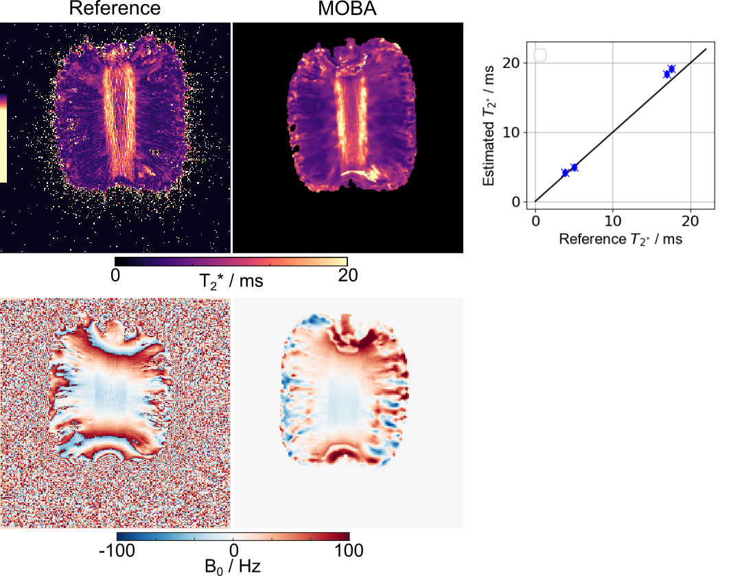
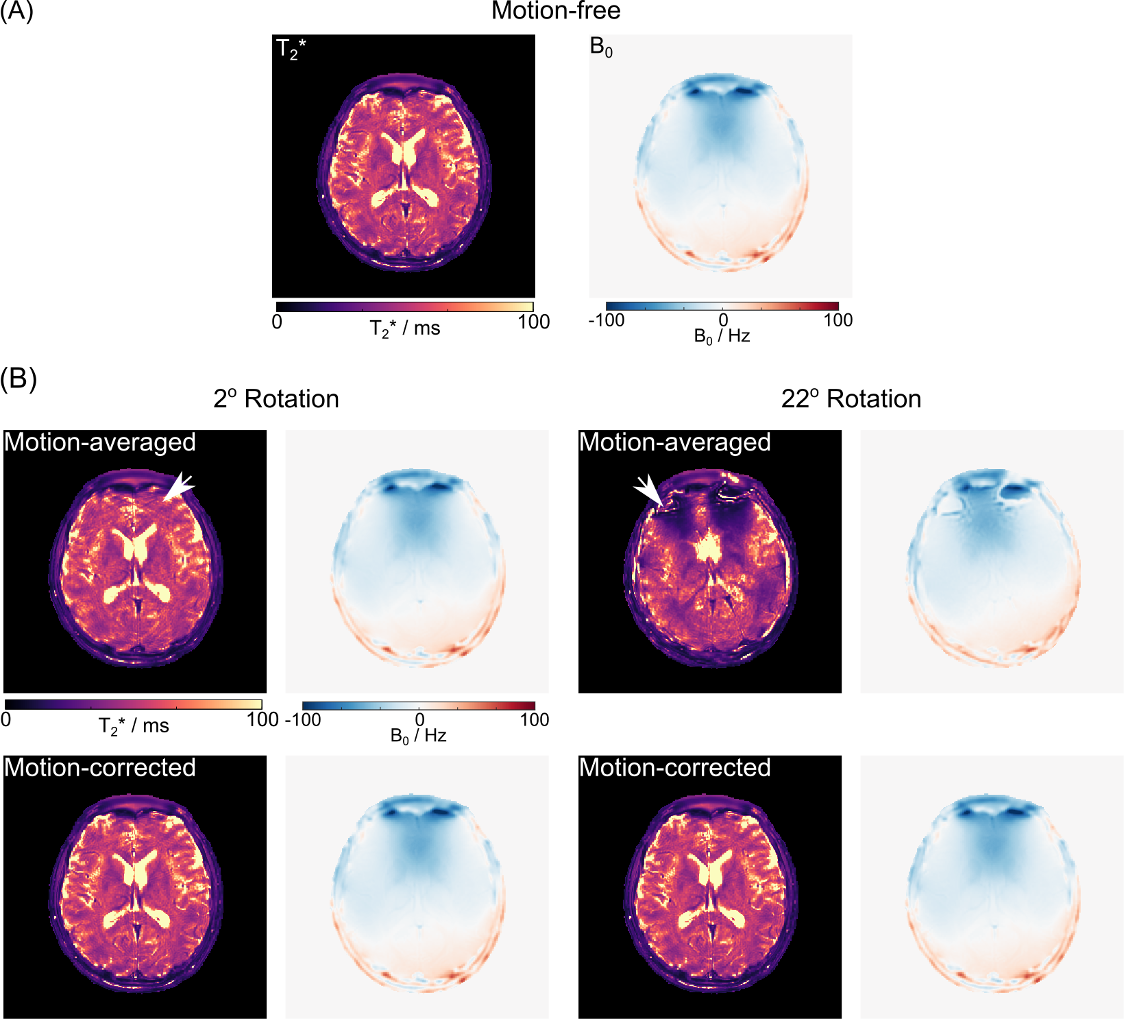
In Figure 1, we compare the quantitative $$$T_{2}^{*}$$$ and $$$B_{0}$$$ maps obtained from a reference approach to our model-based method. While the reference $$$T_{2}^{*}$$$ map appears visually noisy and the $$$B_{0}$$$ map exhibits phase warps, our proposed approach produces high-quality, undistorted $$$T_{2}^{*}$$$ and $$$B_{0}$$$ maps. Despite these differences, the ROI analysis reveals a good correlation between the two methods, with a maximum relative difference of less than $$$5\%$$$.Figure 2(A) displays the adult $$$T_{2}^{*}$$$ and $$$B_{0}$$$ maps estimatedwith 100 spokes. Figure 2(B) shows the impact of motion, particularly rotation. As expected, more severe motion leads to greater artifacts in the maps (indicated by white arrows). However, when incorporating motion in the reconstruction, our method can produce distortion-free quantitative maps (Figure 2(B)).
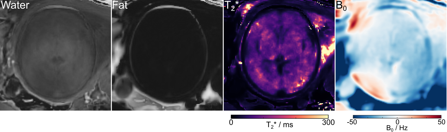
Figure 3 shows the estimated water and fat content, along with the $$$T_{2}^{*}$$$ and $$$B_{0}$$$ maps of a fetal brain, utilizing our proposed reconstruction with 200 spokes. Since minor motion was observed in the case, no motion correction was applied. The ROI-analyzed $$$T_{2}^{*}$$$ values are: frontal white matter: $$$75 \pm 5$$$ ms and $$$81 \pm 6$$$ ms, occipital white matter: $$$83 \pm 3$$$ ms and $$$89 \pm 9$$$ ms. Those values correspond well to the literature findings [2,3,14].
Discussion and Conclusion
We have developed a distortion-free and motion-robust quantitative $$$T_{2}^{*}$$$ and $$$B_{0}$$$ mapping technique using a stack-of-star multi-echo FLASH and model-based Reconstruction. The initial findings indicate accurate $$$T_{2}^{*}$$$ measurements and the motion-corrected reconstruction has effectively minimized motion artifacts. Although our current method has delivered fetal $$$T_{2}^{*}$$$ and $$$B_{0}$$$ mapping without motion correction, further research on advanced motion estimation and optimized sampling might be necessary to enhance more robust quantitative fetal brain imaging.Acknowledgements
No acknowledgement found.References
1. Rivkin M, Wolraich D, Als H, McAnulty G, Butler S, Conneman N, Fischer C, Vajapeyam S, Robertson R, Mulkern R. Prolonged T2* values in newborn versus adult brain: implications for fMRI studies of newborns. Magn Reson Med 2004; 51: 1287–1291.
2. Vasylechko S, Malamateniou C, Nunes RG, Fox M, Allsop J, Rutherford M, Rueckert D, Hajnal J V. T2* relaxometry of fetal brain at 1.5 Tesla using a motion tolerant method. Magn Reson Med 2015; 73: 1795–1802.
3. Blazejewska AI, Seshamani S, Mckown SK, Caucutt JS, Dighe M, Gatenby C, Studholme C. 3D in utero quantification of T2* relaxation times in human fetal brain tissues for age optimized structural and functional MRI. Magn Reson Med 2017; 78: 909–916.
4. Block KT, Chandarana H, Milla S, et al. Towards routine clinical use of radial stack-of-stars 3D gradient-echo sequences for reducing motion sensitivity. J Kor Soc Magn Reson Med 2014; 18: 87–106.
5. Feng L, Grimm R, Block KT, et al. Golden-angle radial sparse parallel MRI: combination of compressed sensing, parallel imaging, and golden-angle radial sampling for fast and flexible dynamic volumetric MRI. Magn Reson Med 2014; 72: 707–717.
6. Armstrong T, Ly KV, Murthy S, et al. Free-breathing quantification of hepatic fat in healthy children and children with nonalcoholic fatty liver disease using a multi-echo 3-D stack-of-radial MRI technique. Pediatr Radiol. 2018; 48: 941–953.
7. Schneider M, Benkert T, Solomon E, et al. Free-breathing fat and R2* quantification in the liver using a stack-of-stars multi-echo acquisition with respiratory-resolved model-based reconstruction. Magn Reson Med. 2020; 84: 2592-2605.
8. Tan Z, Unterberg-Buchwald C, Blumenthal M et al., Free-Breathing Liver Fat, R2* and B0 Field Mapping Using Multi-Echo Radial FLASH and Regularized Model-Based Reconstruction. IEEE Trans on Medical Imaging. 2023 42 (5), 1374-1387.
9. Wang X, Tan Z, Scholand N, Roeloffs V, Uecker M. Physics-based reconstruction methods for magnetic resonance imaging. Philos Trans Royal Soc A. 2021; 379:20200196.
10. McCormick M, Liu X, Jomier J, Marion C, Ibanez L. ITK: enabling reproducible research and open science. Front Neuroinform. 2014; 8:13. doi:10.3389/fninf.2014.00013.
11. Lustig M, Donoho D, Pauly JM. Sparse MRI: the application of compressed sensing for rapid MR imaging. Magn Reson Med 2007; 58: 1182–1195.
12. Uecker M, Hohage T, Block KT, and Frahm J. Image reconstruction by regularized nonlinear inversion-joint estimation of coil sensitivities and image content. Magn Reson Med 2008; 60: 674-682.
13. Uecker M, Holme CHM, Blumenthal M et al., Mrirecon/bart: version 0.7. 00, https://zenodo.org/records/4570601.
Figures