0822
Using TE-Dependent Analysis for Multi-Echo fMRI Analysis of the Fetal Brain1Technical University of Munich, Munich, Germany, 2Biomedical Engineering, King's College London, London, United Kingdom, 3Centre for the Developing Brain, King's College London, London, United Kingdom, 4Women’s Health, Guy's & St.Thomas' Hospital, London, United Kingdom, 5Department of Neuroradiology and TUM-NIC, Klinikum rechts der Isar, Munich, Germany, 6Smart Imaging Lab, Radiological Institute, University Hospital Erlangen, Erlangen, Germany
Synopsis
Keywords: Fetal, Brain, Multi-Echo Analysis
Motivation: fMRI of the fetus in the womb must overcome the challenges of fetal motion and heterogeneous tissue boundaries. Multi-Echo fMRI reduces signal dropout and thermal noise, improving contrast-to-noise ratio of the BOLD signal. This can produce higher quality fetal MRI and allow adequate functional assessment of fetal brain development.
Goal(s): This study aims to denoise ME-fMRI of the fetal brain with TE-dependent analysis (tedana) in a subset of 10 cases (gestational age >35weeks).
Approach: Multi-echo gradient echo EPI scans were acquired in 80 fetuses. The fetal brain is segmented and passed to the analysis.
Results: Credible BOLD components are successfully identified using tedana.
Impact: TE-dependent analysis denoises ME-fMRI data of fetuses which undergo motion during scanning and contain heterogeneous tissue boundaries. This study assesses capabilities of ME-fMRI analysis to determine BOLD components in the fetal brain,paving the way for future research and clinical usage.
Introduction
An optimal intra-uterine environment is crucial for healthy fetal brain development5,13. Neurodevelopment is vulnerable to maternal factors, such as nutrition, stress and infection5,13. These affect the neurodevelopment of a fetus and are associated with emotional, behavioural or cognitive problems in later life including impaired brain growth, delayed verbal abilities and a higher risk of depression, anxiety, ADHD or schizophrenia5,13. Currently, fetal brain development is evaluated via ultrasound, able to detect structural pathologies but not suited to inform on the function12,14,15.Fetal motion and the multi-tissue environment in the uterus pose challenges when analysing BOLD signals in fetuses4,6,7,15. Using multiple echoes to process fMRI data reduces signal dropout and thermal noise, thereby improving the contrast-to-noise ratio of the BOLD signal1,9,15. Exploring the advantages of multi-echo fMRI in the context of the fetal brain fMRI carries the potential to improve our understanding of fetal brain connectivity1,2.
Methods
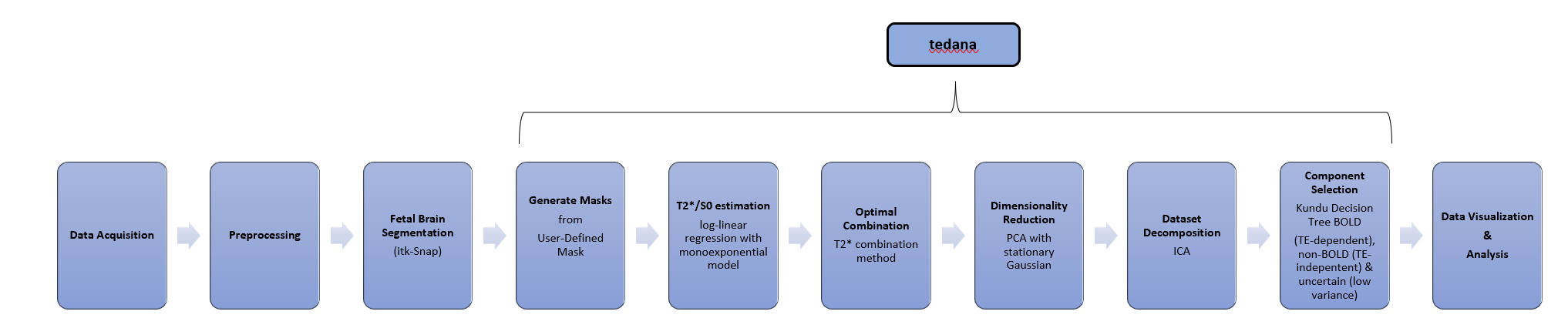
80 fetal MRI scans were performed after informed consent was provided (MEERKAT, REC 21/LO/0742, Dulwich Ethics Committee, 08/12/2021) on a 3T clinical Philips Achieva MRI scanner. MRI data was acquired with a whole uterus single shot 2D multi-echo gradient echo EPI sequence under free breathing. The voxel resolution was 3mm isotropic, TEs=[ 10.2,, 54.3,98.4,142.5, 186.6]ms, flip angle=90. A B0 map was acquired prior to the EPI scan and image-based shimming employed. The data was reconstructed following normal vendor pipelines including EPI ghost correction and gradient waveforms correction.Fetal brain segmentations were performed manually for two control cases in late gestation (36+6, 38 weeks). Two types of masks were obtained, a conservative segmentation of the fetal brain (carefully avoiding amniotic fluid around the fetal brain) and a more liberal mask. TE-dependent analysis (tedana) of the multi-echo fMRI was used to denoise the data. TE-dependence analysis was performed using the tedana workflow3, separately for each mask to assess its dependence. A two-stage masking procedure was applied for optimal combination, T2*/S0 estimation, denoising, and the component classification procedure. A monoexponential model was fit to the data at each voxel using log-linear regression to estimate T2* and S0 maps. Multi-echo data were then optimally combined using the T2* combination method11. Principal component analysis based on the PCA component estimation with a Moving Average process10 was applied to the optimally combined data for dimensionality reduction. The metrics kappa and rho were calculated as measures of TE-dependence and TE-independence, respectively. Independent component analysis was then used to decompose the dimensionally reduced dataset. Next, component selection was performed to identify BOLD (TE-dependent), non-BOLD (TE-independent), and uncertain (low-variance) components using the Kundu decision tree (v2.5)8.
Results
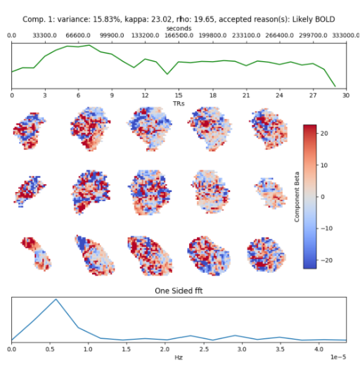
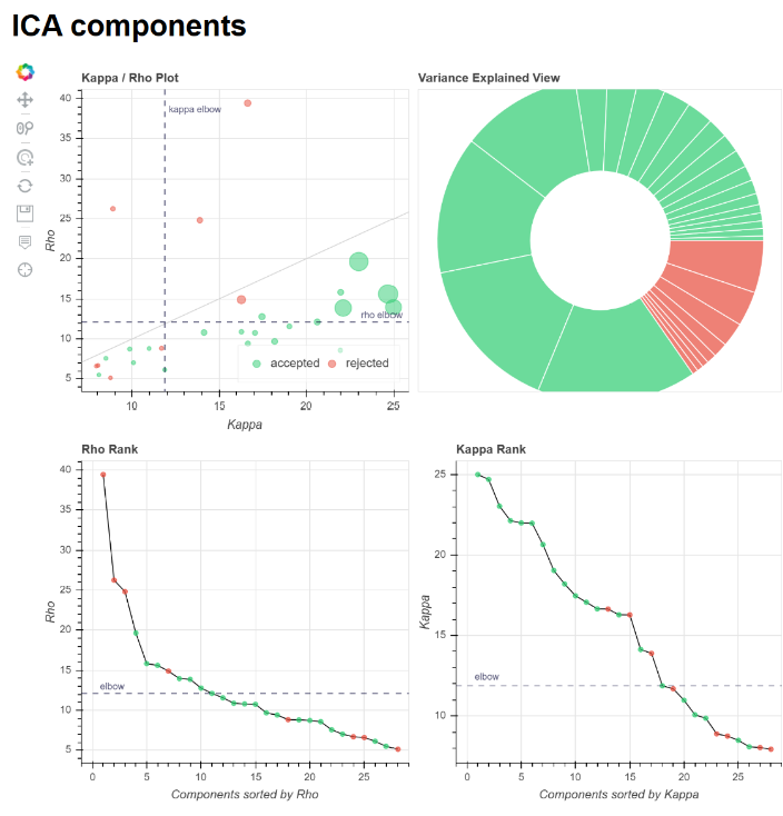
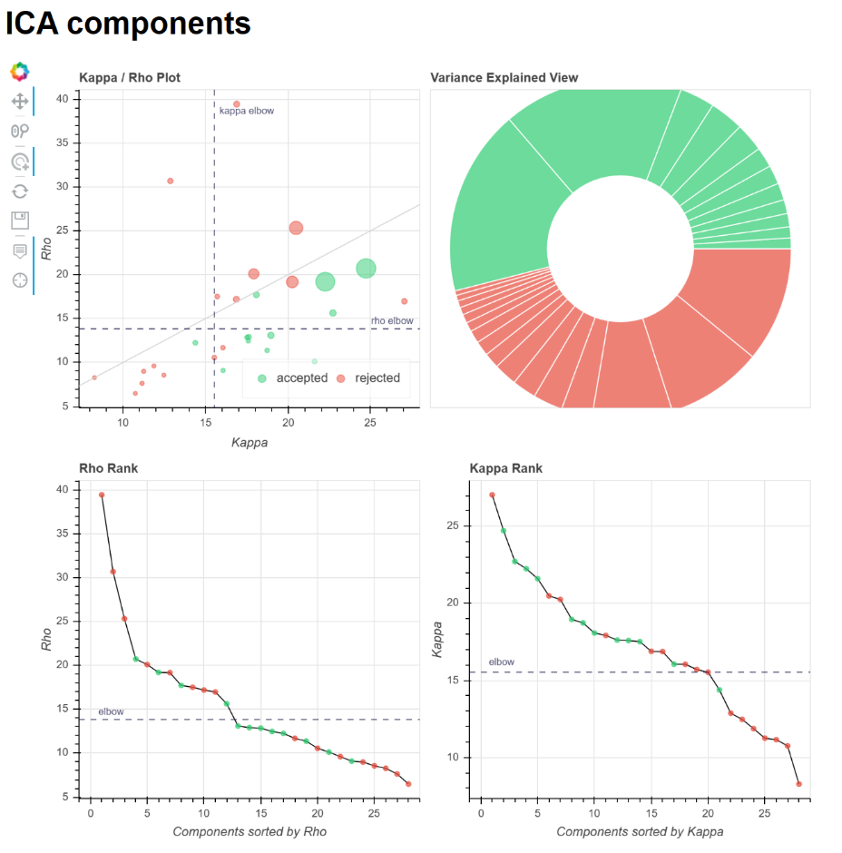
ME-ICA of the fetal brain of case 1 with 38 weeks gestation with a conservative mask yields many accepted BOLD components (20 out of 28) classified by the tedana workflow as “likely BOLD”, with the two top components explaining 15.8% and 15.6% of the variance respectively. ME-ICA with the liberal fetal brain mask detects fewer BOLD components (12 out of 28). Here, the two components with the highest variances are found at 17.7% and 17.1%.ME-ICA of case 2 with 36+6 weeks gestation results in 13 total components, of which 6 are classified as “likely BOLD” using a liberal fetal brain mask. Here, the two highest variances of the accepted components hold the values 23.8% and 21.3%. Applying the conservative mask yields 4 components, none of which were classified as “likely BOLD”.
Conclusion
The tedana workflow successfully decomposes the data into independent components for both, the conservative and liberal masks, and classifies them as likely or unlikely BOLD. Hence, initial results indicate that the choice of brain mask is an important factor.This exploratory study is limited by the number of cases investigated and by fetal motion occurring during scanning. However, application of the tedana workflow shows promising results in determining BOLD components. Hence, multi-echo fMRI has the potential to provide an improved analysis of the fetal brain in its rather inhomogeneous and noisy environment.
The possibilities of multi-echo fMRI and the potential to achieve more specific and higher quality functional analysis of the developing human brain are exciting - including research questions addressing the influence of the intra-uterine environment as well as a potential role in the clinical assessment of complex neuropathologies. In the next steps, temporal realignment of the data could improve the accuracy of the results. Currently, a larger cohort of control cases as well as cases with pathologies will be investigated with the tedana workflow.
Acknowledgements
This research was supported by DFG Heisenberg funding [502024488].References
- Cohen, A. D., Chang, C., & Wang, Y. (2021). Using multiband multi-echo imaging to improve the robustness and repeatability of co-activation pattern analysis for dynamic functional connectivity. NeuroImage, 243. https://doi.org/10.1016/j.neuroimage.2021.118555
- Dosenbach, N. U. F., Nardos, B., Cohen, A. L., Fair, D. A., Power, J. D., Church, J. A., Nelson, S. M., Wig, G. S., Vogel, A. C., Lessov-Schlaggar, C. N., Barnes, K. A., Dubis, J. W., Feczko, E., Coalson, R. S., Pruett, J. R., Barch, D. M., Petersen, S. E., & Schlaggar, B. L. (2010). Prediction of individual brain maturity using fMRI. Science, 329(5997), 1358–1361. https://doi.org/10.1126/science.1194144
- DuPre, E., Salo, T., Ahmed, Z., Bandettini, P., Bottenhorn, K., Caballero-Gaudes, C., Dowdle, L., Gonzalez-Castillo, J., Heunis, S., Kundu, P., Laird, A., Markello, R., Markiewicz, C., Moia, S., Staden, I., Teves, J., Uruñuela, E., Vaziri-Pashkam, M., Whitaker, K., & Handwerker, D. (2021). TE-dependent analysis of multi-echo fMRI with tedana. Journal of Open Source Software, 6(66), 3669. https://doi.org/10.21105/joss.03669
- Ferrazzi, G., Kuklisova Murgasova, M., Arichi, T., Malamateniou, C., Fox, M. J., Makropoulos, A., Allsop, J., Rutherford, M., Malik, S., Aljabar, P., & Hajnal, J. V. (2014). Resting State fMRI in the moving fetus: A robust framework for motion, bias field and spin history correction. NeuroImage, 101, 555–568. https://doi.org/10.1016/j.neuroimage.2014.06.074
- Fitzgerald, E., Hor, K., & Drake, A. J. (2020). Maternal influences on fetal brain development: The role of nutrition, infection and stress, and the potential for intergenerational consequences. Early Human Development, 150. https://doi.org/10.1016/j.earlhumdev.2020.105190
- Karolis, V. R., Fitzgibbon, S. P., Cordero-Grande, L., Farahibozorg, S. R., Price, A. N., Hughes, E. J., Fetit, A. E., Kyriakopoulou, V., Pietsch, M., Rutherford, M. A., Rueckert, D., Hajnal, J. V., Edwards, A. D., O’Muircheartaigh, J., Duff, E. P., & Arichi, T. (2023). Maturational networks of human fetal brain activity reveal emerging connectivity patterns prior to ex-utero exposure. Communications Biology, 6(1). https://doi.org/10.1038/s42003-023-04969-x
- Kundu, P., Benson, B. E., Baldwin, K. L., Rosen, D., Luh, W. M., Bandettini, P. A., Pine, D. S., & Ernst, M. (2015). Robust resting state fMRI processing for studies on typical brain development based on multi-echo EPI acquisition. Brain Imaging and Behavior, 9(1), 56–73. https://doi.org/10.1007/s11682-014-9346-4
- Kundu, P., Brenowitz, N. D., Voon, V., Worbe, Y., Vértes, P. E., Inati, S. J., Saad, Z. S., Bandettini, P. A., & Bullmore, E. T. (2013). Integrated strategy for improving functional connectivity mapping using multiecho fMRI. Proceedings of the National Academy of Sciences of the United States of America, 110(40), 16187–16192. https://doi.org/10.1073/pnas.1301725110
- Kundu, P., Voon, V., Balchandani, P., Lombardo, M. V., Poser, B. A., & Bandettini, P. A. (2017). Multi-echo fMRI: A review of applications in fMRI denoising and analysis of BOLD signals. NeuroImage, 154, 59–80. https://doi.org/10.1016/j.neuroimage.2017.03.033
- Li, Y. O., Adali, T., & Calhoun, V. D. (2007). Estimating the number of independent components for functional magnetic resonance imaging data. Human Brain Mapping, 28(11), 1251–1266. https://doi.org/10.1002/hbm.20359
- Posse, S., Wiese, S., Gembris, D., Mathiak, K., Kessler, C., Grosse-Ruyken, M. L., Elghahwagi, B., Richards, T., Dager, S. R., & Kiselev, V. G. (1999). Enhancement of BOLD-contrast sensitivity by single-shot multi-echo functional MR imaging. Magnetic Resonance in Medicine, 42(1), 87–97. https://doi.org/10.1002/(SICI)1522-2594(199907)42:1<87::AID-MRM13>3.0.CO;2-O
- Prayer, D., Malinger, G., De Catte, L., De Keersmaecker, B., Gonçalves, L. F., Kasprian, G., Laifer-Narin, S., Lee, W., Millischer, A. E., Platt, L., Prayer, F., Pugash, D., Salomon, L. J., Sanz Cortes, M., Stuhr, F., Timor-Tritsch, I. E., Tutschek, B., Twickler, D., & Raine-Fenning, N. (2023). ISUOG Practice Guidelines (updated): performance of fetal magnetic resonance imaging. Ultrasound in Obstetrics and Gynecology, 61(2), 278–287. https://doi.org/10.1002/uog.26129
- Rees, S., & Harding, R. (2004). Brain development during fetal life: Influences of the intra-uterine environment. Neuroscience Letters, 361(1–3), 111–114. https://doi.org/10.1016/j.neulet.2004.02.002
- Story, L., & Rutherford, M. (2015). Advances and applications in fetal magnetic resonance imaging. The Obstetrician & Gynaecologist, 17(3), 189–199. https://doi.org/10.1111/tog.12203
- Stout, J. N., Bedoya, M. A., Grant, P. E., & Estroff, J. A. (2021). Fetal Neuroimaging Updates. In Magnetic Resonance Imaging Clinics of North America (Vol. 29, Issue 4, pp. 557–581). W.B. Saunders. https://doi.org/10.1016/j.mric.2021.06.007
Figures