0821
Evaluation of high-resolution fetal brain anatomical imaging with a reduced field of view using outer volume suppression1Department of Radiology, Weill Cornell Medicine, New York, NY, United States, 2Department of Obstetrics and Gynecology, Weill Cornell Medicine, New York, NY, United States
Synopsis
Keywords: Fetal, Fetus
Motivation: Conventional anatomical imaging of the fetal brain is limited by low resolution due to an inherently large field-of-view and a restricted matrix size.
Goal(s): To achieve high-resolution fetal brain imaging and evaluate the image quality compared to conventional fetal brain imaging.
Approach: Fetal brain anatomical imaging was performed using optimized outer volume suppression for higher resolution. Image quality was scored by neuroradiologists, and image sharpness was calculated using gradient norms.
Results: High-resolution anatomical images acquired using our approach demonstrated improved image quality both quantitatively and qualitatively, without an increased scan time.
Impact: High-resolution fetal brain anatomical imaging with a reduced field-of-view achieved by optimized outer volume suppression demonstrates improved image quality compared to conventional imaging methods. This approach may help increase diagnostic accuracy in identifying brain abnormalities in utero.
Introduction
Single-shot fast-spin-echo (SSFSE) is a standard method for fetal brain anatomical imaging due to its short scan time and T2 contrast. However, the image quality is suboptimal and limited by low spatial resolution because the field-of-view (FOV) is inherently large in fetal MRI to avoid aliasing from maternal tissue, and the acquisition matrix size is restricted by T2 decay that occurs during SSFSE readout. We have previously proposed high-resolution SSFSE imaging with a reduced FOV using outer volume suppression (OVS)1. Here, we evaluated the image quality of the proposed approach quantitatively and qualitatively, in comparison to the conventional SSFSE.Methods
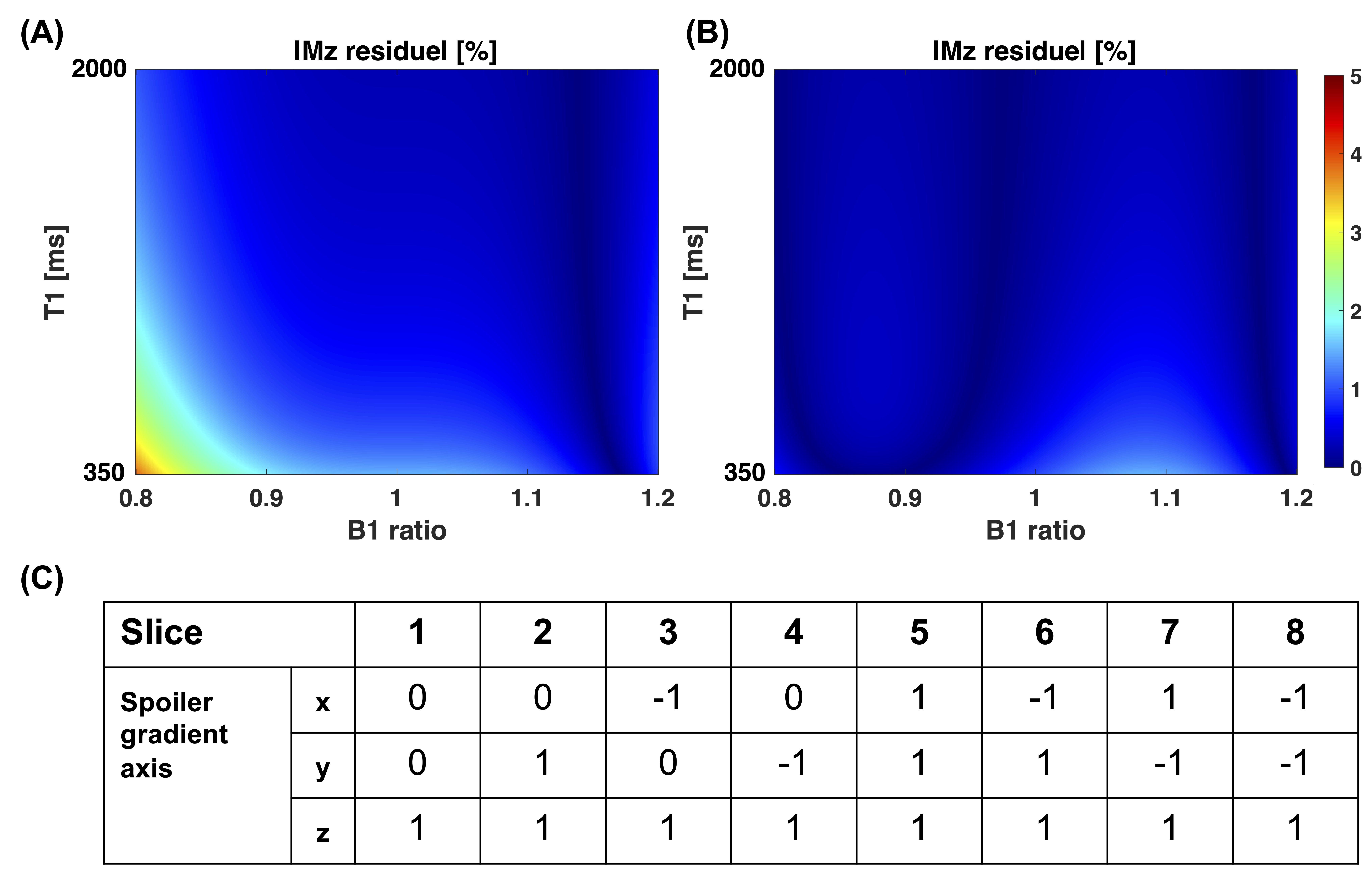
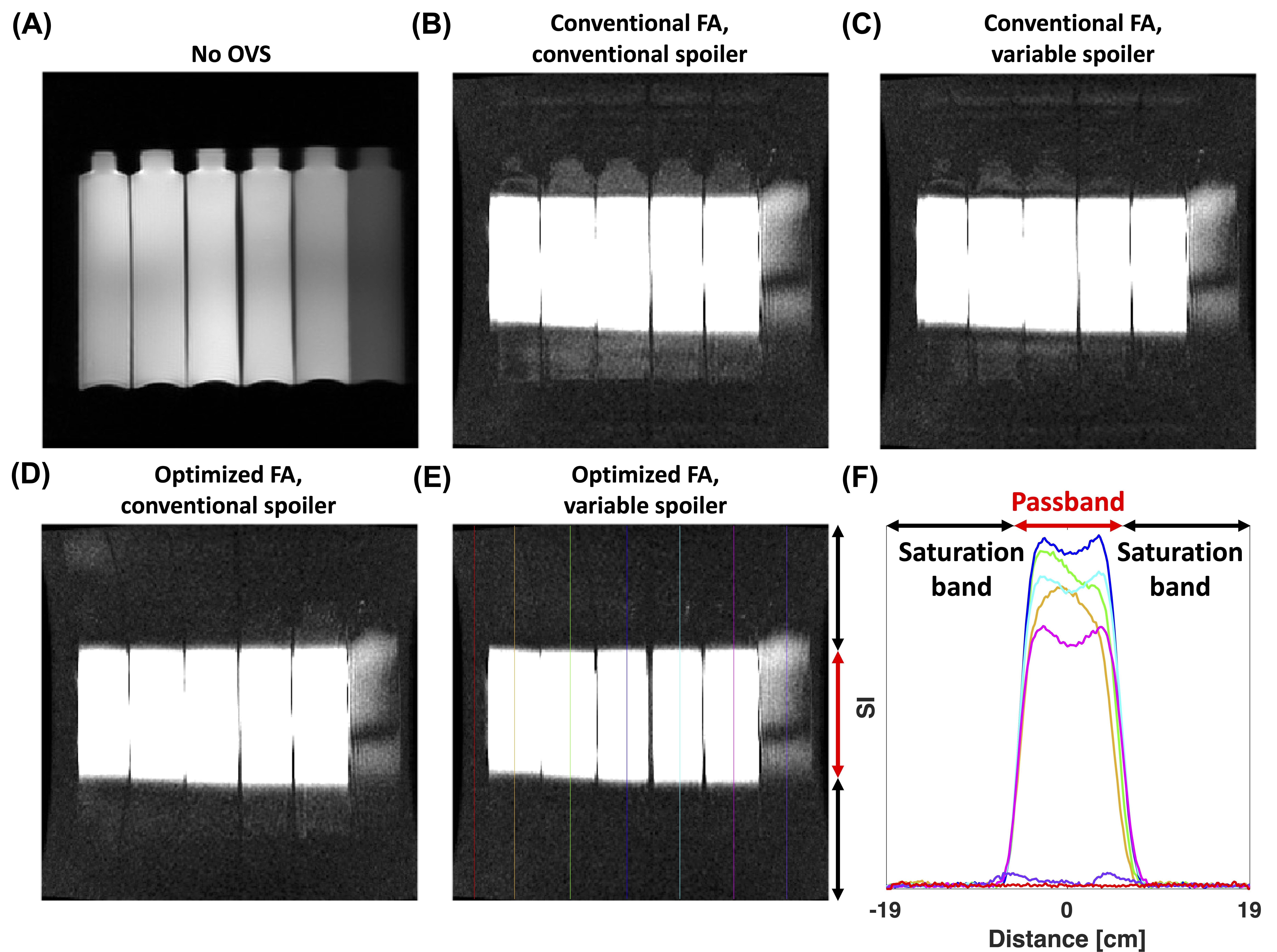
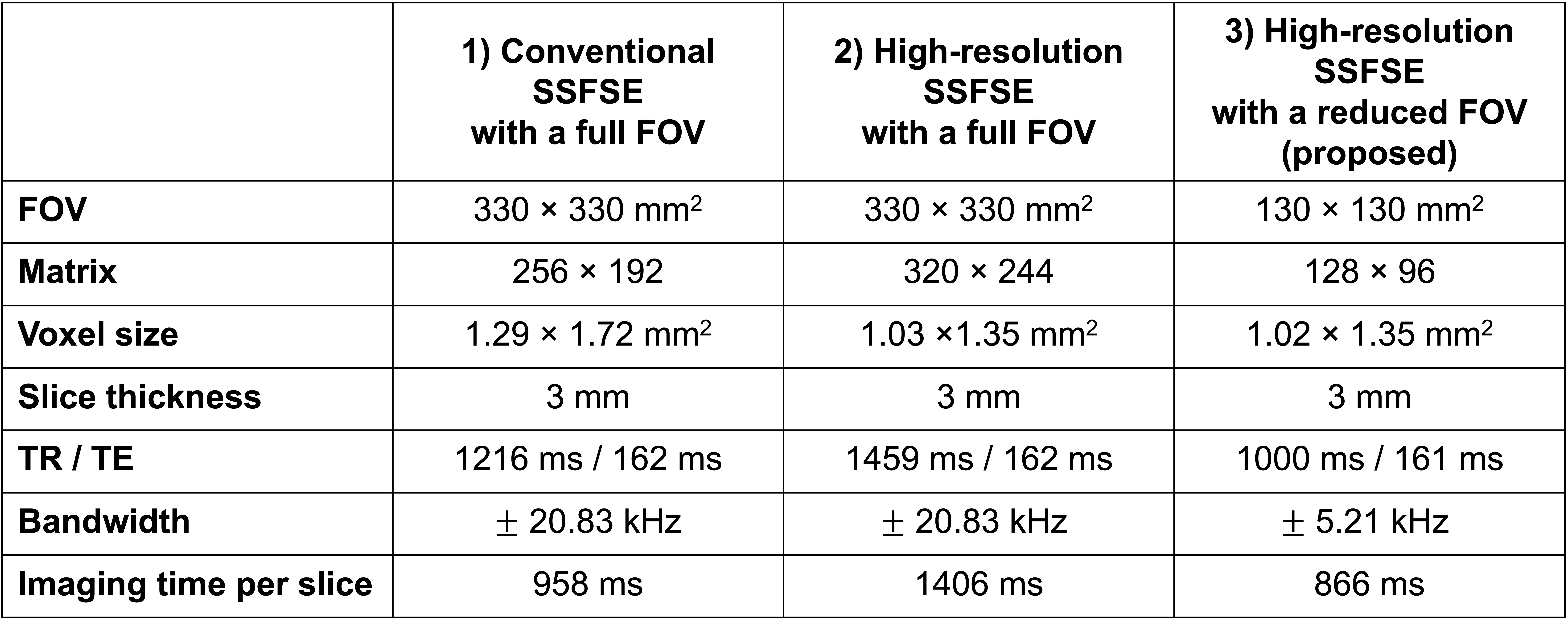
Pulse sequence: We have further improved the previously proposed OVS pulses as follows. We designed the OVS pulse by modulating a Hamming-windowed Shinnar-Le Roux RF pulse2,3 with a cosine waveform. The designed RF pulse followed by a gradient spoiler was repeated three times to improve the overall OVS results. Instead of three 90° flip angles (FAs), we used a combination of 83°-83°-117° that was found by an exhaustive search to minimize the maximum |Mz| in the saturation bands in the ranges of B1 ratio (0.8–1.2) and T1 (350–2000 ms) (Fig. 1A-B)4,5. Furthermore, we varied the directions and areas of spoiler gradients across repetitions to avoid stimulated echoes6. The gradient directions were varied across imaging slices as shown in Fig. 1C, and the ratio of three gradient areas within each slice was 4:2:1.Image acquisition: We performed OVS-SSFSE in T1 phantoms with and without optimized FAs and variable spoiler gradients. In 7 healthy pregnant women (gestational age: 31.41±2.78 weeks), we performed in-vivo scans using three different imaging methods: (1) conventional SSFSE with a full FOV, (2) high-resolution SSFSE with a full FOV, and (3) high-resolution SSFSE with a reduced FOV using OVS (proposed). Scan parameters are summarized in Table 1. All experiments were performed on a 1.5T GE scanner.
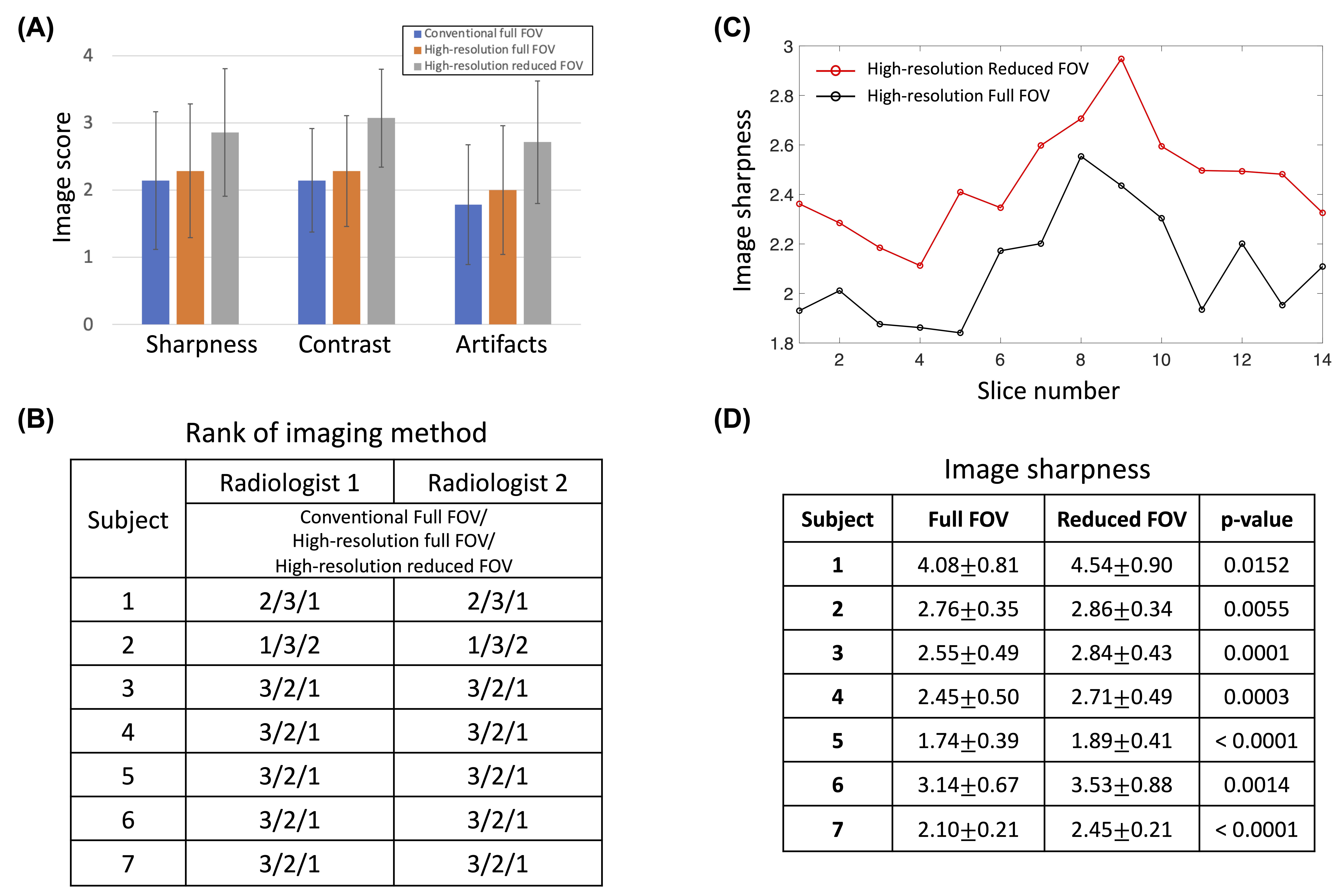
Qualitative analysis: Two experienced neuroradiologists independently graded the image quality of each acquisition method for each subject in three criteria of sharpness, contrast, and artifacts using a 4-point scale (1=poor; 2=fair; 3=good; 4=excellent), blinded to the acquisition method. Furthermore, the neuroradiologists determined the rank of each method based on overall image quality (1=best; 2=intermediate; 3=worst).
Quantitative analysis: We calculated image sharpness of the brain structures to compare the high-resolution images with a full FOV and those with a reduced FOV. To this end, we matched the matrix size between the two datasets using interpolation and manually segmented the whole brain. We then estimated the image sharpness within the segmented brain for each slice using the following equation: $$$\frac{1}{n}\sum_{1}^{n}\sqrt{\left ( \frac{\partial f}{\partial x} \right )^{2}+\left ( \frac{\partial f}{\partial y} \right )^{2}}$$$, where n is the number of pixels and f is the signal intensity.
Results
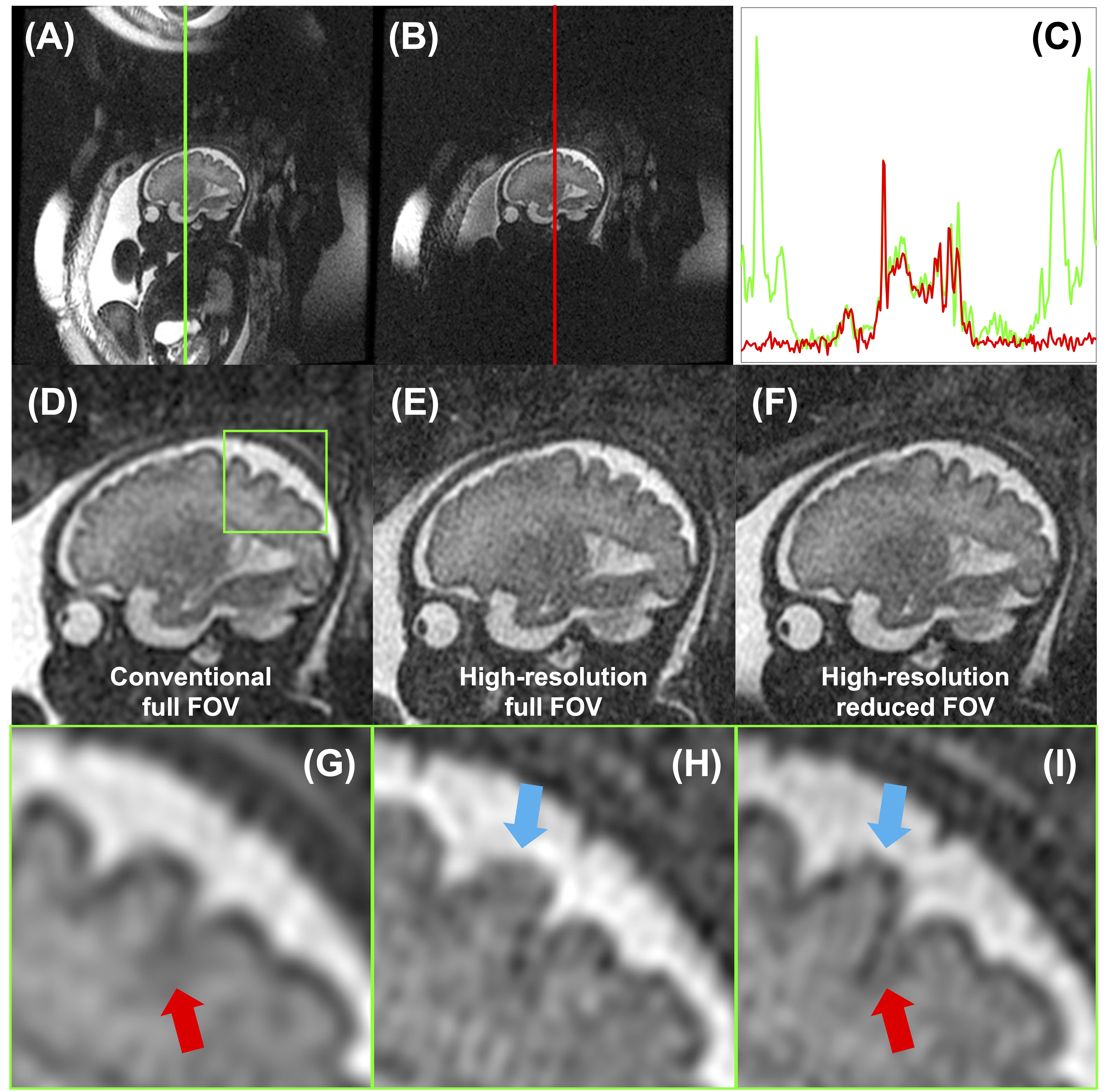
In phantom scans, the residual signals in the saturation bands were found to be the lowest when both optimized FAs and variable spoilers were used among the four combinations (Fig. 2). Excellent signal suppression was demonstrated for various T1 values (161-2199 ms). The optimized OVS pulses were also used for in-vivo scans. Maternal tissue and amniotic fluid signals were well suppressed in the saturation bands, and the signals in the passband were comparable to those without OVS pulses (Fig. 3A-C). Visualization of the brain structures was improved in high-resolution SSFSE images with a reduced FOV (Fig. 3D-I). Across all subjects, high-resolution imaging with a reduced FOV achieved higher mean image scores than the other methods in all three criteria. Both neuroradiologists rated the proposed approach as the best method in all subjects except one (Fig. 4A-B). Calculated image sharpness of high-resolution images was significantly higher with a reduced FOV than full FOV in all subjects (p<0.0152) (Fig. 4C-D).Discussion and Conclusion
Simply increasing the acquisition matrix size of conventional SSFSE may not be an effective approach to obtaining high-resolution fetal brain images because of greater T2 decay and increased susceptibility to fetal motion associated with a prolonged imaging time. We achieved high-resolution imaging even with a decreased matrix size and scan time afforded by a reduced FOV, and demonstrated improved image quality compared to the conventional methods. The proposed approach may be easily translated into the clinical setting, given that the neuroradiologists determined that the proposed method outperforms the current standard method in most of our subjects; no extra effort is required from the operator in prescribing the proposed sequence; and the scan time is shorter than that of the conventional methods. The diagnostic accuracy of the proposed method in identifying brain abnormalities must be validated in future studies in comparison with the conventional methods.Acknowledgements
R01HD100012References
1. Jang MJ, Zun Z, High-resolution fetal brain anatomical imaging using a reduced field-of-view with outer volume suppression. ISMRM, 2023. p 2333.
2. Pauly J, Le Roux P, Nishimura D, Macovski A. Parameter relations for the Shinnar-Le Roux selective excitation pulse design algorithm (NMR imaging). IEEE transactions on medical imaging. 1991;10:53-65.
3. Holbrook AB, Santos JM, Kaye E, Rieke V, Pauly KB. Real-time MR thermometry for monitoring HIFU ablations of the liver. Magnetic Resonance in Medicine. 2010;63:365-373.
4. Chow K, Kellman P, Spottiswoode BS, et al. Saturation pulse design for quantitative myocardial T1 mapping. Journal of Cardiovascular Magnetic Resonance. 2015;17:1-5.
5. Sung K, Nayak KS. Design and use of tailored hard-pulse trains for uniformed saturation of myocardium at 3 Tesla. Magnetic Resonance in Medicine. 2008;60:997-1002.
6. Zun Z, Hargreaves BA, Rosenberg J, Zaharchuk G. Improved multislice perfusion imaging with velocity‐selective arterial spin labeling. Journal of Magnetic Resonance Imaging. 2015;41:1422-1431.
Figures