0819
Normative growth models for T2w 0.55T fetal brain and body MRI: population-averaged 4D atlases and volumetry centiles1Department of Biomedical Engineering, King's College London, London, United Kingdom, 2Centre for the Developing Brain, King's College London, London, United Kingdom, 3Department of Women and Children’s Health, King's College London, London, United Kingdom, 4Fetal Medicine Unit, Guy’s and St Thomas’ NHS Foundation Trust, London, United Kingdom, 5Radiological Institute, University Hospital Erlangen, Erlangen, Germany
Synopsis
Keywords: Fetal, Fetus
Motivation: Low field MRI is a promising direction for fetal imaging. Yet, there are no reported models of fetal development specific to 0.55T.
Goal(s): We aim to formalise normal fetal growth models for structural 0.55T MRI.
Approach: We use registration-based approach for generation of spatio-temporal templates from 3D D/SVR reconstructed images of the fetal brain and body and apply deep learning segmentation to parcellate organs for volumetry from >100 control subjects.
Results: This work introduces the first T2w fetal atlases and volumetry growth charts for 0.55T brain and body MRI depicting normal development across 22-38 weeks gestation. All models are publicly available online.
Impact: This work is the first step toward formalisation of analysis protocols for normal fetal brain and body development and optimisation of segmentation methods for low field strength fetal MRI.
Introduction
Recently, low field strength MRI at 0.55T has emerged as a promising new direction for fetal imaging due to reduced susceptibility artefacts and improved accessibility by enabling a larger bore size, thereby facilitating imaging of women with a raised BMI, whilst maintaining good field homogeneities. It provides clinically acceptable image information for the fetal brain and body1 with standard quantitative measurements showing good correlation with 1.5-3T ranges2.T2-weighted single-shot turbo spin echo (ssTSE, HASTE) imaging is the work horse for structural fetal examination. However, due to the novelty of low field fetal MRI, there are yet no reported population-averaged atlases or volumetry growth charts of fetal development specific to 0.55T.
This work presents the first formalisation of growth models for structural 0.55T fetal MRI created from 131 normal control datasets across the 21-39 weeks gestational age (GA) range.
Methods
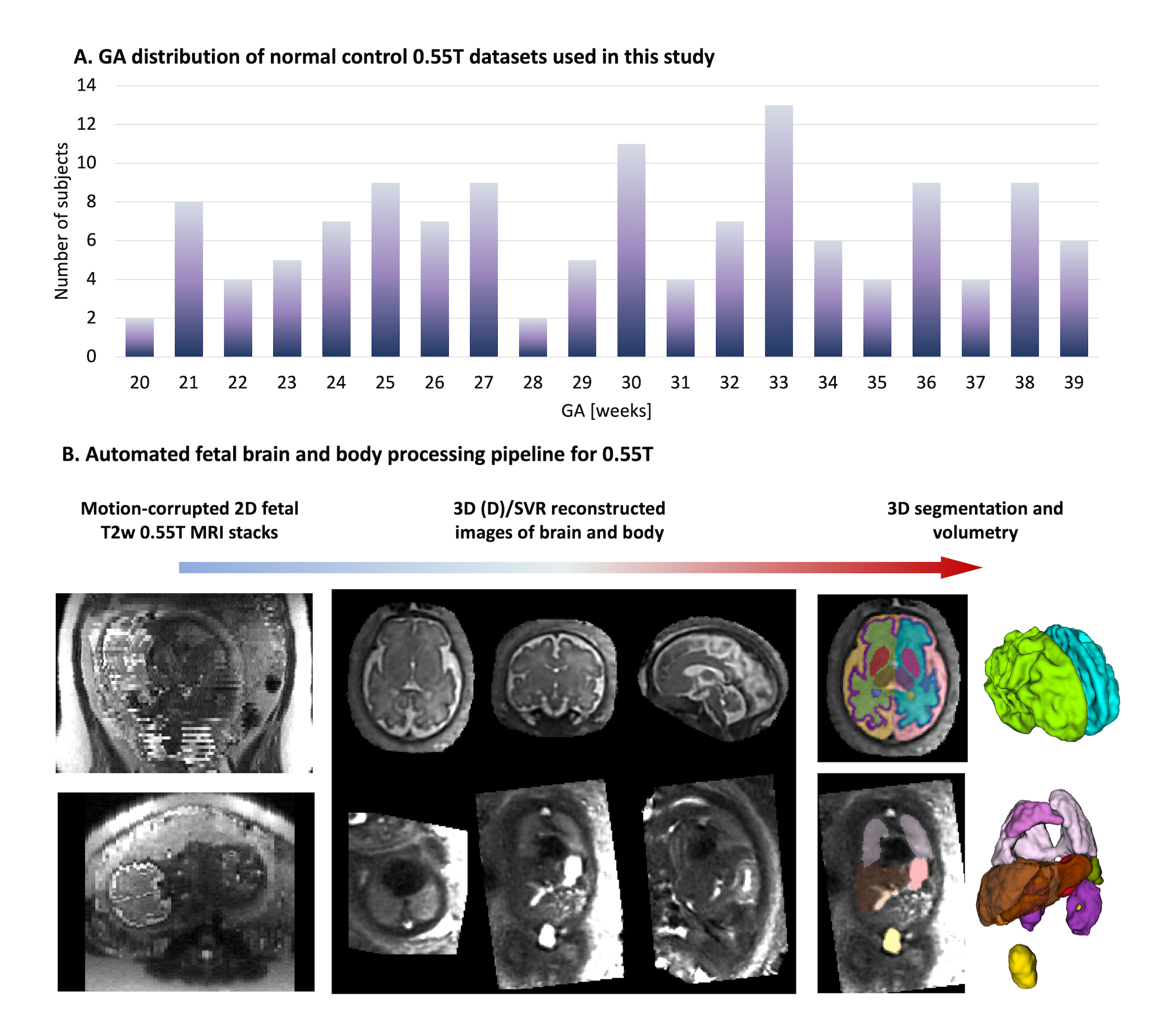
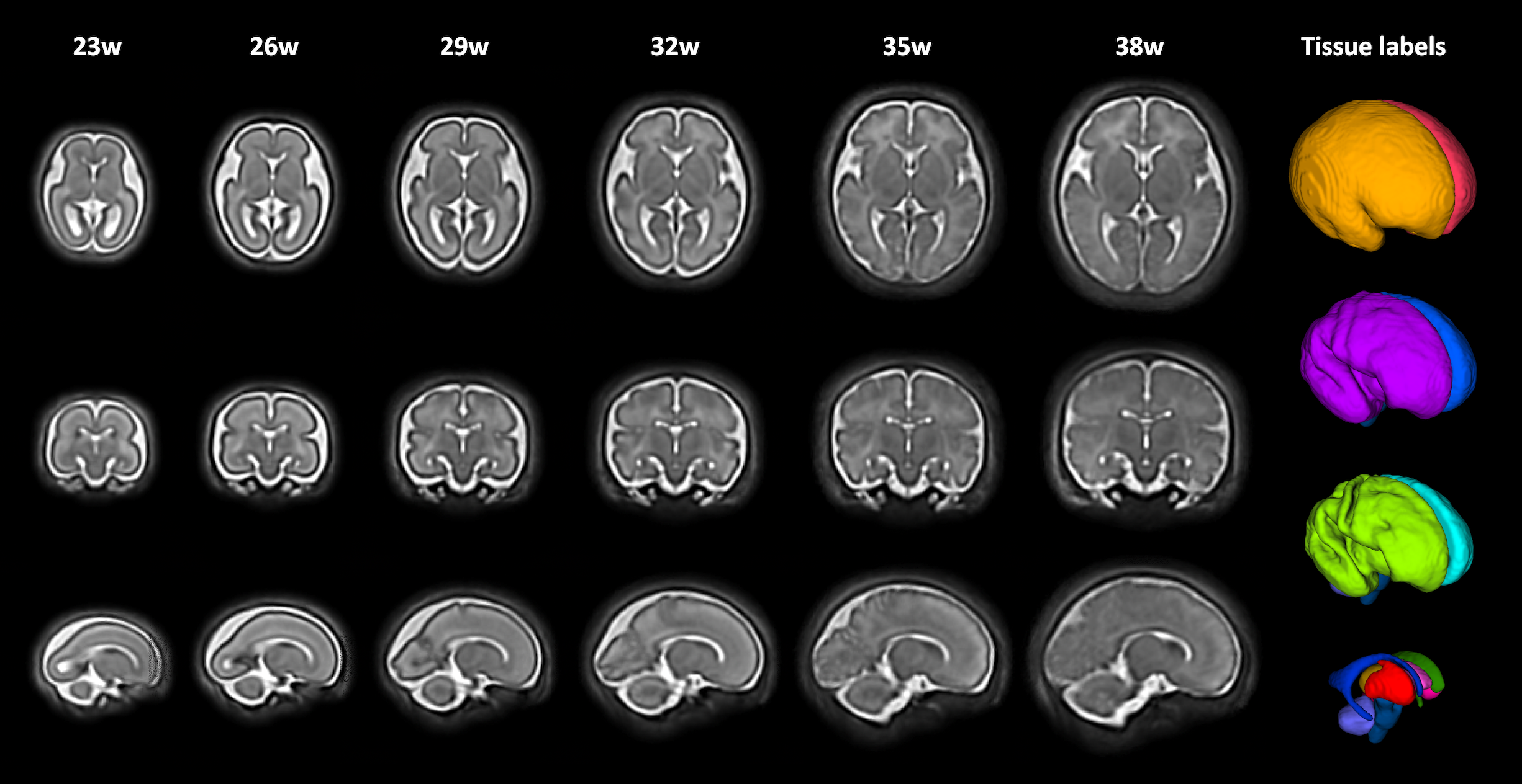
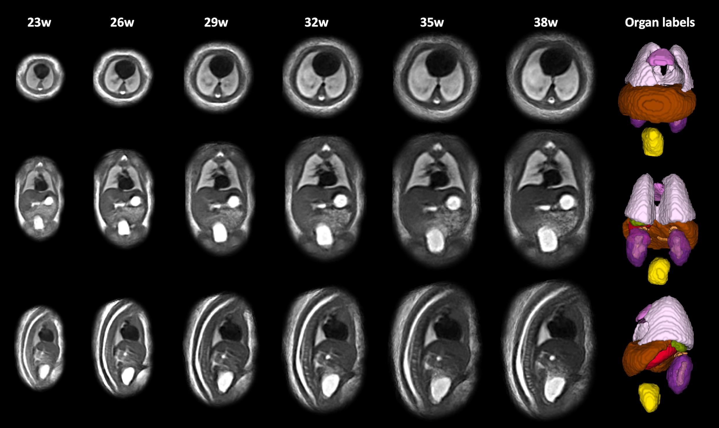
Datasets and preprocessing: The fetal MRI data includes 131 datasets of normal control fetuses (20-39 weeks GA, Fig.1A) acquired at St. Thomas’s Hospital, London as part of the MEERKAT study [REC: 21/LO/0742] on a contemporary clinical 0.55T scanner (MAGNETOM Free.Max, Siemens Healthcare, Erlangen, Germany) using a HASTE sequence with TE=105–106ms optimised for low field fetal MRI3. Exemplary images are shown in Fig.1B. Each dataset includes 6-11 stacks with 1.48mm in-plane resolution, 4.5mm slice thickness. The 3D reconstructions of the fetal brain and body (1.2mm resolution, standard radiological space) were performed using the in-house automated rigid and deformable slice-to-volume registration (D/SVR) pipelines4,5 in SVRTK6,7 adapted to the 0.55T protocol (retrained 3D localisation and reorientation networks). The inclusion criteria were: no reported fetal or placental anomalies, no extreme deviations in the anatomy and acceptable SNR and reconstruction quality (sufficient visibility for differentiation of organ boundaries and tissue texture).Generation of population-averaged 4D atlases: The atlases were created using MIRTK8 registration tools. After affine alignment to the existing T2w fetal brain9 and body10 atlases at 3T field strength, the transformed 3D brain (N=108) and body (N=106) images were averaged with a 1 week temporal sigma kernel into 17 timepoints with 0.8mm resolution. This was followed by 3 iterations of non-rigid registration and averaging with Laplacian sharpening. Automated segmentation of the fetal brain and body: In order to segment 3D brain and body images for volumetry, we employed the already existing in-house networks for 3D fetal brain tissue11 and body organ10 that were additionally trained for 1000 iterations with blurring and histogram matching augmentation in MONAI12 in order to adapt to 0.55T contrast appearance.
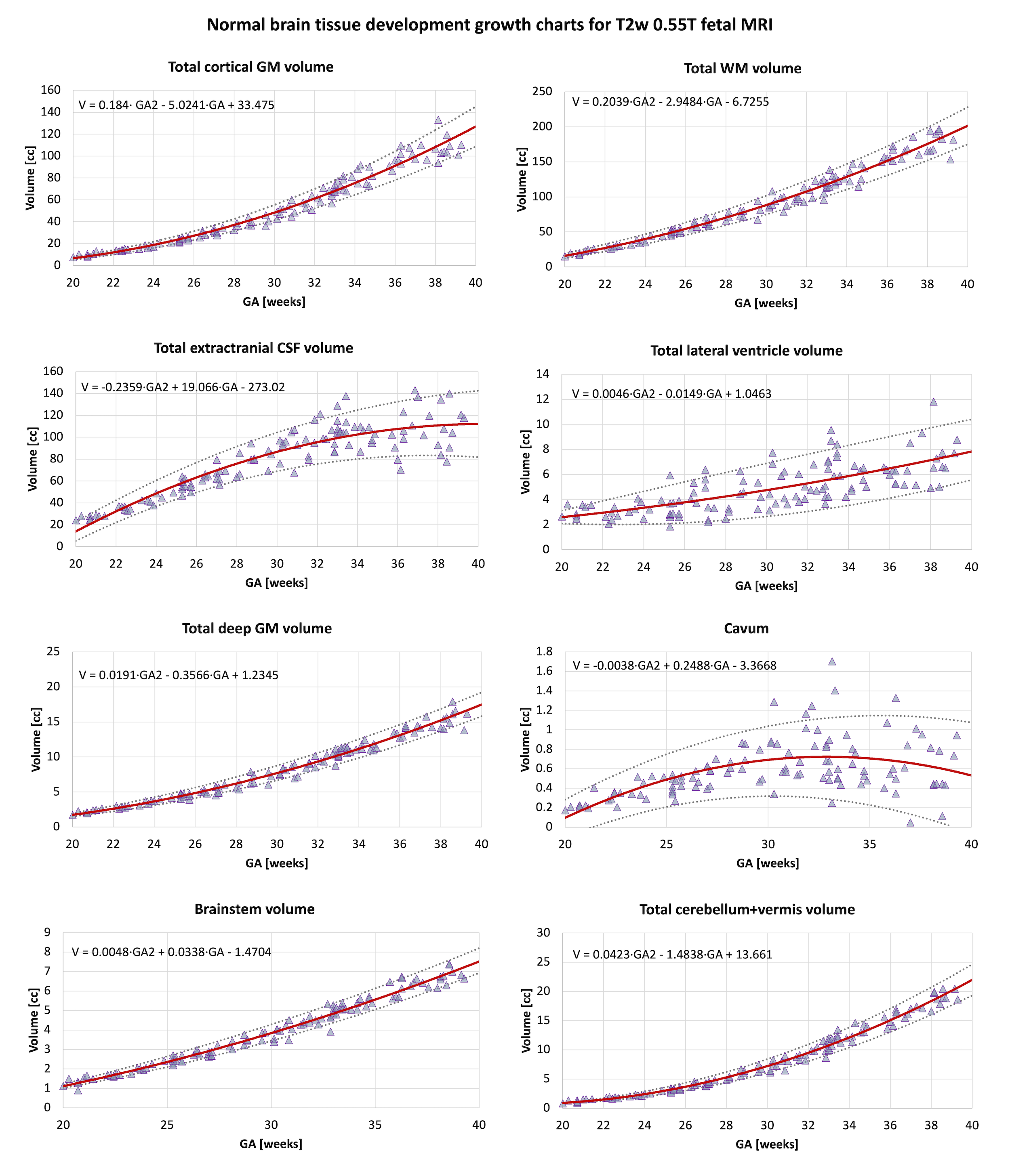
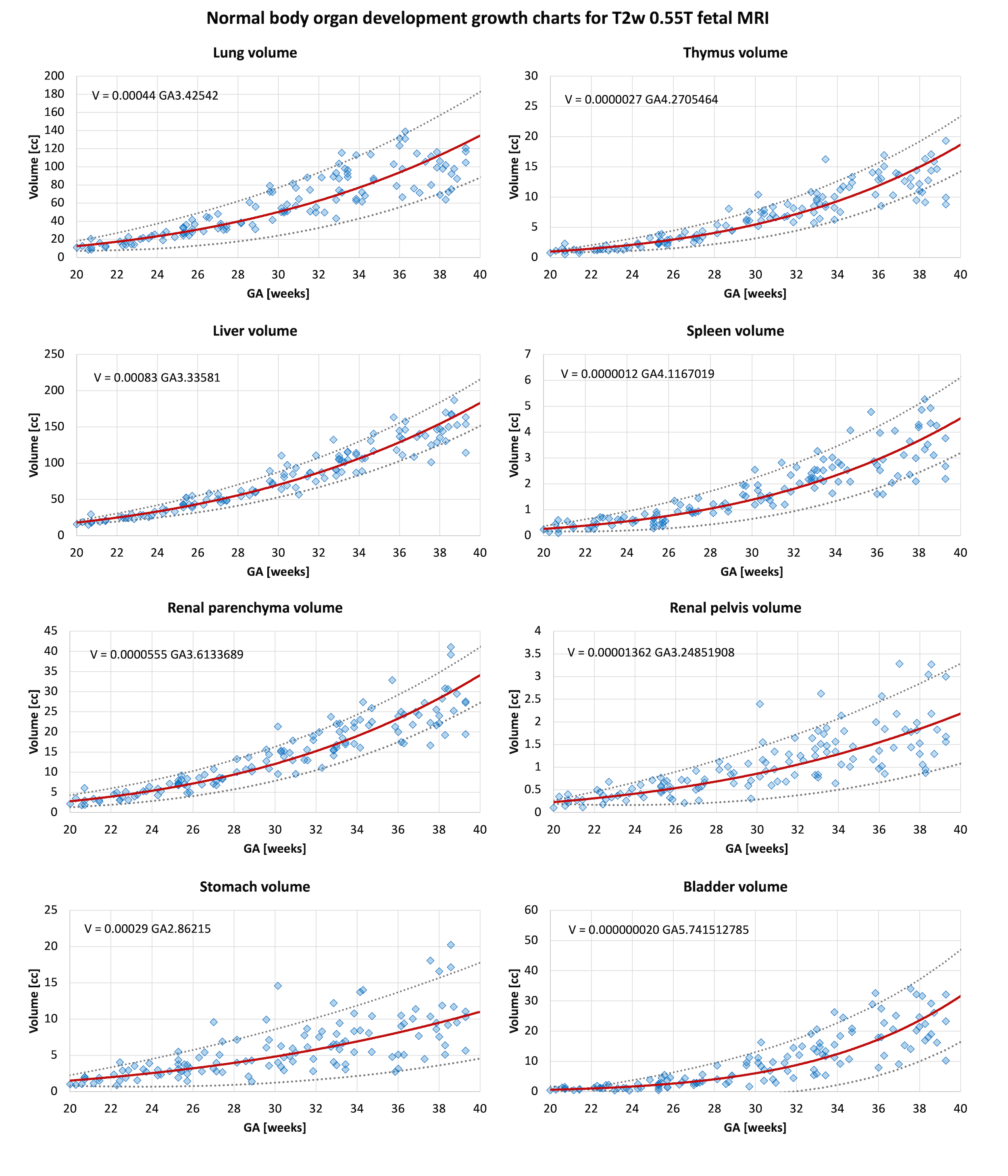
Volumetry growth charts: The brain tissue and body organ segmentation for all normal control subjects were reviewed by AU. In total, 115 brain and 125 body label files were selected based on acceptable segmentation quality. Additional minor manual refinements were performed in ~34% of the brain labels (primarily early < 22w and late > 33w) at the cortex and white matter interface and ~42% of body labels (lungs, liver and kidney ROIs). The growth charts were created using 2nd order polynomial and power fitting with 0.95, 0.5, and 0.05 centiles.
Results and Discussion
Population-averaged 4D fetal brain and body atlases: Fig.2-3 present the created 4D brain and body atlases covering the 22-38 weeks GA showing the expected increase in size as well as changes in cortical folding. While the image quality (in terms of sharpness and visibility of small features) is lower than to the recently published 3T atlases9,10 (with lower visibility and segmentation quality for the cortex ROI at later GA), they provide the baseline information about T2w appearance of fetal organs at 0.55T. The atlases were inspected by experienced fetal MRI clinicians (LS, MH) and were confirmed to have physiologically realistic anatomical features corresponding to normal fetal development.Growth charts of normal fetal development: The growth charts generated from 115 brain and 125 body segmentations show the expected increase in brain tissue and body organ volumes with GA with value ranges like the previously reported fetal MRI volumetry studies9,10,14. Future combined analysis with datasets from high field strength acquisitions will still require image harmonisation and potentially further optimisation of the segmentation pipelines.The atlases and centiles are publicly available online at KCL CDB data repository: https://gin.g-node.org/kcl_cdb/055t_fetal_mri_atlases 13.
Conclusions
This work introduces the first set of 4D population-averaged T2w fetal atlases and volumetry growth charts for 3D 0.55T brain and body MRI depicting normal development across 22-38 weeks GA range. All models are publicly available online. Our future work will focus on optimisation and harmonisation of automated segmentation methods for different acquisition parameters and anomalies.Acknowledgements
We thank everyone who was involved in acquisition and analysis of the datasets at the Department of Perinatal Imaging and Health at Kings College London and St Thomas' Hospital. We thank all participants and their families.
This work was supported by NIHR Advanced Fellowship awarded to Lisa Story [NIHR30166], by the Wellcome Trust, Sir Henry Wellcome Fellowship [201374/Z/16/Z], UKRI FLF [MR/T018119/1], and Heisenberg funding to JH [502024488], MRC grant [MR/W019469/1], the Wellcome/ EPSRC Centre for Medical Engineering at King’s College London [WT 203148/Z/16/Z], the NIHR Clinical Research Facility (CRF) at Guy’s and St Thomas’ and by the National Institute for Health Research Biomedical Research Centre based at Guy’s and St Thomas’ NHS Foundation Trust and King’s College London.
The views expressed are those of the authors and not necessarily those of the NHS, the NIHR or the Department of Health.
References
- [1] Ponrartana, S., Nguyen, H. T. N., Cui, S. X., Tian, Y., Kumar, P., Wood, J. C., & Nayak, K. S. (2023). Low-field 0.55 T MRI evaluation of the fetus. Pediatric Radiology, 53(7), 1469–1475. https://doi.org/10.1007/s00247-023-05604-x
- [2] Aviles Verdera, J., Story, L., Hall, M., Finck, T., Egloff, A., Seed, P. T., Malik, S. J., Rutherford, M. A., Hajnal, J. v, Tomi-Tricot, R., & Hutter, J. (2023). Reliability and Feasibility of Low-Field-Strength Fetal MRI at 0.55T during Pregnancy. Radiology, 309(1), e223050. https://doi.org/10.1148/radiol.223050
- [3] Aviles, J., Colford, K., Hall, M., Marenzana, M., Uus, A., Giles, S., Bridgen, P., Rutherford, M. A., Malik, S. J., Hajnal, J. v, Tomi-Tricot, R., & Hutter, J. (2022). A Fast Anatomical and Quantitative MRI Fetal Exam at Low Field. In MICCAI 2023 PIPPI Workshop (pp. 13–24). https://doi.org/10.1007/978-3-031-17117-8_2
- [4] Uus, A. U., Hall, M., Payette, K., Hajnal, J. v, Deprez, M., Rutherford, M. A., Hutter, J., & Story, L. (2023). Combined Quantitative T2* Map and Structural T2-Weighted Tissue-Specific Analysis for Fetal Brain MRI: Pilot Automated Pipeline. In MICCAI 2024 PIPPI Workshop. https://doi.org/10.1007/978-3-031-45544-5_3
- [5] Uus, A. U., Grigorescu, I., van Poppel, M. P. M., Steinweg, J. K., Roberts, T. A., Rutherford, M. A., Hajnal, J. v., Lloyd, D. F. A., Pushparajah, K., & Deprez, M. (2022). Automated 3D reconstruction of the fetal thorax in the standard atlas space from motion-corrupted MRI stacks for 21–36 weeks GA range. Medical Image Analysis, 80. https://doi.org/10.1016/j.media.2022.102484
- [6] “SVRTK Auto Reconstruction package.” [Online]. Available: https://github.com/SVRTK/auto-proc-svrtk. [Accessed: 01-Nov-2023].
- [7] Uus, A., Zhang, T., Jackson, L. H., Roberts, T. A., Rutherford, M. A., Hajnal, J. v., & Deprez, M. (2020). Deformable Slice-to-Volume Registration for Motion Correction of Fetal Body and Placenta MRI. IEEE Transactions on Medical Imaging, 29(9), 2750–2759. https://doi.org/10.1109/TMI.2020.2974844
- [8] “MIRTK package.” [Online]. Available: https://github.com/BioMedIA/MIRTK/. [Accessed: 01-Nov-2023].
- [9] Uus, A., Kyriakopoulou, V., Cordero-Grande, L., Christiaens, D., Pietsch, M., Price, A., Wilson, S., Patkee, P., Karolis, S., Schuh, A., Gartner, A., Williams, L., Hughes, E., Arichi, T., O’Muircheartaigh, J., Hutter, J., Robinson, E., Tournier, J. D., Rueckert, D., … Edwards, A. D. (2023). Multi-channel spatio-temporal MRI atlas of the normal fetal brain development from the developing Human Connectome Project. G-Node Open Data.
- [10] Uus, A. U., Hall, M., Grigorescu, I., Zampieri, C. A., Collado, A. E., Payette, K., Matthew, J., Kyriakopoulou, V., Hajnal, J. v, Hutter, J., Rutherford, M. A., Deprez, M., & Story, L. (2023). 3D T2w fetal body MRI: automated organ volumetry, growth charts and population-averaged atlas. MedRxiv, 2023.05.31.23290751. https://doi.org/10.1101/2023.05.31.23290751
- [11] Uus, A. U., Kyriakopoulou, V., Makropoulos, A., Fukami-Gartner, A., Cromb, D., Davidson, A., Cordero-Grande, L., Price, A. N., Grigorescu, I., Williams, L. Z. J., Robinson, E. C., Lloyd, D., Pushparajah, K., Story, L., Hutter, J., Counsell, S. J., Edwards, A. D., Rutherford, M. A., Hajnal, J. v, & Deprez, M. (2023). BOUNTI: Brain vOlumetry and aUtomated parcellatioN for 3D feTal MRI. ELife. 12:RP88818. https://doi.org/10.7554/elife.88818.1
- [12] Cardoso, M. J., Li, W., Brown, R., Ma, N., Kerfoot, E., Wang, Y., Murrey, B., Myronenko, A., Zhao, C., Yang, D., Nath, V., He, Y., Xu, Z., Hatamizadeh, A., Myronenko, A., Zhu, W., Liu, Y., Zheng, M., Tang, Y., … Feng, A. (2022). MONAI: An open-sourceframework for deep learning in healthcare. arXiv:2211.02701. http://arxiv.org/abs/2211.02701
- [13] “KCL CDB 0.55T fetal MRI data repository.” [Online]. Available: https://gin.g-node.org/kcl_cdb/055t_fetal_mri_atlases. [Accessed: 01-Nov-2023].
- [14] Meyers, M. L., Garcia, J. R., Blough, K. L., Zhang, W., Cassady, C. I., & Mehollin-Ray, A. R. (2018). Fetal Lung Volumes by MRI: Normal Weekly Values From 18 Through 38 Weeks’ Gestation. AJR. American Journal of Roentgenology, 211(2), 432–438. https://doi.org/10.2214/AJR.17.19469
Figures