0817
EX VIVO 11,7T MR MESOSCOPIC MULTIMODAL IMAGING OF FETAL BRAIN DEVELOPMENT1UMR 1141 NeuroDiderot, Eq inDEV, INSERM, Univeristé Paris Cité, Hôpital Robert Debré, Paris, France, 2UNIACT, NeuroSpin, CEA, Université Paris-Saclay, Gif-sur-Yvette, France, 3BAOBAB, NeuroSpin, Université Paris-Saclay, CNRS, CEA, Gif-sur-Yvette, France, Metropolitan, 4Unité fonctionnelle de fœtopathologie, AP-HP, Hôpital Universitaire Robert-Debré, Paris, France, Metropolitan, 5Service d'imagerie pédiatrique, AP-HP, Hôpital Robert-Debré, Paris, France, 6Service d'Anatomie Pathologique, AP-HP, Hôpital Lariboisière, Paris, France, Metropolitan, 7UNIACT, NeuroSpin, CEA, Université Paris-Saclay, Gif-sur-Yvette, France, Metropolitan, 8UNIACT, NeuroSpin, CEA, Université Paris-Saclay, Paris, France, Metropolitan
Synopsis
Keywords: Data Acquisition, Multimodal, Quantitative imaging, Mesoscopic
Motivation: White matter injuries are common in very premature babies and carry a significant risk of lifelong motor/cognitive disabilities.
Goal(s): Create mesoscopic resolution anatomical imaging, relaxometries, and diffusion MRI data to collect full 3D coverage of multiparametric information on tissue composition and connectivity, and compare to co-registered histology.
Approach: Each brain is imaged in situ at 3T shortly after death for registration purposes. After sample preparation, brains are imaged at 7T, then at 11.7T.
Results: We develop a unique multimodal mesoscopic (~100µm isotropic) post-mortem MRI atlas of brain development during the prenatal period (from 20 to 41 gestational weeks -GW) using 11,7T MRI.
Impact: The premature Human Connectome Project (p-HCP) provides the first mesoscopic multimodal quantitative MRI data at 11.7T of the anatomy, connectivity, cytoarchitecture, and microstructure of normal prenatal neurodevelopment during the second and third trimester of pregnancy.
Motivation & Goals
During the perinatal period, high vulnerability may lead to damage of cerebral tissues and carry significant risks of lifelong motor/cognitive disabilities.To finely characterize these lesions, we develop a unique multimodal mesoscopic (~100µm) post-mortem MRI atlas of brain development during the prenatal period (from 20 to 41 gestational weeks -GW) using a pre-clinical small bore 11,7T MRI [1].
Material & Methods
Currently, the cohort is composed of 5 postmortem fetal pseudo-typical brains of 20, 22, 23, 29 and 33 GW (with moderate ventricular hemorrhage, 22q11 deletion, focal abnormalities in diffusion imaging respectively for the last three).3T in situ MRI- Each brain was imaged in situ at 3T shortly after death. (3D T1w and T2w anatomical imaging with anisotropic resolution 250x250x800µm)
Sample preparation
- Fixation : Immersion in formalin (12% formaldehyde) for 2 months.
- Hydration : immersion in PBS (0.1M)/ Gadolinium (2mM)/ Azide (0.01%) for 1 month.
MRI protocol : Given the great variability in brain shape and MRI contrast of cerebral structures during prenatal brain development [2], parameters must be ajusted to each specimen age.
Protocol 7T clinical MRI – A Siemens Magnetom scanner with a 32 channel head coil was used with a T2w 3D Turbo Spin Echo sequence (TE/TR = 45/500 ms, ETL = 7, resolution = 400µm isotropic).
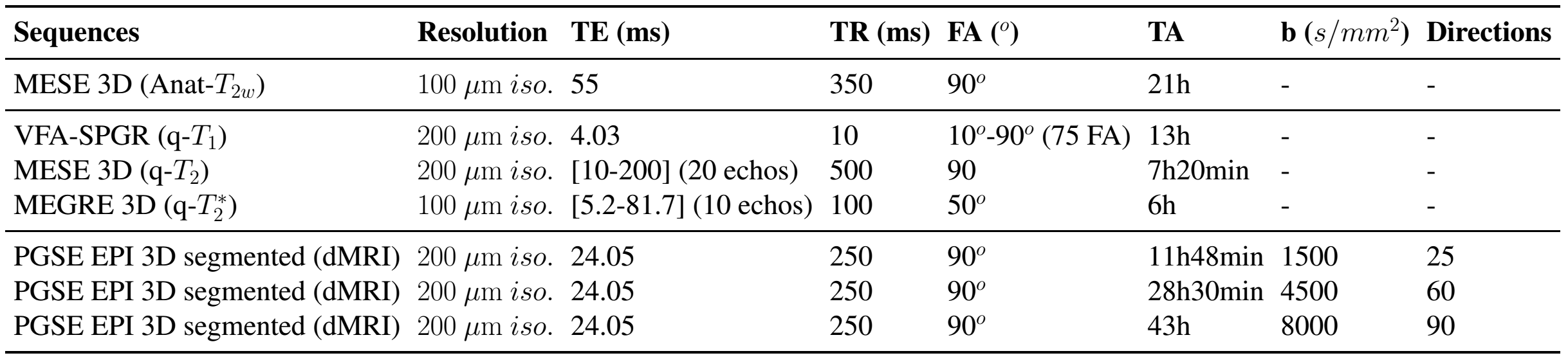
Protocol 11.7T pre-clinical MRI - A Bruker BioSpec 117/60 scanner with a 60 mm quadrature coil. The modalities included i. 3D Multi Echo Spin Echo (MESE) T2w for high resolution anatomical imaging, ii. a 3D Variable Flip Angle Spoiled Gradient Recalled (VFA-SPGR) sequence, a 3D MESE sequence, and a 3D Multi Echo Gradient Echo (MEGRE) sequence, for T1, T2, and T2* relaxometry respectively. Taking advantage of the strong gradients (760mT/m) of the 11.7T MRI, we included Diffusion Weighted Imaging following Pulsed Gradient Spin Echo (PGSE) scheme with a 3D Echo Planar Imaging (EPI) segmented reader module on 3 shells (see table 1 for details). The total acquisition time for a block was 150h.
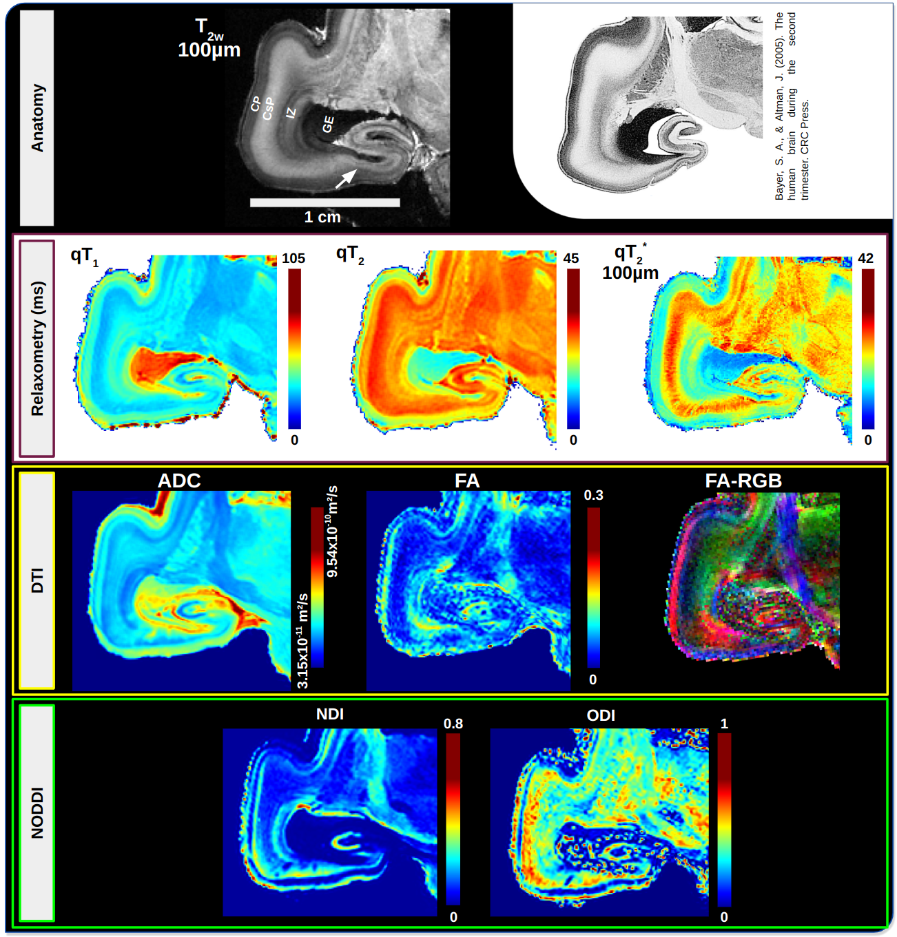
Post-processing - Quantitative T1, T2 and T2* maps were reconstructed from log-linear regression of VFA-SPGR, MESE and MEGRE data. Apparent Diffusion Coefficient (ADC) and Fractional Anisotropy (FA) were computed by fitting the Diffusion Tensor Imaging (DTI) model on the b = 4500 s/mm² shell. Neurite Density Index (NDI) and Orientation Dispersion Index (ODI) were obtained with the NODDI model [3] whose parameters were optimized using residuals minimization [4] (d//=6.48x10-10 m²/s). In the case of large specimens cut into several blocks, the whole hemispheres will be reconstructed based on a registration approach [5].
Results & Discussion
Thanks to our dedicated methodology, our data exhibit high SNR, and exemplary images on a 20 GW fetal brain (fig 1) reveal details seen so far on histology only. In this presentation, we focus on four anatomical structures : the ganglionic eminence (GE), the intermediate zone (IZ), the Cortical Subplate (CsP), and the Cortical Plate (CP) (see figure first line).The GE showed strong T2 hyposignal (T2w) with heterogeneous quantitative values (T1=80-90 ms, T2=20-28 ms, T2* =14-20 ms) and restricted diffusion (ADC=5,2-6,3x10⁻¹⁰m²/s), which is consistent with high cell density (neuronal progenitors). In the IZ, we could observe the microvasculature (qT2*) and a gradient of fiber orientation dispersion increasing up to the CsP (ODI from 0.4 to 0.8 and FA from 0.15 to 0.03). Also, we distinguished 3 waves of neuronal migration with quantitative values similar to the Cortical Plate (CP). We could also highlight the transition from 5 visible layers (future neocortex) to 3 layers (archeocortex) in the subiculum area. The transition from CsP to the CP was characterized by the fall of the fiber orientation dispersion (ODI from 0.8 to 0.2 and FA from 0.03 to 0.2) with a loss of axonal density (NDI from 0.4 to 0.06).
Perspectives & Conclusion
Thanks to this tailored methodology of acquisition and image processing, our mesoscopic imaging protocol shows details seen so far in histology and provides additional information on tissue composition and connectivity combined with full 3D coverage. The prenatal brain tissue maturation along gestational weeks will be studied through a systematic multiparametric study of dedicated ROIs. These unprecedented data however require to adapt and create new processing tools. Improving data processing and expending our dataset will allow us to describe neurodevelopmental mechanisms of both grey and white matter.These data will be confronted to co-registered histology data and made available in open access.
Acknowledgements
No acknowledgement found.References
[1] Dawood, Y., Strijkers, G. J., Limpens, J., Oostra, R. J., & De Bakker, B. S. (2020). Novel imaging techniques to study postmortem human fetal anatomy: a systematic review on microfocus-CT and ultra-high-field MRI. European Radiology, 30, 2280-2292.
[2] Vasung, L., Turk, E. A., Ferradal, S. L., Sutin, J., Stout, J. N., Ahtam, B., ... & Grant, P. E. (2019). Exploring early human brain development with structural and physiological neuroimaging. Neuroimage, 187, 226-254.
[3] Zhang, H., Schneider, T., Wheeler-Kingshott, C. A., & Alexander, D. C. (2012). NODDI: practical in vivo neurite orientation dispersion and density imaging of the human brain. Neuroimage, 61(4), 1000-1016.
[4] Alexander, D. C. (2008). A general framework for experiment design in diffusion MRI and its application in measuring direct tissue‐microstructure features. Magnetic Resonance in Medicine: An Official Journal of the International Society for Magnetic Resonance in Medicine, 60(2), 439-448.
[5] Beaujoin, J., Popov, A., Hot, R. Y., Poupon, F., Mangin, J. F., Destrieux, C., & Poupon, C. (2019). CHENONCEAU: towards a novel mesoscopic (100/200μm) post-mortem human brain MRI atlas at 11.7 T. In OHBM (Organization for Human Brain Mapping).
Figures