0812
Simultaneous Off-Resonance Correction and Fat-Water Separation From Center-Out Spiral Acquisition Using a Physics-Informed DL Framework1UC Berkeley, Berkeley, CA, United States, 2Radiology, Stanford University, Palo Alto, CA, United States, 3Computer Science and Engineering, University of Michigan, Michigan, MI, United States
Synopsis
Keywords: AI/ML Image Reconstruction, Fat, Off-Resonance
Motivation: Accelerated MRI protocols and fat/water separation are critical in clinical imaging but are compromised by off-resonance artifacts from B0 inhomogeneities, particularly in non-Cartesian trajectories with longer readouts.
Goal(s): We aim to develop a deep learning framework that enables off-resonance correction from Center-Out Spiral acquisitions, enhancing scan efficiency and image fidelity without extended acquisition times, with the added value of performing fat/water separation.
Approach: Our physics-informed framework employs a multi-frequency bin model trained on synthetic noise data, enabling off-resonance deblurring and extraction of fat and water components without additional acquisition steps.
Results: We showcase our model's efficacy through phantom and in-vivo reconstructions.
Impact: Our physics-informed deep learning framework offers off-resonance correction in Non-Cartesian Spiral MRI, enabling rapid imaging. Our model handles partial volume effects, with the added value of providing fat/water image separation.
Introduction
Rapid imaging and fat/water separation in MRI are essential for improving clinical workflow and image quality but pose significant challenges. Non-Cartesian trajectories, while advantageous in speed1 and motion robustness2, are sensitive to off-resonance artifacts due to $$$B_0$$$ inhomogeneities and chemical properties of the object, a challenge accentuated with longer readouts. Additionally, these inhomogeneities often compromise traditional fat/water separation methods, leading to increased scan times and lower sequence efficiency3. In Spiral MRI, off-resonance manifests as a spatially varying image blurring proportional to the amount of local off-resonance.Traditional off-resonance correction methods such as Autofocus4 and conjugate phase reconstruction5 do not handle partial volume effects and either rely on oversimplified models or require extra scans for field map acquisition. Emerging deep learning approaches on non-Cartesian MRI aim to deblur the image directly or predict a fieldmap, but often require anatomical training data and neglect partial volume effects6-8.
Our previous work introduced a physics-informed deep learning framework capable of off-resonance correction using a Spiral-Out trajectory trained on synthetic noise data9,10. We demonstrate its capabilities to handle partial volume effects for concurrent off-resonance blur correction and fat/water separation from a single echo time Spiral acquisition. This approach promises rapid imaging, reduced scan complexity, and enhanced generalization across various anatomies and contrasts without the need for retraining.
Methods
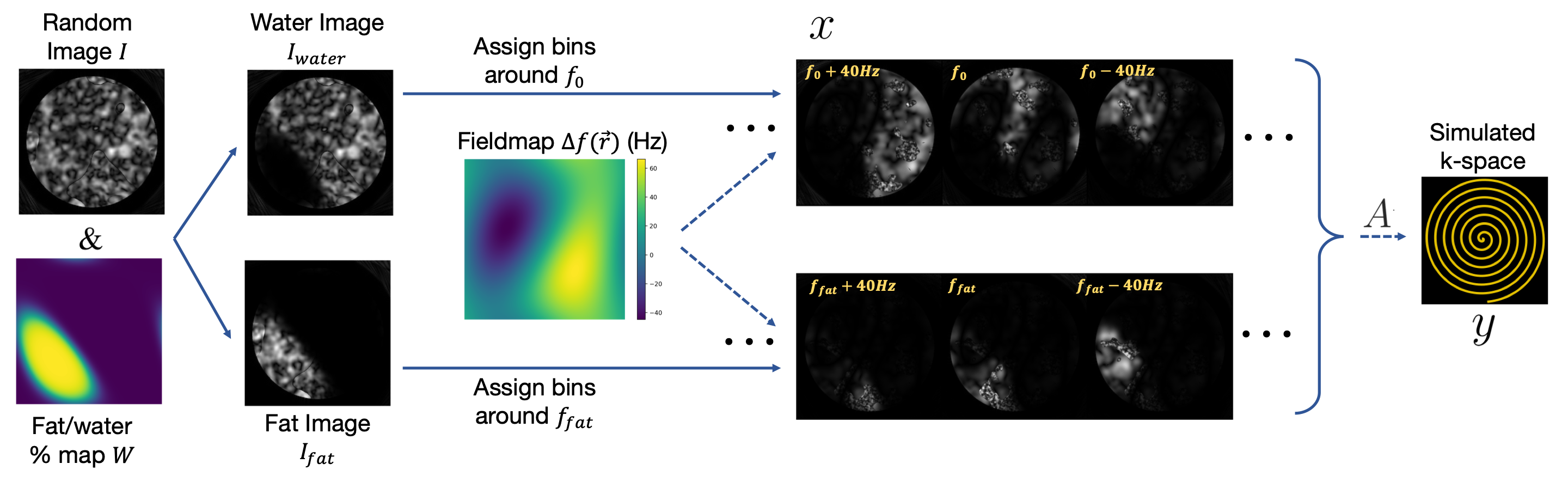
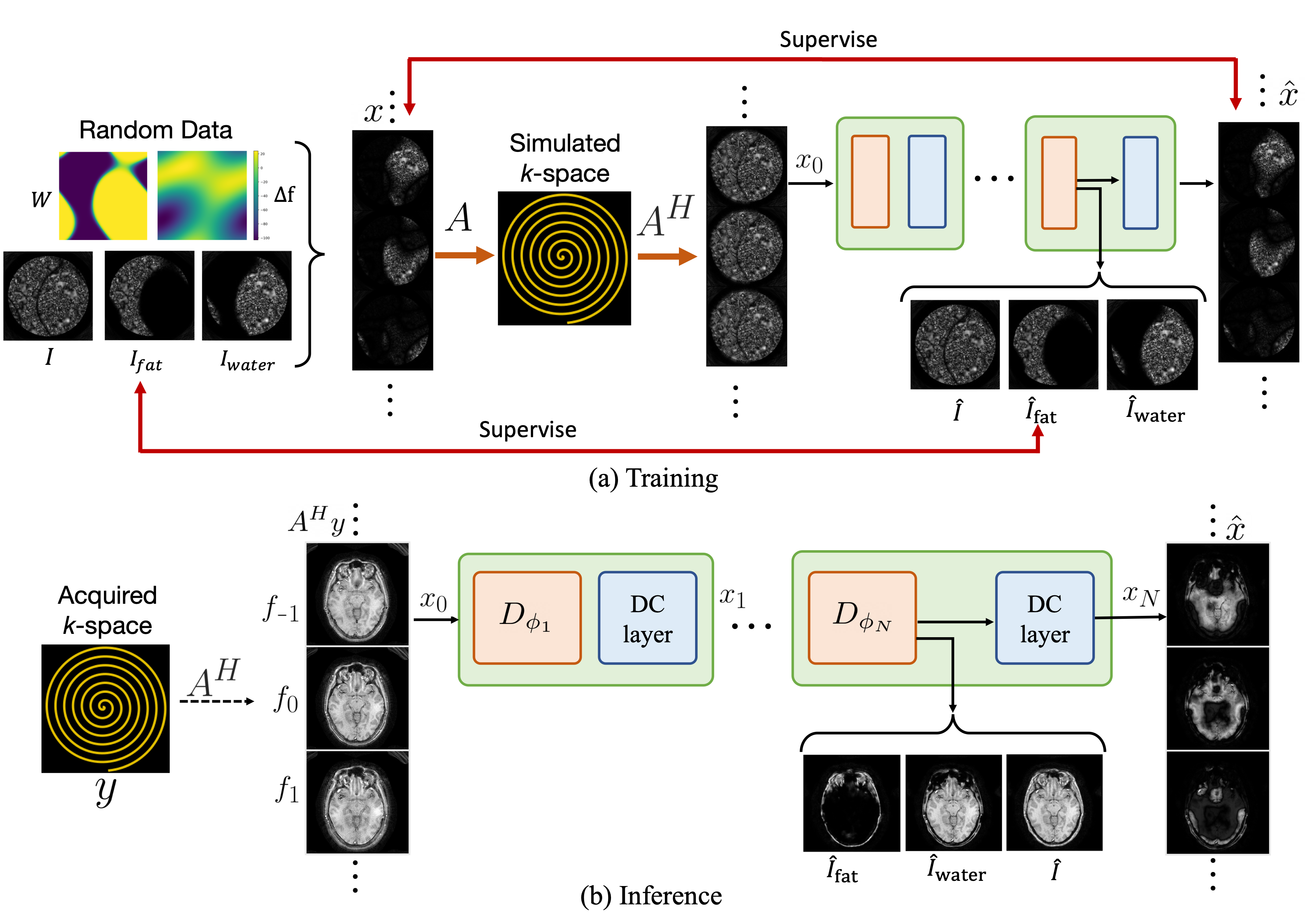
Using our previous framework9-10, we model the image as a series of frequency bins representing sharp image components at different resonant frequencies $$$f_i$$$. Our forward model, $$A = \Sigma_i M_{f_i} E,$$ is a summation over all frequency bins, where E is an encoding matrix encompassing density-compensated non-uniform Fourier transforms and coil sensitivities, and $$$M_{f_i}$$$ is a diagonal matrix with a frequency modulations $$$e^{-j2\pi f_i t}$$$ for each bin.Training data is generated by simulating random spatially varying field maps, fat/water partial-volume effects, coil sensitivities, and noise-like images with marginal statistics mimicking natural signals (inspiration from Hu et al.12). We model fat-water partial volume effects with a random weight map $$$W$$$, which sets the proportion of fat to water at each voxel, obtaining separate fat and water images. These fat and water images are then assigned to different frequency bins based on the generated field map $$$(\Delta f(\vec{r}))$$$, the fat chemical shift frequency offset $$$(f_\mathrm{fat})$$$, and the bin frequencies $$$(f_i)$$$, resulting in the multi-bin image representation $$$x$$$. This representation is then projected through the forward model $$$A$$$ to produce the simulated k-space data $$$y$$$ (Figure 1).
Using the simulated data, we train an unrolled neural network11 to reconstruct image bins $$$\hat{x}$$$ from the adjoint operation on measured multi-coil k-space data, $$$A^Hy$$$. Each unroll consists of a convolutional neural network module and a data consistency module. The network is also tasked with combining $$$\hat{x}$$$ into distinct fat and water images, along with the final combined image (Figure 2).
Experiments and Results
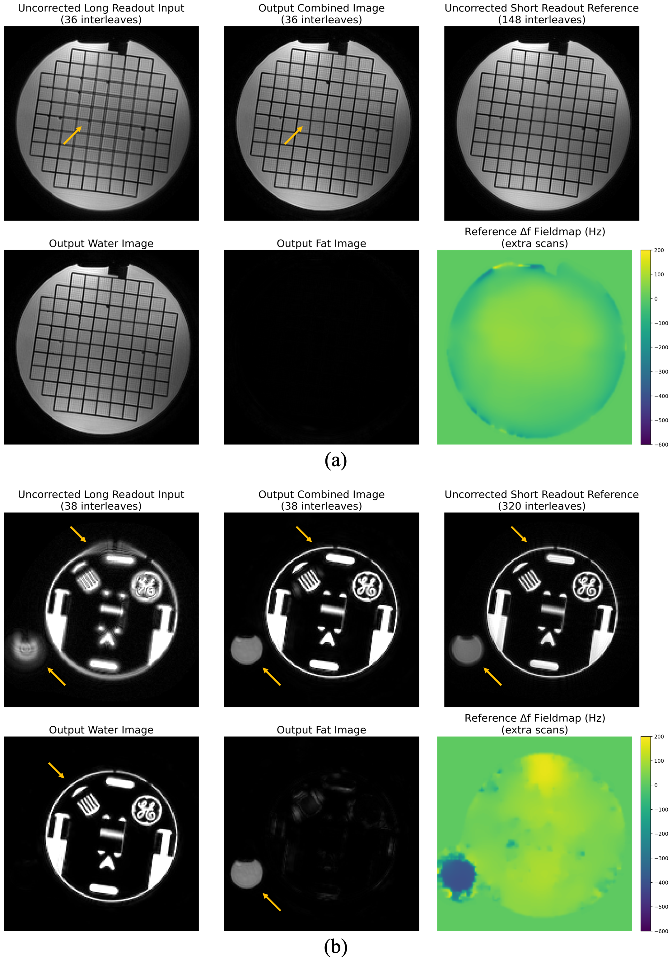
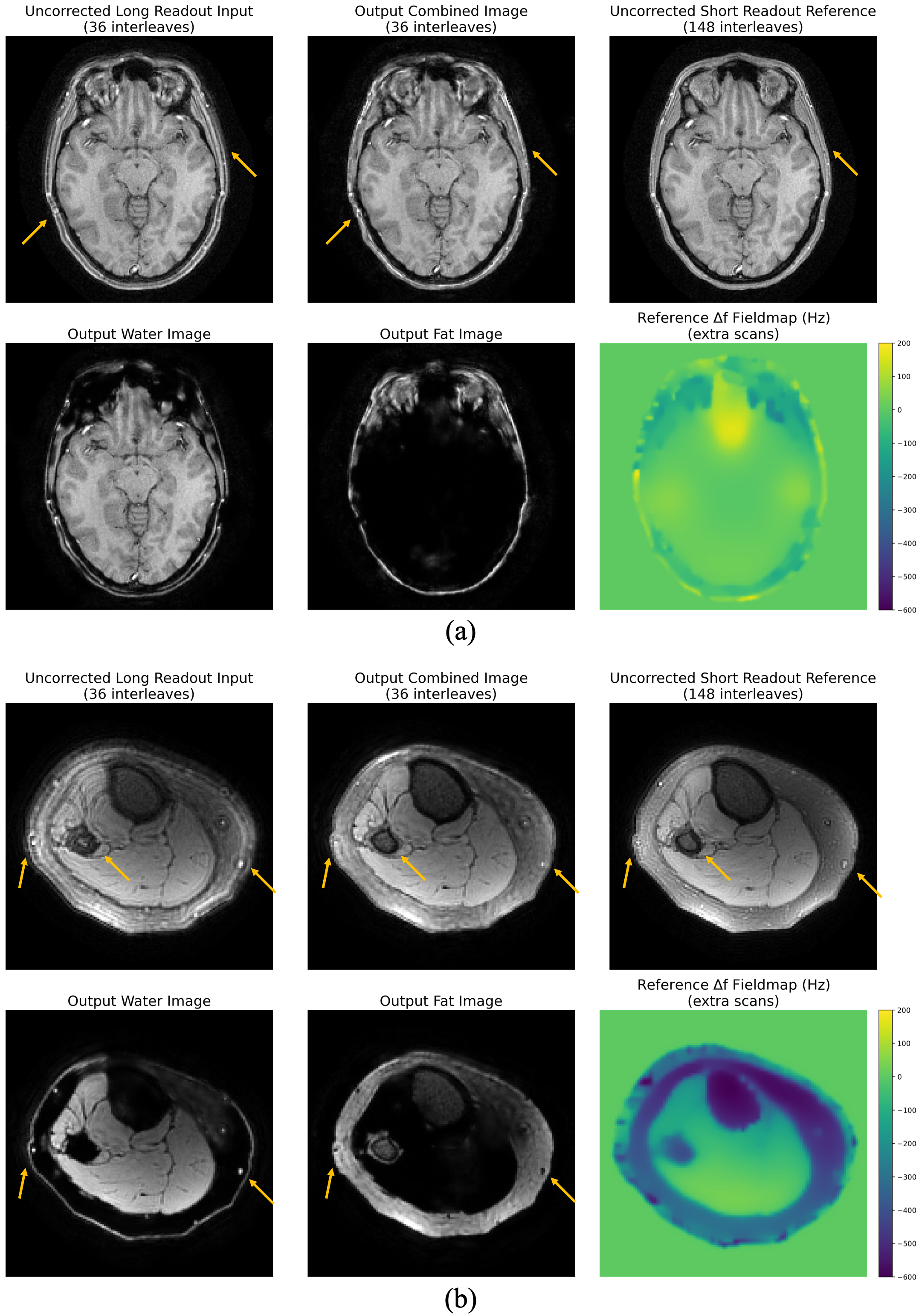
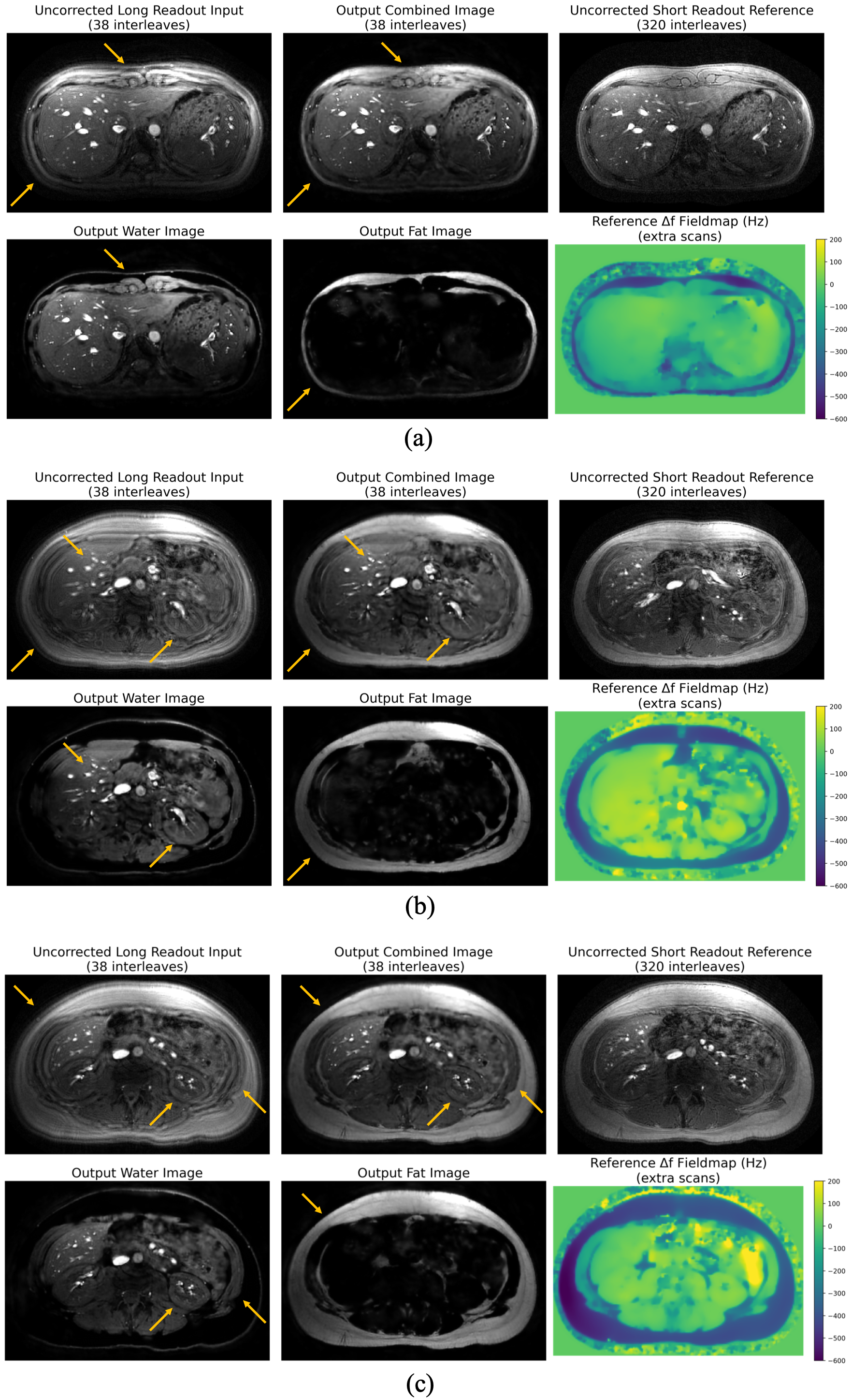
We trained two distinct models for two different Spiral trajectories, each of 1mm resolution. The first model, for a 23cm FOV with 36 interleaves of 5.2ms readout, consisted of four unrolls and was trained with 21 frequency bins between -600 to 200Hz with simulated off-resonance to address up to $$$\pm180$$$Hz field variation. The second model, accommodating a 35cm FOV with 38 interleaves with 10.4ms readout, consisted of three unrolls and was trained with 31 bins between -800 and 400Hz with simulated off-resonance of $$$\pm380$$$Hz.Qualitative evaluations were performed using ACR Phantom, Resolution Phantom, and in-vivo T1-weighted brain, PD-weighted knee, and T1-weighted abdominal scans. All imaging was conducted on a GE 3T MR750W System using SpinBench-designed pulse sequences and RTHawk v2.5.2 acquisition software (Vista Inc., Palo Alto, CA). Sensitivity maps for the acquired data were estimated with ESPIRiT13 using BART14.
Figures 3a, 4a, and 4b display results for ACR phantom, brain, and knee scans, respectively, corrected for off-resonance and fat/water separation using the first trajectory and model (23cm FOV, 36 interleaves, 5.2ms readout). The resolution phantom and abdominal scans, illustrated in Figures 3b and 5, employed the second trajectory and trained model (35cm FOV, 38 interleaves, 10.2ms readout). Each figure includes the original uncorrected image demodulated at the water frequency, the corrected combined, water-only, and fat-only images, plus a reference short readout scan and fieldmap.
Conclusion
We demonstrate the capabilities of our framework to perform off-resonance correction deblurring and fat/water separation from a single echo spiral acquisition. It generalizes effectively across different anatomies and contrasts without the need for retraining or additional data collection. This advancement makes progress toward clinical integration of non-Cartesian sampling trajectories, thus enabling more efficient, rapid, and motion-robust MRI exams.Acknowledgements
We acknowledge support from GE Healthcare and NIH grants R01EB009690 and U01EB029427.References
Bernstein M. A., King K. F., Zhou X. J.. Handbook of MRI Pulse Sequences. Elsevier; 2004.
Yang Y., Glover G. H., van Gelderen P., Patel A. C., Mattay V. S., Frank J. A., Duyn J. H. A comparison of fast MR scan techniques for cerebral activation studies at 1.5 Tesla. Magn Reson Med. 1998 Jan;39(1):61-67.
Bley TA, Wieben O, François CJ, Brittain JH, Reeder SB. Fat and water magnetic resonance imaging. J Magn Reson Imaging. 2010;31:4-18. https://doi.org/10.1002/jmri.21895
Noll DC, Pauly JM, Meyer CH, Nishimura DG, Macovski A. Deblurring for non-2D Fourier transform magnetic resonance imaging. Magn Reson Med. 1992 Jun;25(2):319-33. doi: 10.1002/mrm.1910250210. PMID: 1614315.
Noll DC, Fessler JA, Sutton BP. Conjugate phase MRI reconstruction with spatially variant sample density correction. IEEE Trans Med Imaging. 2005 Mar;24(3):325-36. doi: 10.1109/TMI.2004.842452. PMID: 15754983.
- Zeng D. Y., Shaikh J., Holmes S., Brunsing R. L., Pauly J. M., Nishimura D. G., Vasanawala S. S., Cheng J. Y. Deep residual network for off-resonance artifact correction with application to pediatric body MRA with 3D cones. Magn Reson Med. 2019;82(4):1398-1411. Available from: https://onlinelibrary.wiley.com/doi/abs/10.1002/mrm.27825
Lim Y., Bliesener Y., Narayanan S., Nayak K. S. Deblurring for spiral real-time MRI using convolutional neural networks. Magn Reson Med. 2020;84(6):3438-3452. Available from: https://onlinelibrary.wiley.com/doi/abs/10.1002/mrm.28393
Haskell M. W., Cao A. A., Noll D. C., Fessler J. A. Deep learning field map estimation with model-based image reconstruction for off-resonance correction of brain images using a spiral acquisition. Presented at: ISMRM Workshop on Data Sampling & Image Reconstruction; January 26-29, 2020; Sedona, AZ, USA.
De Goyeneche A, Ramachandran S, Wang K, Karasan E, Yu S, Lustig M. ResoNet: Physics Informed Deep Learning based Off-Resonance Correction Trained on Synthetic Data. Proceedings of the 31st ISMRM Annual Meeting; 2022.
De Goyeneche A., Ramachandran S., Wang K., Karasan E., Cheng J. Y., Yu S. X., Lustig M. ResoNet: a Physics-Informed DL Framework for Off-Resonance Correction in MRI Trained with Noise. In: Proceedings of the Thirty-seventh Conference on Neural Information Processing Systems; 2023.
Aggarwal H. K., Mani M. P., Jacob M. MoDL: Model-based deep learning architecture for inverse problems. IEEE Trans Med Imaging. 2019 Feb;38(2):394-405. Available from: http://dx.doi.org/10.1109/TMI.2018.2865356
Hu B. S., Cheng J. Y. System and method for noise-based training of a prediction model. March 2020.
Uecker M., Lai P., Murphy M. J., Virtue P., Elad M., Pauly J. M., Vasanawala S. S., Lustig M. ESPIRiT—an eigenvalue approach to autocalibrating parallel MRI: Where SENSE meets GRAPPA. Magn Reson Med. 2014;71.
Uecker M., Ong F., Tamir J. I., Bahri D., Virtue P., Cheng J. Y., Zhang T., Lustig M. Berkeley advanced reconstruction toolbox. In Proc. Intl. Soc. Mag. Reson. Med. 2015;23:2486.
Figures