0804
Aberrant interstitial fluids may be associated with cognitive impairment in patients on chronic kidney disease1Department of Radiology, Beijing Friendship Hospital, Capital Medical University, Beijing, China, 2MR Research, GE Healthcare, Beijing, China
Synopsis
Keywords: Neurofluids, Neurofluids
Motivation: Chronic kidney disease (CKD) leads to cognitive impairment; however, the pathophysiology remains unclear.
Goal(s): This study aims to evaluate differences in interstitial fluids (ISF) between patients with CKD and healthy controls, and investigate correlation between ISF circulation with cognitive impairment.
Approach: Spectral diffusion analysis was applied to measure the ISF fraction (fint).
Results: A significant difference in fint was detected between the HD and HC groups in the right basa gangliaand biliteral centrum semiovale(CSO). In the CKD group, MoCA scores were negatively correlated with fint in the biliteral CSO.
Impact: A novel approach to measure ISF exhibits the potential for detecting brain glymphatic dysfunction in patients with CKD, which provides unique insights into the pathological mechanisms of patients on CKD with cognitive impairment.
Introduction
Chronic kidney disease (CKD) is defined as a decreased glomerular filtration rate of less than 60 mL/min/1.73 m2 that is often accompanied by cognitive impairment1. Since the vascular and neurodegenerative processes related to clinical dementia cause cell loss which induces an increase in interstitial fluid (ISF), it can be expeceted that increased ISF may be present in patients with CKD. In order to maintain homeostasis of the interstitial space, the glymphatic system is responsible for the exchange of cerebrospinal fluids (CSF) and ISF2. Enlargement of the perivascular spaces filled by ISF are characteristic of glymphatic system disorder, but the relationship between cognitive impairment induced by CKD and ISF remains unclear. ISF can be examined using the non-negative least squares (NNLS) methodology applied to the intravoxel incoherent motion (IVIM) data3. Recent studies revealed that changes in the ISF are associated with cerebral small vessel disease and degenerative conditions4,5. Our study aims to evaluate differences in ISF between patients with CKD and healthy controls, and investigate correlation between ISF circulation with cognitive impairment.Methods
We performed an IVIM imaging assessment and a Montreal Cognitive Assessment (MoCA) evaluation on 19 patients with CKD. In the study, 17 healthy controls (HC) underwent brain IVIM imaging.All participants underwent imaging on a 3T MRI system (Discovery MR750W, General Electric, Milwaukee, Wisconsin, USA) with an eight-channel phased array coil.IVIM imaging was performed using a single-shot spin-echo echo planar imaging sequence (TE/TR=77.2ms/10000ms; acquisition matrix=160×160; number of slices=16; voxel size=1.5×1.5×3.3mm3), after applying a cerebrospinal fluid suppression pulse (TI=2230ms). Thirteen b-values were employed (b=0, 10, 15, 20, 30, 40, 50, 60, 100, 200, 400, 700, 1000s/mm2).
IVIM images were denoised using local principal component analysis (dipy version 1.5.0) and corrected for head displacements and eddy current distortions (eddy_correct, FSL version 6.0.7.4). Voxel-wise spectral analysis using NNLS was conducted to analyze the IVIM data. Following previous studies 4,5, A basis set of 200 logarithmically spaced functions (diffusivity range of 0.1×10⁻³ to 1000.0×10−3 mm2/s) was used. The obtained spectrum was subsequently divided into the parenchymal diffusion range (Dpar, 0.1×10⁻³ to 1.5×10⁻³ mm2/s), the intermediate diffusion range (Dint, proxy for ISF volume, 1.5×10⁻³ to 4.0×10⁻³ mm2/s), and the microvascular pseudo-diffusion range (Dmv, 4.0×10⁻³ to 1000.0×10−3 mm2/s). The ISF fraction (fint) was quantified by determining the contribution of the intermediate component to the signal, while correcting for T1- and T2-relaxation effects. 4,5
The biliteral basa ganglia (BG) and biliteral centrum semiovale (CSO) were selected as regions of interests (ROIs), where enlarged PVS are likely to be present. The ROIs were carefully drawn by one neuroradiologist with 3-years of experience and further checked by a senier neuroradiologist with 10-years of experience. Mean values of fint, were extracted for four ROIs. In Figure 1, diffusion spectrum of voxels in the left BG in patients with CKD are displayed.
Two independent sample t-tests were conducted to assess the differences of fint between the CKD and HC groups in these four ROIs. To investigate the relationship between MoCA scores and fint, Pearson's correlation analysis was employed for both the CKD and HC groups within these four ROIs.
Results
In the left BG, no significant difference in fint was found between the two groups (P= 0.41). A significant difference in fint was detected between the CKD and HC groups in the right BG, left CSO and right CSO (P = 0.04, 0.02, 0.04). (Figure 2) In the CKD group, MoCA scores were negatively correlated with fint in both the left CSO (r = -0.643, P = 0.003) and the right CSO (r = -0.555, P = 0.014). No significant correlation were found between MoCA scores and the biliteral BG. (Figure 3)Discussion
In the study, Significant differences between the CKD group and the HC group were observed in the majority of ROIs, suggesting that the increased ISF presents in CKD patients. Our findings may be attributed to factors such as small molecule toxicants and oxidative stress impacting ISF circulation. The fint in the bilateral CSO were correlated with MoCA scores, implying that the aberrant amounts of ISF in the CSO may have a more significant impact on cognitive impairment in patients with CKD. For further research into glymphatic system disorder in CKD patents, it will be necessary to expand the sample size and conduct a more comprehensive study using multimodal MR techniques.Conclusion
CKD may lead to aberrant ISF, potentially contributing to cognitive impairment.Acknowledgements
This work was supported by the Beijing Municipal Administration of Hospitals Clinical Medicine Development of Special Funding Support (contract grant numbers: ZYLX201824 and ZYLX202101), Beijing Municipal Administration of Hospital’s Mission Plan (contract grant number: SML20150101), Beijing Scholars Program (contract grant number:[2015] 160), Beijing Friendship Hospital, Capital Medical University (contract grant number: seed project YYZZ202129), Training Fund for Open Projects at Clinical Institutes and Departments of Capital Medical University (CCMU2022ZKYXY011), and Natural Science Foundation of China (82202099).References
1. Drew DA, Weiner DE, Sarnak MJ. Cognitive Impairment in CKD: Pathophysiology, Management, and Prevention. American Journal of Kidney Diseases. 2019;74(6):782-790.
2. Klostranec JM, Vucevic D, Bhatia KD, et al. Current Concepts in Intracranial Interstitial Fluid Transport and the Glymphatic System: Part II-Imaging Techniques and Clinical Applications. Radiology. 2021;301(3):516-532.
3. van der Thiel MM, Backes WH, Ramakers I, Jansen JFA. Novel developments in non-contrast enhanced MRI of the perivascular clearance system: What are the possibilities for Alzheimer's disease research? Neurosci Biobehav Rev. 2023;144:104999.
4. Wong SM, Backes WH, Drenthen GS, et al. Spectral Diffusion Analysis of Intravoxel Incoherent Motion MRI in Cerebral Small Vessel Disease. J Magn Reson Imaging. 2020;51(4):1170-1180.
5. van der Thiel MM, Freeze WM, Verheggen ICM, et al. Associations of increased interstitial fluid with vascular and neurodegenerative abnormalities in a memory clinic sample. Neurobiol Aging. 2021;106:257-267.
Figures
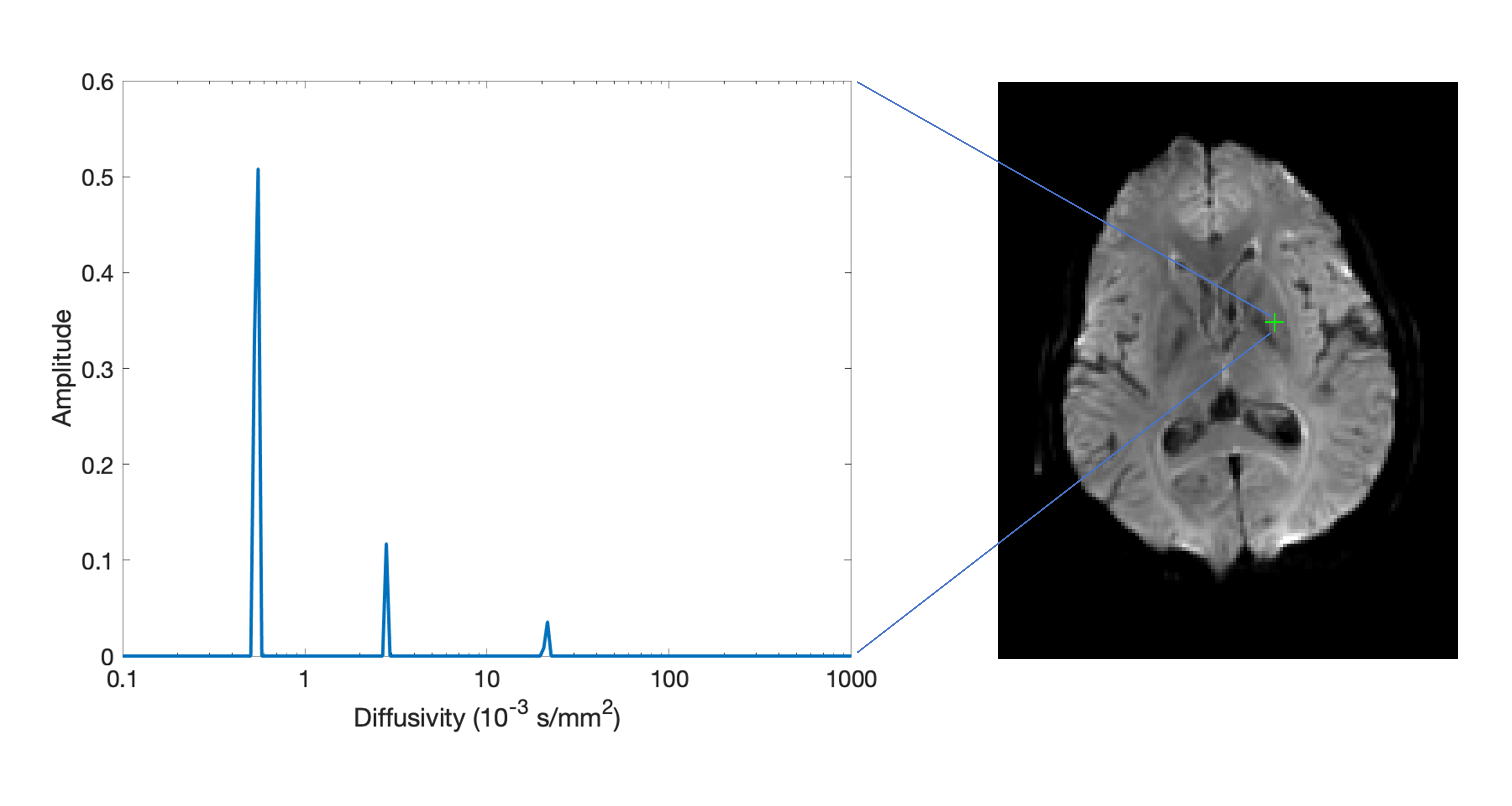
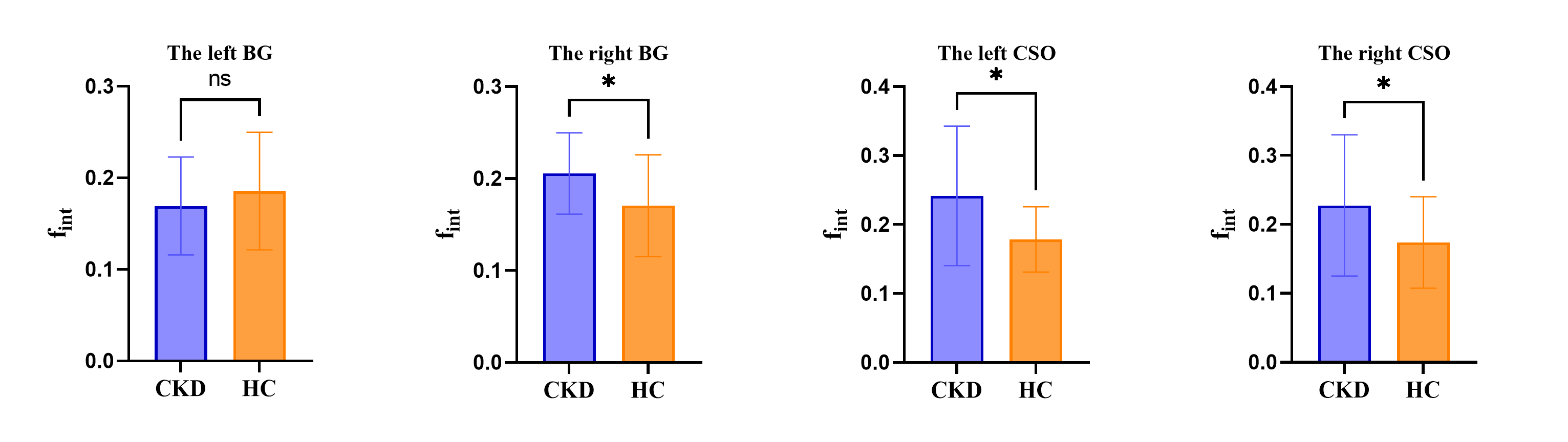
Figure 2 Differences of the intermediate diffusion volume fraction (fint) between the CKD and HC groups in four ROIs (*P<0.05).
Chronic kidney disease, CKD; healthy controls, HC; basa ganglia, BG; centrum semiovale, CSO; ns, not significant.
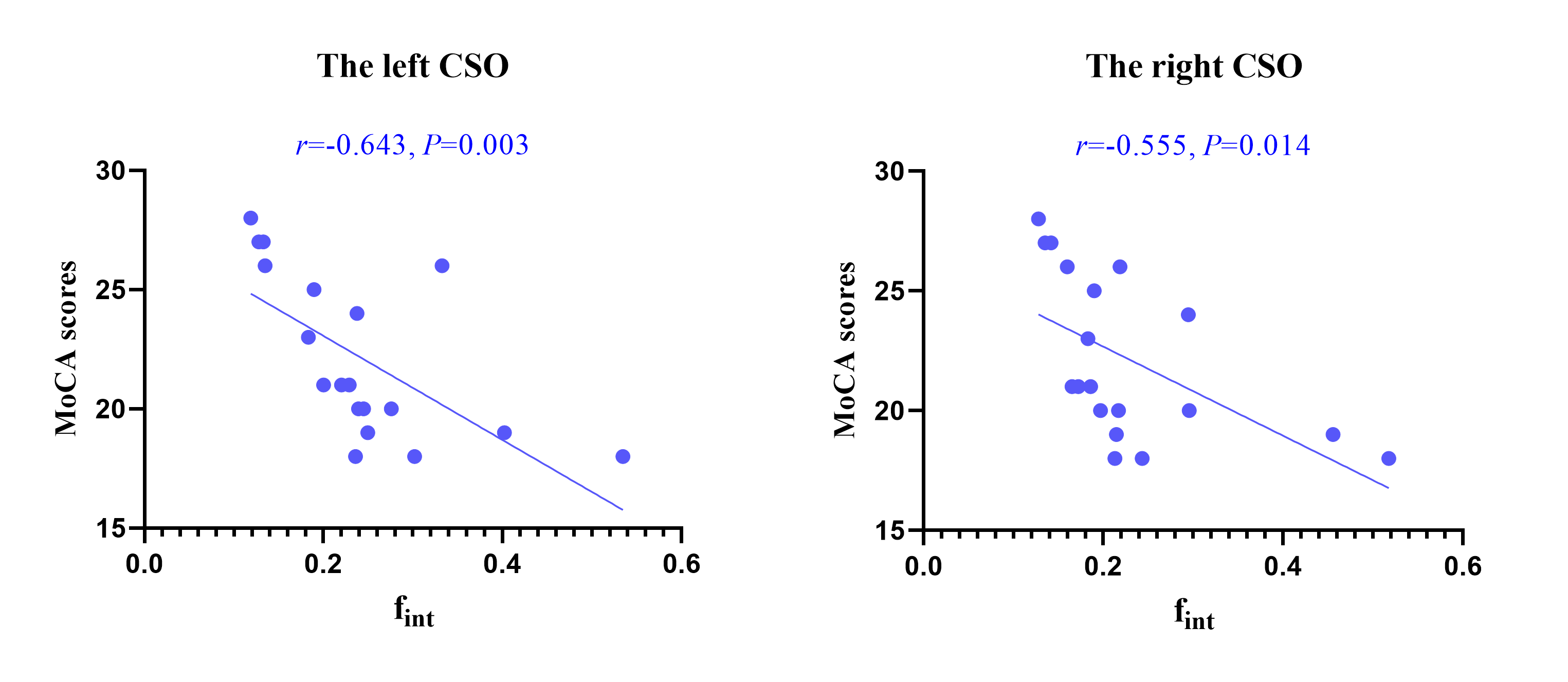
Figure 3 In the CKD group, there were significant correlations between MoCA scores and intermediate diffusion volume fraction (fint) in both the left CSO (r = -0.643, P = 0.003) and the right CSO (r = -0.555, P = 0.014).
Montreal Cognitive Assessment, MoCA; Chronic kidney disease, CKD; healthy controls, HC; centrum semiovale, CSO.