0802
Locus Coeruleus Influence on Cognitive Function via the Glymphatic System in Parkinson's Disease1Radiology, Ruijin Hospital, Shanghai Jiao Tong University School of Medicine, Shanghai, China, 2Wayne State University, Detroit, MI, United States, 3Radiology, Shanghai Jiao Tong University School of Medicine, Shanghai, China, 4Shanghai Jiao Tong University School of Medicine, Shanghai, China, 5Philips Healthcare, Shanghai, China, 6College of Health Science and Technology, Shanghai Jiao Tong University School of Medicine, Shanghai, China
Synopsis
Keywords: Parkinson's Disease, Parkinson's Disease
Motivation: The underlying mechanism of locus coeruleus (LC) in cognitive function of Parkinson’s disease (PD) has not been clearly elucidated.
Goal(s): To investigate the relationship among LC degeneration, cognitive function, and the glymphatic system in PD.
Approach: All participants underwent neuromelanin-sensitive magnetic resonance imaging (NM-MRI) and diffusion tensor image scanning. The whole brain glymphatic activity was measured using diffusion along the perivascular space (ALPS) index, while LC degeneration was estimated using the NM contrast-to-noise ratio of LC (CNRLC).
Results: Mediation analysis demonstrated that the ALPS index acted as a significant mediator between CNRLC and the MoCA score in PD subjects.
Impact: These findings enhance our grasp of how the LC noradrenergic system influences cognitive function through the glymphatic system. This research offers a promising starting point for exploring potential therapies and further research into cognitive impairment in Parkinson's disease.
Introduction
Parkinson’s disease (PD) is a progressive neurodegenerative disease characterized by core motor symptoms and various non-motor symptoms [1]. Cognitive impairment is a prevalent non-motor symptom that can occur at any stage of the disease [1]. Locus coeruleus (LC) is the primary source of noradrenergic neurons in the brain, with implications for cognitive impairment in PD [2]. However, the mechanism by which alterations in the LC impact cognition in PD remains unclear. Previous research has underscored the significance of the glymphatic system in the range of neurological disorders including PD [3]. Notably, the function of the glymphatic system can be influenced by the sleep stage in humans [4]. The noradrenaline system plays a crucial role in the neurovascular coupling in the brain, contributing to the function of brain immune cells essential for glymphatic system operation [5]. Furthermore, LC is also implicated in the regulation of wakefulness and sleep[6]. However, until now, the specific relationship between glymphatic system function and the noradrenergic LC system in regulating cognitive function remains unknown in PD. Evaluation of the function of the glymphatic system can be performed in vivo using diffusion tensor image analysis along the perivascular space (DTI-ALPS) index [7]. In the metabolism of noradrenaline, neuromelanin as a by-product was generated which can be detected by neuromelanin-sensitive magnetic resonance imaging (NM-MRI). By using NM-MRI, the LC integrity and function in PD have been investigated [8]. In this study, we aim to evaluate the relationship between LC degeneration, glymphatic system, and cognitive function to determine the potential role of glymphatic system in modulating cognitive function.Methods
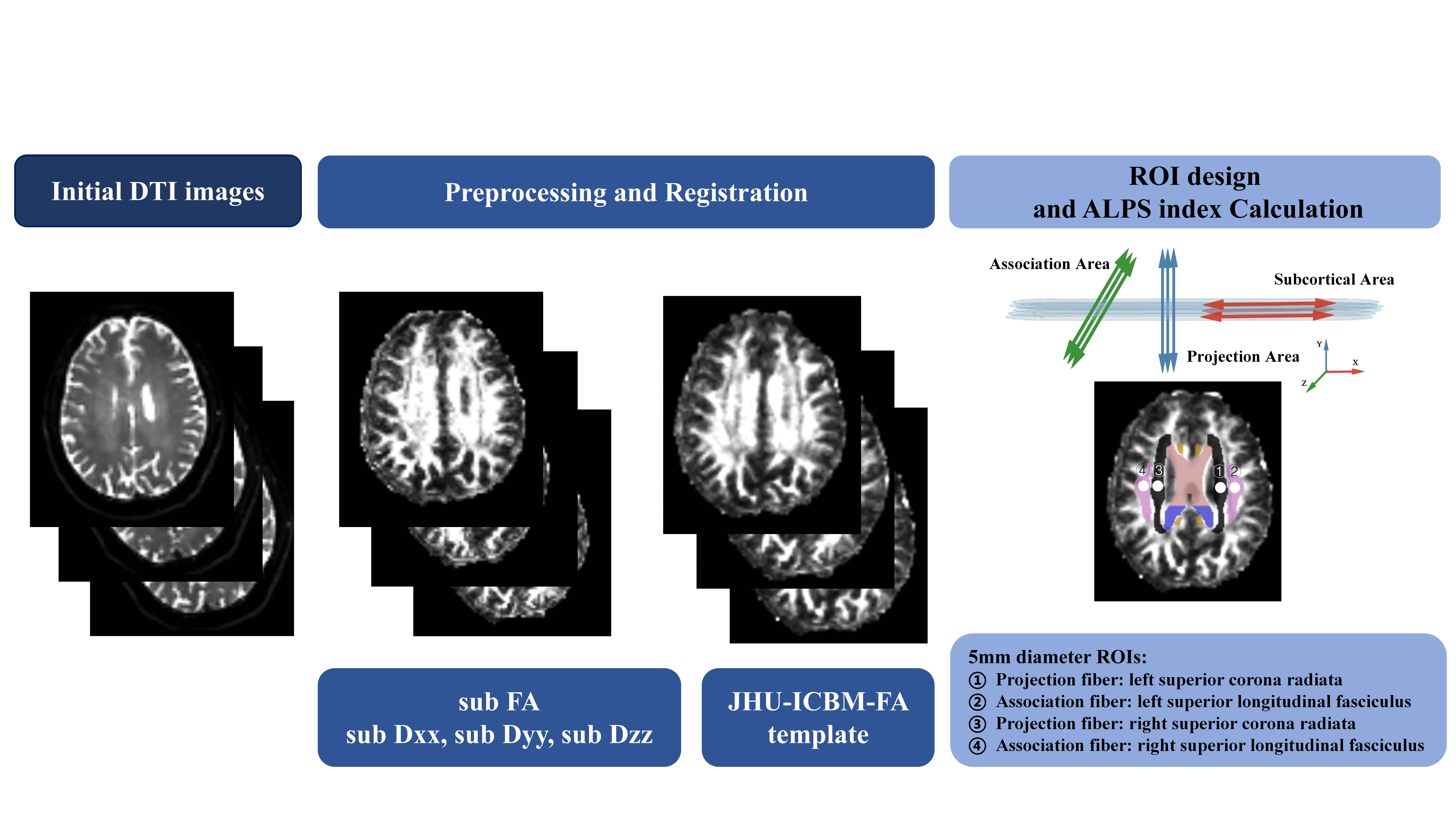
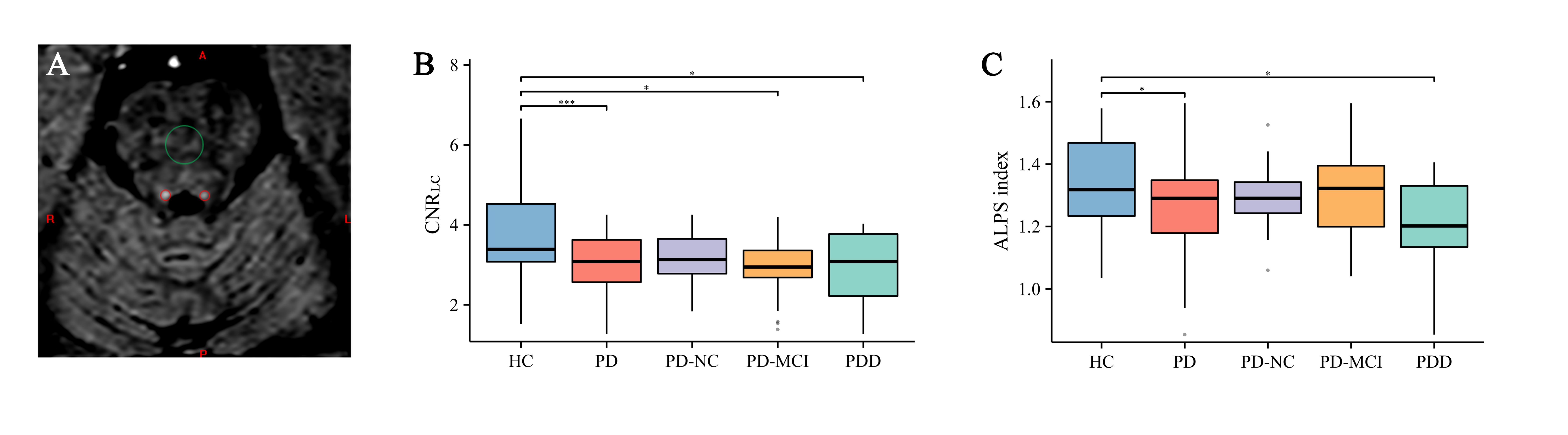
In this retrospective study, 71 PD subjects (21 with normal cognition; 29 with cognitive impairment (PD-MCI); 21 with dementia (PDD)) and 26 healthy controls were included. All participants underwent neuromelanin-sensitive magnetic resonance imaging (NM-MRI) and diffusion tensor image scanning on a 3.0 T scanner (Ingenia, Philips Healthcare, Netherlands) with a 15-channel phased coil. The imaging parameters of the 3D multi-echo gradient recalled echo sequence with a magnetization transfer contrast pulse were as follows: voxel size = 0.67×1×1.34 mm3, first echo time (TE) = 7.5ms (with 4 more echoes each with a separation of 7.5ms), repeat time (TR) = 62ms, flip angle =30°, pixel bandwidth =174 Hz/pixel, slice thickness =2 mm, number of slices = 48, a sense factor of 2. The parameters of diffusion tensor images were: field of view = 256 × 192 × 142 mm3, acquisition voxel size = 2 × 2 mm2, slice thickness = 2 mm, 71 axial slices, TE = 85 ms, TR = 8223 ms, diffusion direction = 48, b = 0 and 800 s/mm2, phase encoding direction = posterior-anterior. Another b = 0 image with the same imaging parameters except an opposite phase encoding direction was acquired to correct EPI distortions. The whole brain glymphatic activity was measured using diffusion along the perivascular space (ALPS) index and was calculated by using FSL software automatically (version 6.0.1, FMRIB Software Library; http://www.fmrib.ox.ac.uk/fsl) (Figure 1). LC degeneration was estimated using the NM contrast-to-noise ratio of LC (CNRLC), and was manually measured by using the SPIN software (SpinTech, Inc., Bingham Farms, MI, USA).Results
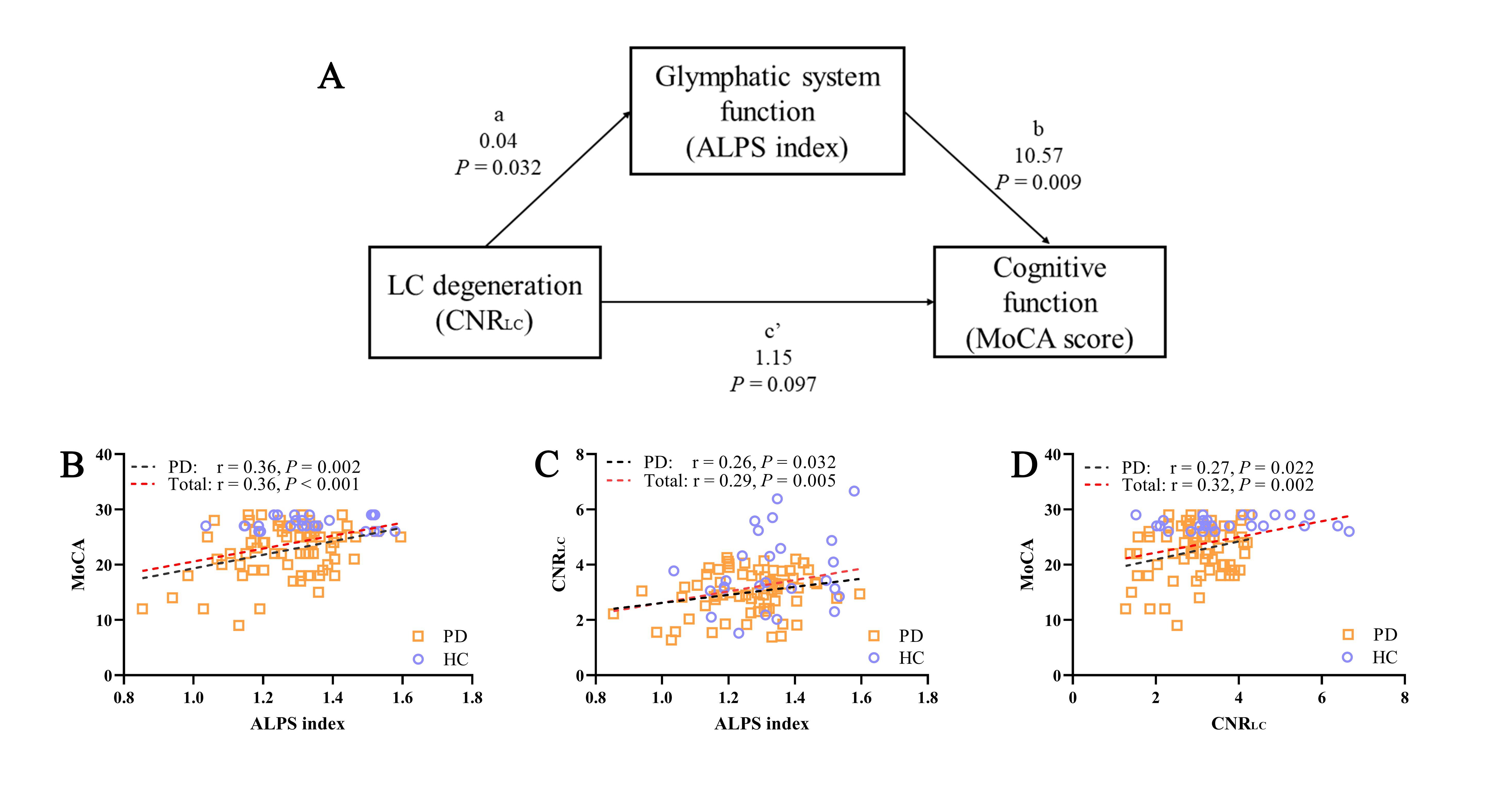
The baseline of subjects is shown in Table 1. The ALPS index was significantly lower in both the whole PD group (P = 0.04) and PDD subgroup (P = 0.02) when compared to the controls. Similarly, the CNRLC was lower in the whole PD group (P < 0.001) when compared to the controls (Figure 2). In the PD group, a positive correlation was found between the ALPS index and both the Montreal Cognitive Assessment (MoCA) score (r = 0.36; P = 0.002) and CNRLC (r = 0.26; P = 0.03). Mediation analysis demonstrated that the ALPS index acted as a significant mediator between CNRLC and the MoCA score in PD subjects (Figure 3).Discussion
This study employed multimodal neuroimaging MRI approaches to explore how cognitive function, glymphatic system function, and LC degeneration in PD may interact. Additionally, the study also compared LC degeneration and glymphatic system function between HC and PD subgroups. The ALPS index served as a significant mediator in the relationship between LC degeneration and cognitive function. Therefore, based on our study findings and aforementioned literature, particularly regarding the crucial role of the LC in the development of PD cognitive impairment, it is plausible to propose that LC stimulation influences the glymphatic system, subsequently affecting changes in the ALPS index and ultimately impacting cognitive function.Conclusion
Impaired glymphatic function serves as a mediator between LC degeneration and cognitive function in PD.Acknowledgements
This work was supported, in part, by the National Natural Science Foundation of China (grant number: 82271954, 81971576); Chinese National Science & Technology Pillar Program (grant number: 2022YFC2009900/2022YFC2009905) and the Innovative Research Team of High-level Local Universities in Shanghai.References
[1] D. Aarsland, L. Batzu, G.M. Halliday, G.J. Geurtsen, C. Ballard, K. Ray Chaudhuri, D. Weintraub, Parkinson disease-associated cognitive impairment, Nat Rev Dis Primers. 7 (2021) 47. https://doi.org/10.1038/s41572-021-00280-3.
[2] Y. Li, C. Wang, J. Wang, Y. Zhou, F. Ye, Y. Zhang, X. Cheng, Z. Huang, K. Liu, G. Fei, C. Zhong, M. Zeng, L. Jin, Mild cognitive impairment in de novo Parkinson’s disease: A neuromelanin MRI study in locus coeruleus, Mov Disord. 34 (2019) 884–892. https://doi.org/10.1002/mds.27682.
[3] T. Shen, Y. Yue, F. Ba, T. He, X. Tang, X. Hu, J. Pu, C. Huang, W. Lv, B. Zhang, H.-Y. Lai, Diffusion along perivascular spaces as marker for impairment of glymphatic system in Parkinson’s disease, Npj Parkinsons Dis. 8 (2022) 174. https://doi.org/10.1038/s41531-022-00437-1.
[4] N.E. Fultz, G. Bonmassar, K. Setsompop, R.A. Stickgold, B.R. Rosen, J.R. Polimeni, L.D. Lewis, Coupled electrophysiological, hemodynamic, and cerebrospinal fluid oscillations in human sleep, Science. 366 (2019) 628–631. https://doi.org/10.1126/science.aax5440.
[5] S. Sugama, Y. Kakinuma, Noradrenaline as a key neurotransmitter in modulating microglial activation in stress response, Neurochem Int. 143 (2021) 104943. https://doi.org/10.1016/j.neuint.2020.104943.
[6] M.E. Carter, A. Adamantidis, H. Ohtsu, K. Deisseroth, L. de Lecea, Sleep homeostasis modulates hypocretin-mediated sleep-to-wake transitions, J Neurosci. 29 (2009) 10939–10949. https://doi.org/10.1523/JNEUROSCI.1205-09.2009.
[7] T. Taoka, Y. Masutani, H. Kawai, T. Nakane, K. Matsuoka, F. Yasuno, T. Kishimoto, S. Naganawa, Evaluation of glymphatic system activity with the diffusion MR technique: diffusion tensor image analysis along the perivascular space (DTI-ALPS) in Alzheimer’s disease cases, Jpn J Radiol. 35 (2017) 172–178. https://doi.org/10.1007/s11604-017-0617-z.
[8] D. García-Lorenzo, C. Longo-Dos Santos, C. Ewenczyk, S. Leu-Semenescu, C. Gallea, G. Quattrocchi, P. Pita Lobo, C. Poupon, H. Benali, I. Arnulf, M. Vidailhet, S. Lehericy, The coeruleus/subcoeruleus complex in rapid eye movement sleep behaviour disorders in Parkinson’s disease, Brain. 136 (2013) 2120–2129. https://doi.org/10.1093/brain/awt152.Figures
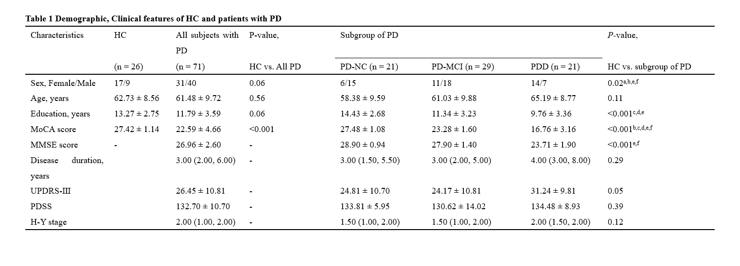
Table 1: HC = Healthy controls; PD = Parkinson’s disease; PD-NC = PD with normal cognition; PD-MCI: PD with cognitive impairment; PDD: PD with dementia; MoCA = Montreal Cognitive Assessment; MMSE = Mini-mental State Examination; UPDRS = Unified Parkinson’s disease Rating Scale; PDSS = Parkinson’s disease sleep scale; H-Y stage = Hoehn-Yahr scale. Data are expressed as mean ± SD, median (interquartile range) or number;
a = HC vs. PD-NC; b = HC vs. PD-MCI; c = HC vs. PDD; d = PD-NC vs. PD-MCI; e = PD-NC vs. PDD; f = PD-MCI vs. PDD.