0801
Brain Clearance Assessment Using the DTI-ALPS Index and Intrathecal Contrast Enhanced Magnetic Resonance Imaging1Division of Radiology and Nuclear Medicine, Department of Physics and Computational Radiology, Oslo University Hospital, Oslo, Norway, 2Institute of Clinical Medicine, Faculty of Medicine, University of Oslo, Oslo, Norway, 3Department of Neurosurgery, Oslo University Hospital-Rikshospitalet, Oslo, Norway, 4Department of Radiology, Oslo University Hospital-Rikshospitalet, Oslo, Norway
Synopsis
Keywords: Neurofluids, Neurofluids
Motivation: To investigate if DTI-ALPS as a non-invasive method compared to intrathecal contrast enhanced magnetic resonance (gMRI) imaging can be used for evaluating human brain clearance function.
Goal(s): To investigate whether or not DTI-ALPS can be used as a non-invasive alternative to gMRI.
Approach: This study compared the ALPS index with brain clearance parameters derived from gMRI in a reference group (REF).
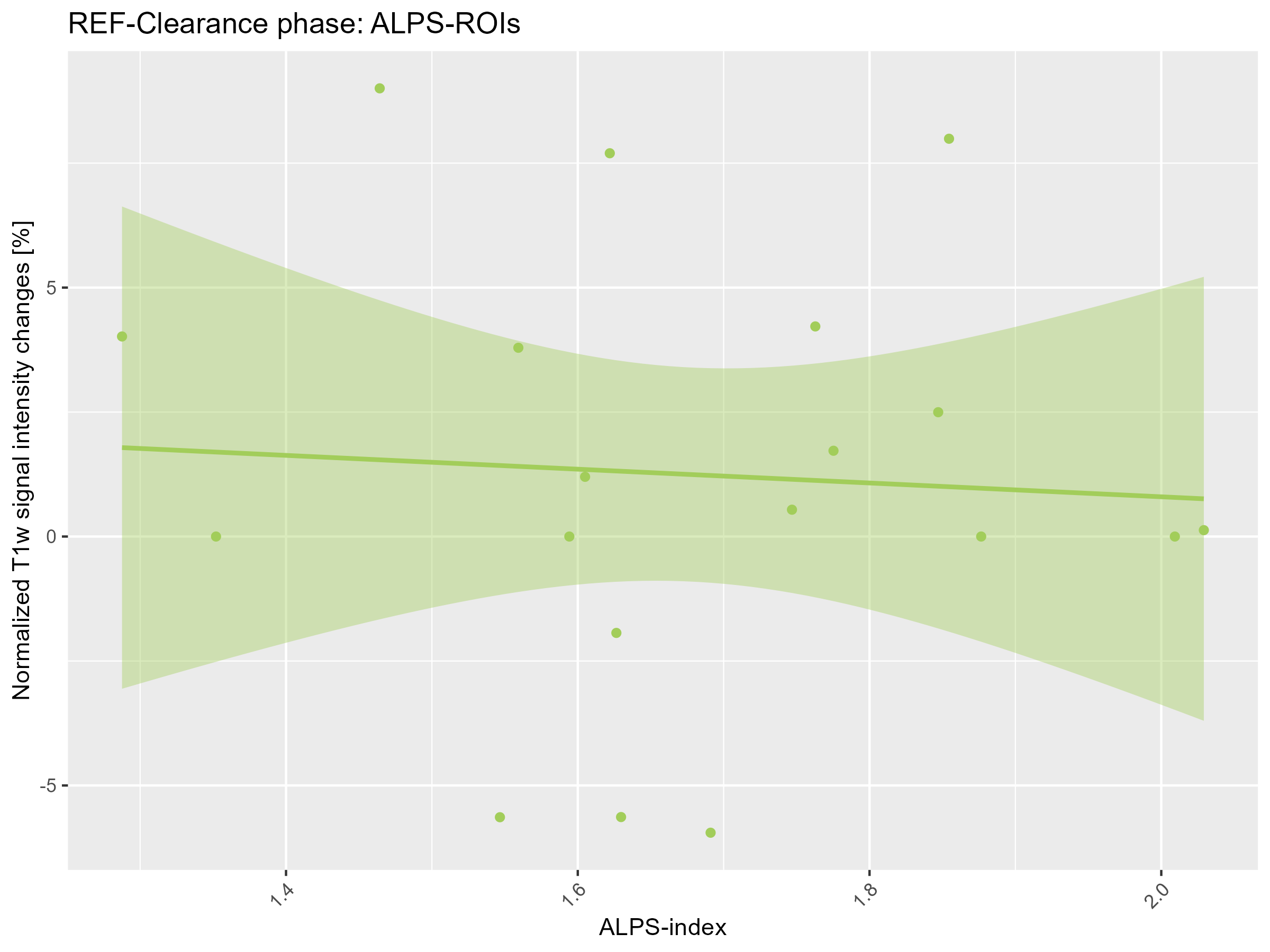
Results: No significant relationships were found between the ALPS index and change in normalized T1w signal intensity from the gMRI data in both the distribution phase and clearance phase.
Impact: The proposed DTI-ALPS index may not be a valid marker of glymphatic function.
Background
Diffusion tensor image analysis along the perivascular spaces (DTI-ALPS) [1] has been proposed to non-invasively evaluate human glymphatic clearance function. However, the methodology awaits validation against intrathecal contrast enhanced magnetic resonance imaging (gMRI), which can currently be considered the gold standard for brain clearance assessment based on exchange between cerebrospinal fluid (CSF) and interstitial fluid (ISF) (glymphatic function).Purpose
This study compared the DTI-ALPS index with brain clearance parameters derived from gMRI in a reference group (REF).Methods
This study includes data from the 2015-2019 gMRI research project in Oslo, Norway, where patients with various cerebrospinal fluid (CSF) disorders were included as part of their clinical work-up [2]. The study was approved by The Regional Committee for Medical and Health Research Ethics (REK) of Health Region South-East, Norway (2015/96), The Institutional Review Board of Oslo University Hospital (2015/1868) and The National Medicines Agency of Norway (15/04932-7). Data from 19 reference (REF) participants was used. They were characterized into the REF group if no apparent evidence of CSF disturbances was found. MRI scans were acquired before and after intrathecal injection of the contrast agent gadobutrol (0.5 mL of 1 mol/L), including scans at 24 and 48 hours. DTI images (EPI/axial, b=0, 1000 s/mm2, MPG = 15, resolution= 2.5x2.5x2.5mm3) were corrected for susceptibility, eddy currents, and motion using FSL 6.0.5. All MRI scans were registered to the baseline T1w image and segmented using FreeSurfer. ALPS-index values from DTI images at baseline were compared with intensity changes in whole white matter and cortex in normalized T1w images from baseline to 24 hours (distribution phase) and from 24 hours to 48 hours (clearance phase). We also compared the ALPS-index with enrichment and clearance in the DTI-ALPS ROI.Results
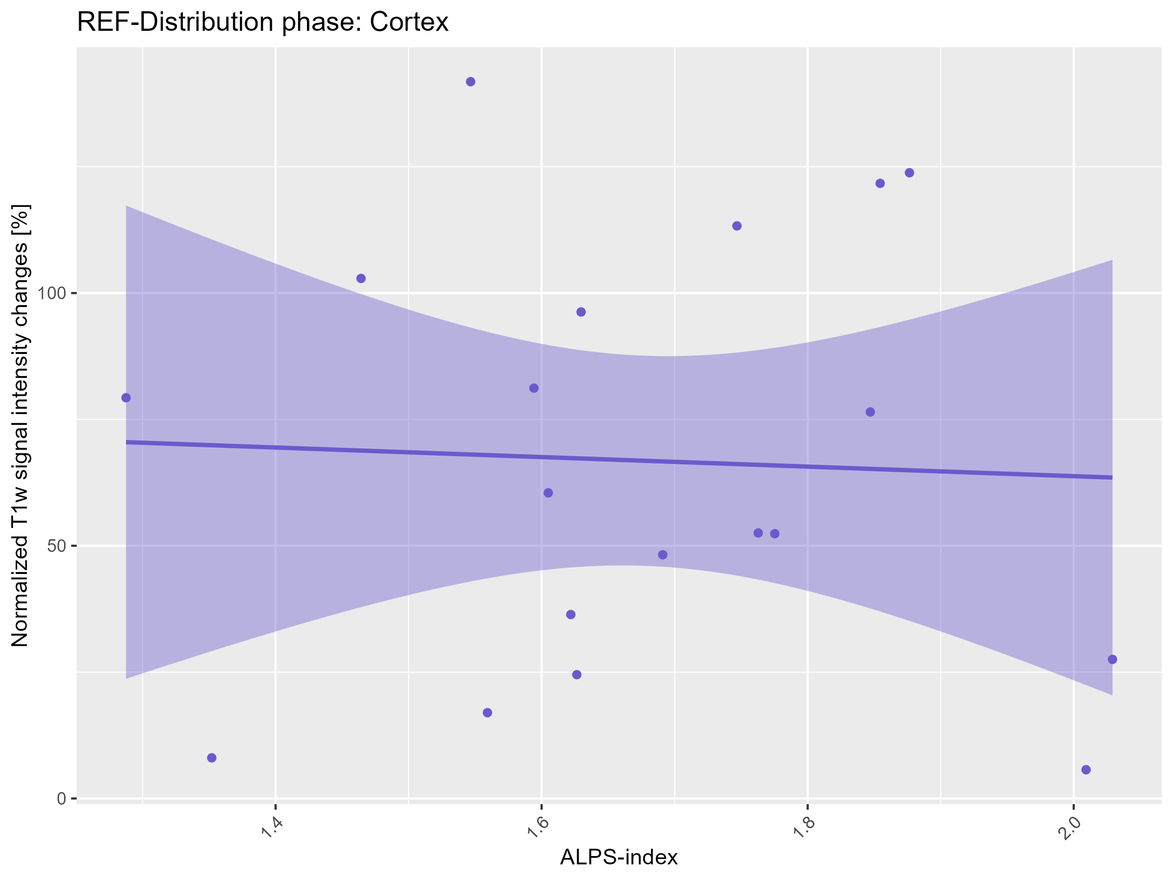
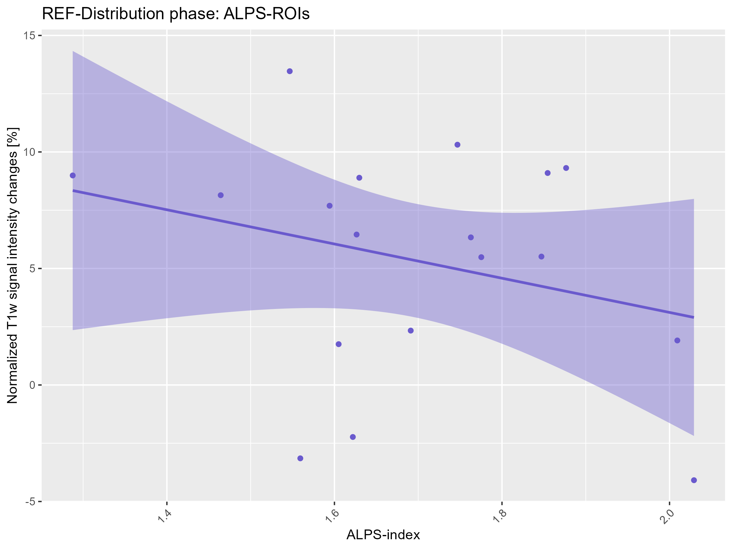
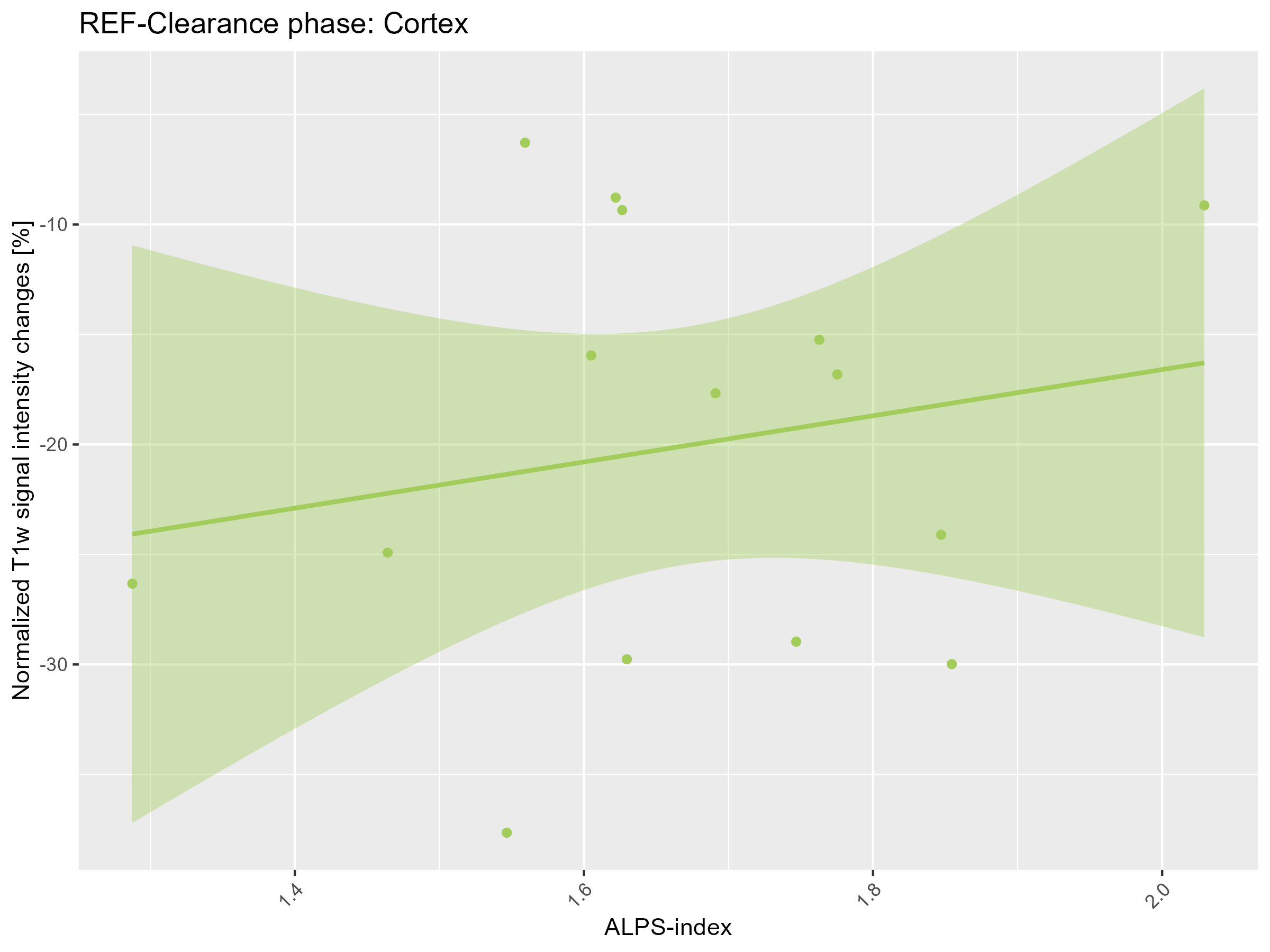
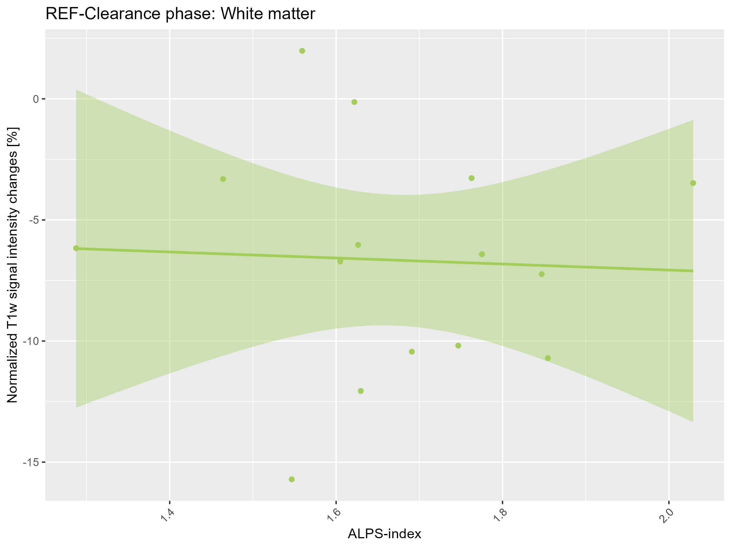
No significant relationships were found between the ALPS index and change in normalized T1w signal intensity from the gMRI data in either the distribution phase or clearance phase. It can be seen that the ALPS index is not associated with the clearance of tracer in both cortex and white matter (see Figure 1, 3, and 4). The enrichment in the ALPS ROI (mean of the projection area and association area) is also very sparse, and some of the reference participants have close to zero enrichment in this area (see Figure 2 and 5).Conclusion
CSF-ISF tracer exchange in the DTI-ALPS ROI was subtle, and in some cases close to zero. Moreover, ALPS index was not associated with local tracer clearance or clearance from cortex nor white matter as whole.Acknowledgements
No acknowledgement found.References
[1] T. Taoka et al., “Evaluation of glymphatic system activity with the diffusion MR technique: diffusion tensor image analysis along the perivascular space (DTI-ALPS) in Alzheimer’s disease cases,” Jpn J Radiol, vol. 35, no. 4, pp. 172–178, Apr. 2017, doi: 10.1007/s11604-017-0617-z.
[2] G. Ringstad et al., “Brain-wide glymphatic enhancement and clearance in humans assessed with MRI,” JCI insight, vol. 3, no. 13, 2018, doi: 10.1172/jci.insight.121537.
Figures