0797
Exploring radial asymmetry in MR diffusion tensor imaging and its impact on the interpretation of glymphatic mechanisms1Biomedical Engineering, Purdue University, West Lafayette, IN, United States, 2Department of Radiology and Imaging Sciences, Indiana University School of Medicine, Indianapolis, IN, United States, 3Stark Neurosciences Research Institute, Indiana University School of Medicine, Indianapolis, IN, United States, 4Department of Biomedical Engineering, University of Arizona, Tucson, AZ, United States
Synopsis
Keywords: Neurofluids, Neurofluids
Motivation: Researchers have used diffusion tensor imaging along the perivascular space (DTI-ALPS) to investigate glymphatic function, but the influence of white matter properties on the ALPS-index remains unstudied.
Goal(s): Establish whether a reduction in the ALPS-index could be influenced by axonal changes.
Approach: A key assumption underlying the ALPS-index is that axons demonstrate symmetric radial diffusivities, such that eigenvalue-2 and eigenvalue-3 are equal (λ2=λ3). We investigated this assumption and evaluated λ2/λ3 changes in white matter tracts.
Results: Contrary to the DTI-ALPS assumption, widespread radial asymmetry (λ2/λ3≈1.5) was observed within all white matter tracts, the extent of which decreased with aging and neurodegeneration.
Impact: This study unveils widespread radial asymmetry of white matter tracts — a phenomenon that has been overlooked in DTI studies. The results provide evidence of axonal contributions to the ALPS-index, prompting researchers to consider axonal influences when interpreting this metric.
Introduction
Diffusion imaging holds great potential for the non-invasive assessment of the glymphatic system in humans. One technique, diffusion tensor imaging along the perivascular space (DTI-ALPS), has introduced the ALPS-index for evaluating diffusivity within the perivascular space1. In recent years, the ALPS-index has been widely applied to study glymphatic function2–4. Nevertheless, it has not been firmly established that the observed reduction in the ALPS-index seen in neuropathologies is specific to changes in perivascular diffusivity.Study Rationale:
The interpretation of the ALPS-index relies on two assumptions: (1) white matter tracts exhibit symmetric radial diffusivities, where the second and third eigenvalues of diffusion tensors are approximately equal (λ2≈λ3), and (2) radial symmetry remains constant in neurodegenerative diseases. However, our own observations of diffusion tensor properties have raised questions about these assumptions.
In this study, we formulated and tested two hypotheses concerning the radial asymmetry of white matter tracts (λ2/λ3), with direct implications for the underlying ALPS assumptions.
Hypothesis 1: Diffusion tensor imaging (DTI) will reveal widespread radial asymmetry in white matter tracts, characterized by λ2/λ3>1. This hypothesis tests whether the ALPS-index>1 is linked to white matter radial asymmetry, rather than solely related to perivascular space diffusivity (Figure 1).
Hypothesis 2: Radial asymmetry (λ2/λ3) will decrease ubiquitously in white matter with neurodegeneration regardless of the fibers’ orientations. This hypothesis evaluates whether a reduction in the ALPS-index could be attributed to changes in the intrinsic properties of the white matter tracts (Figure 2).
Methods
DTI acquisition: diffusion MRI (dMRI) employed a single-shot spin-echo echo-planar imaging sequence that contained three zero diffusion-weighting and five concentric diffusion-weighting shells (b-values = 0, 250, 1000, 2000, 3250, 5000 s/mm2) with 142 diffusion-weighting gradient directions.DTI fitting and tract-wise analysis: Maps of DTI metrics were computed utilizing the first (b-value = 250 s/mm2, 6 directions) and second shells (b-value = 1000 s/mm2, 21 directions) of the dMRI data (FSL: DTIFIT). Twenty-four white matter tracts were delineated using all five shells (FSL: autoPtx5). The projection and association fibers were considered ALPS fibers (containing discernable perivascular space), and the forceps were considered control fibers (indiscernible perivascular space).
Radial asymmetry: This study focused on the ratio of radial eigenvalues (λ2/λ3) within white matter tracts. Radial asymmetry was defined as λ2>λ3, or λ2/λ3>1.
Participant information: The cohort consisted of 78 cognitively normal (CN) participants and 22 participants with mild cognitive impairments (MCI), outlined in Figure 3a.
Analysis: For each white matter tract in the CN cohort, the median, 75th percentile, and 90th percentile were measured. Linear regression was used to investigate the relationship between λ2/λ3 and age, as well as the association between λ2/λ3 and cognitive function (measured with Rey Auditory Verbal Learning Test, RAVLT6), while accounting for age. Subsequently, Pearson correlation analyses were conducted on the residuals obtained from the linear regression analyses.
Results
Evidence of widespread radial asymmetry: Brain maps of fractional anisotropy (FA) and λ2/λ3 measurements for two representative subjects aged 50 and 90 suggest widespread radial asymmetry (λ2/λ3 > 1) in white matter tracts (Figure 3). The median, 75th, and 90th percentile distributions of λ2/λ3 for each of the 24 white matter tracts revealed a consistent trend. The median value of λ2/λ3 hovered around or exceeded 1.5 for all tracts, indicating a notable discrepancy in diffusivity between λ2 and λ3 with λ2 being approximately 50% greater than λ3 (Figure 4).Radial asymmetry was associated with increasing age and decreased recall memory function: Among cognitively normal participants, a decrease was observed in λ2/λ3 with advancing age within the association tract (Pearson's r = -0.39) and in the forceps tracts (r = -0.29), (Figure 5b). Furthermore, when considering memory function across all participants, while correcting for age, a reduced λ2/λ3 was associated with worse performance on the RAVLT in the association fibers (r = 0.24) and in the forceps tracts (r = 0.27), (Figure 5c).
Discussion
This study reveals previously unexplored radial asymmetry within white matter tracts, a phenomenon often overlooked in previous diffusion imaging studies. It also indicates that the ALPS-index may be influenced by this radial asymmetry of white matter tracts. The data demonstrate that radial asymmetry decreases with aging and neurodegeneration, mirroring the effects that would occur in the ALPS-index due to alterations of the diffusivity within the perivascular space. Collectively, these findings suggest a potential white matter contribution to the ALPS-index, underscoring the importance of considering white matter radial asymmetry in diffusion studies.Acknowledgements
This work was supported by the National Institutes of Health [F30 AG084336, RF1 AG083762, R01 AG053993, P30 AG010133].
References
1. Taoka T, Masutani Y, Kawai H, et al. Evaluation of glymphatic system activity with the diffusion MR technique: diffusion tensor image analysis along the perivascular space (DTI-ALPS) in Alzheimer’s disease cases. Jpn J Radiol. 2017;35(4):172-178. doi:10.1007/s11604-017-0617-z
2. Carotenuto A, Cacciaguerra L, Pagani E, Preziosa P, Filippi M, Rocca MA. Glymphatic system impairment in multiple sclerosis: relation with brain damage and disability. Brain. 2022;145(8):2785-2795. doi:10.1093/brain/awab454
3. Tang J, Zhang M, Liu N, et al. The Association Between Glymphatic System Dysfunction and Cognitive Impairment in Cerebral Small Vessel Disease. Front Aging Neurosci. 2022;14:916633. doi:10.3389/fnagi.2022.916633
4. Hsu JL, Wei YC, Toh CH, et al. Magnetic Resonance Images Implicate That Glymphatic Alterations Mediate Cognitive Dysfunction in Alzheimer Disease. Ann Neurol. 2023;93(1). doi:10.1002/ana.26516
5. Wakana S, Jiang H, Nagae-Poetscher LM, van Zijl PCM, Mori S. Fiber Tract–based Atlas of Human White Matter Anatomy. Radiology. 2004;230(1):77-87. doi:10.1148/radiol.2301021640
6. Rey A. L’examen clinique en psychologie [The clinical examination of psychology]: Press Universitaire de France. Paris, France. Published online 1964.
Figures
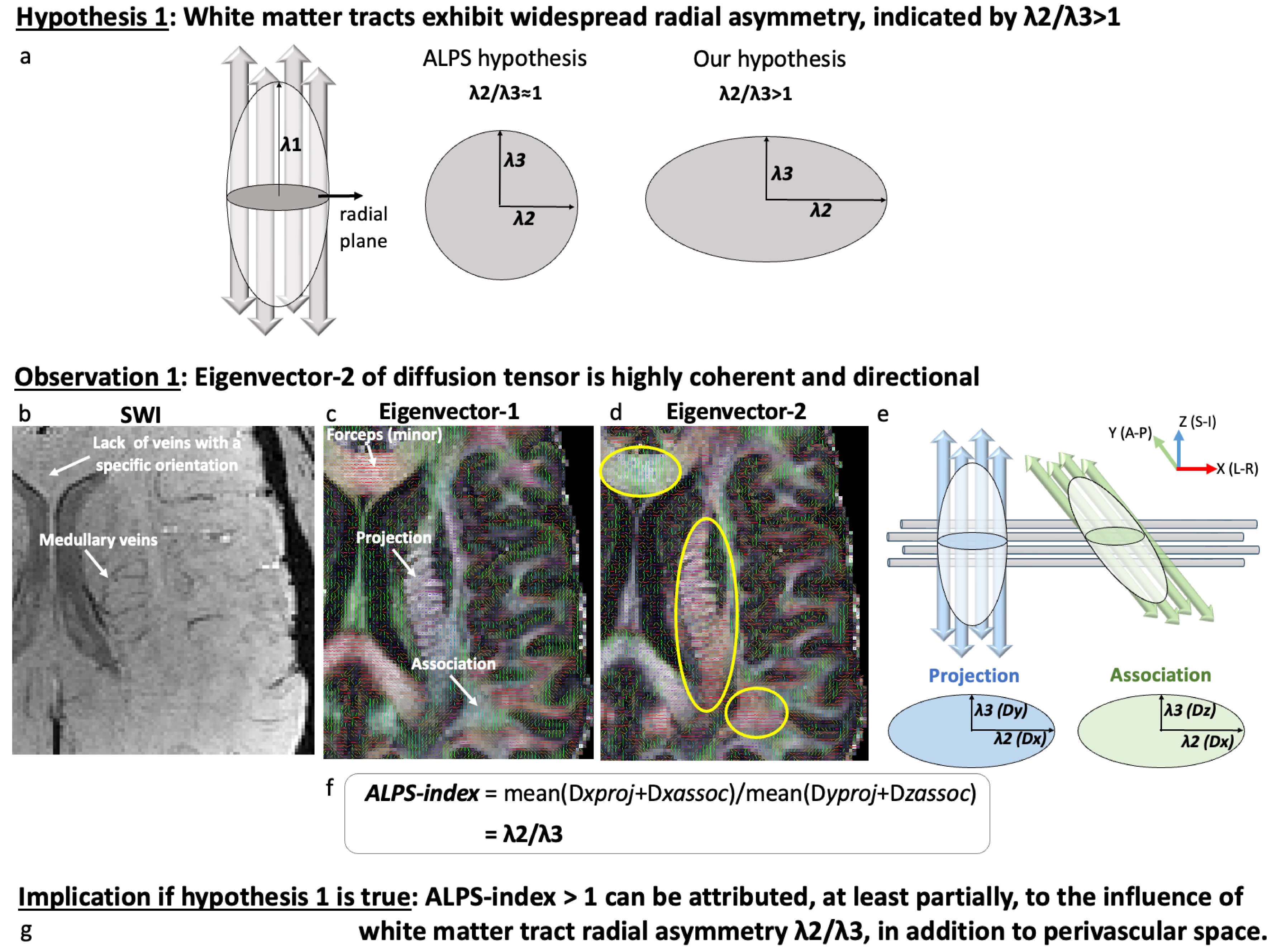
Figure 1: Conceptual representation of hypothesis 1 and its implication on the ALPS-index. (a) Hypothesis 1 posits that there exists widespread radial asymmetry within white matter tracts, indicated by the homogeneous alignment of the second eigenvector (eigenvector-2). (b-f) Observations supporting hypothesis 1. Illustration of the high coherence and directional alignment of eigenvector-2 of the diffusion tensor in both ALPS fibers (projection and association) and control fibers (forceps).
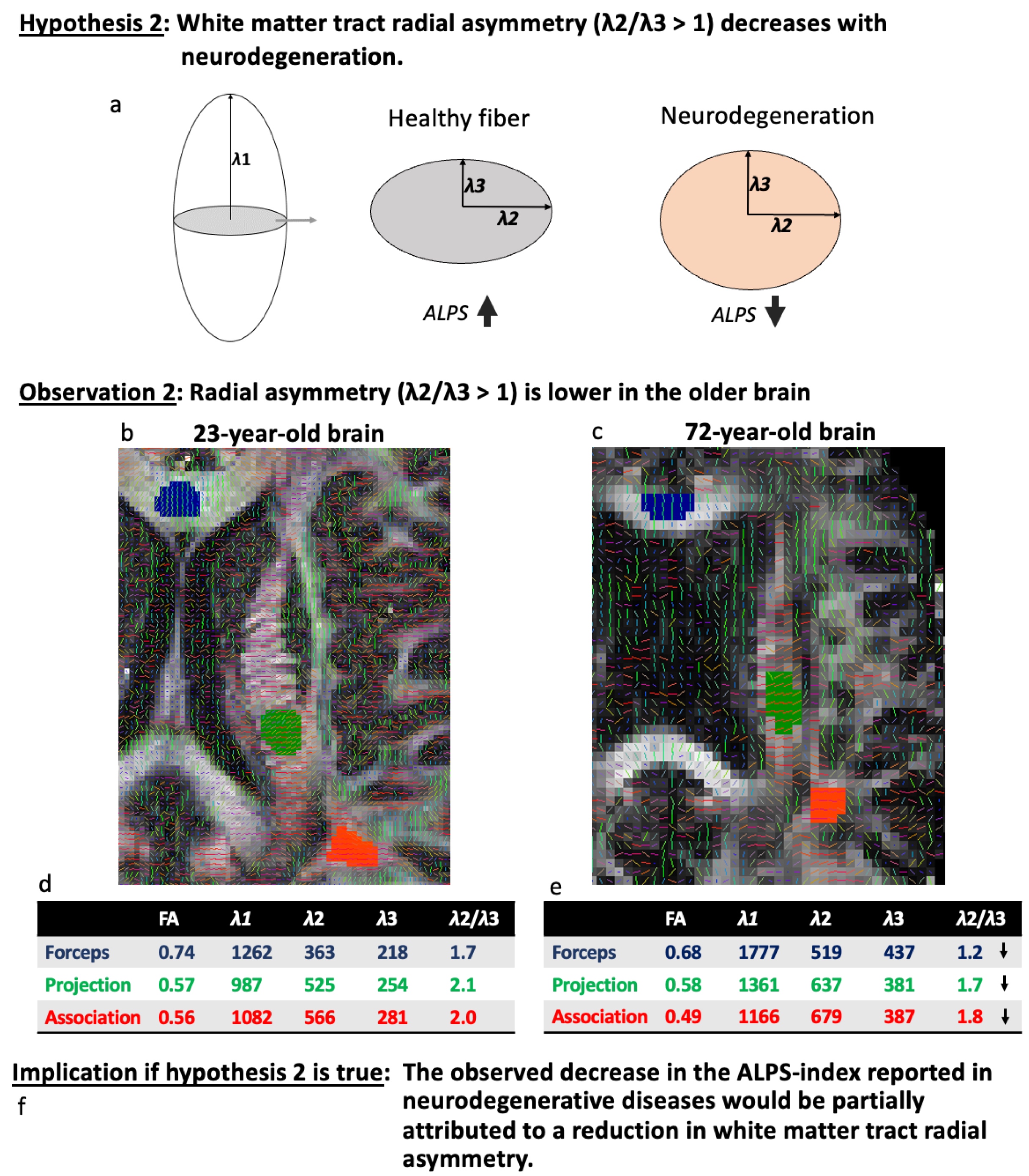
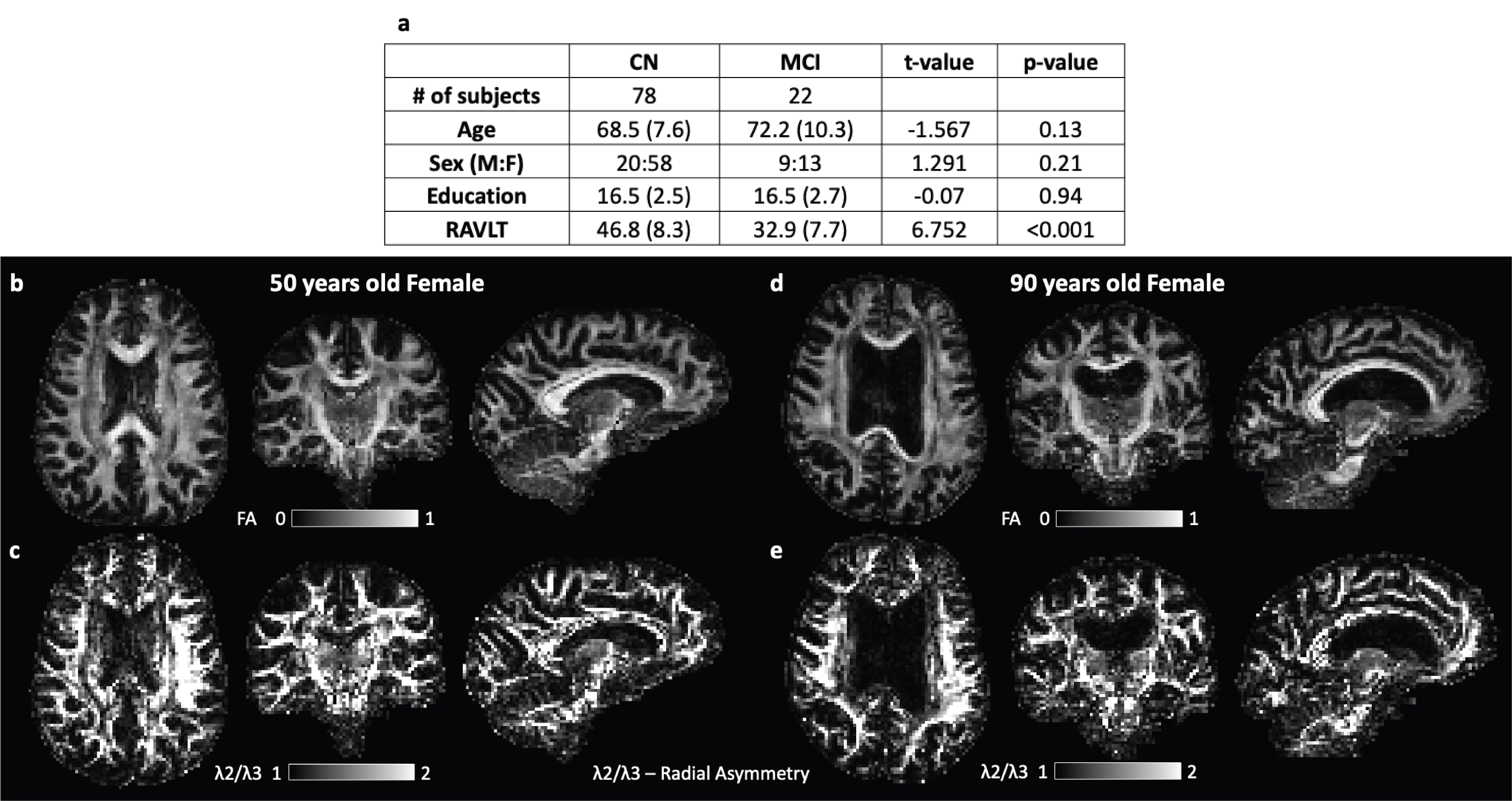
Figure 3: Participant demographics and an FA and λ2/λ3 map for two representative subjects. (a) Demographic and cognitive characteristics include the mean (standard deviation) for each group. The (b) FA map and (c) radial asymmetry map of a 50-year-old female. The (d) FA map and (e) radial asymmetry map of a 90-year-old female. These maps demonstrate that radial diffusion asymmetry is present in both ages. Abbreviations: FA, fractional anisotropy.
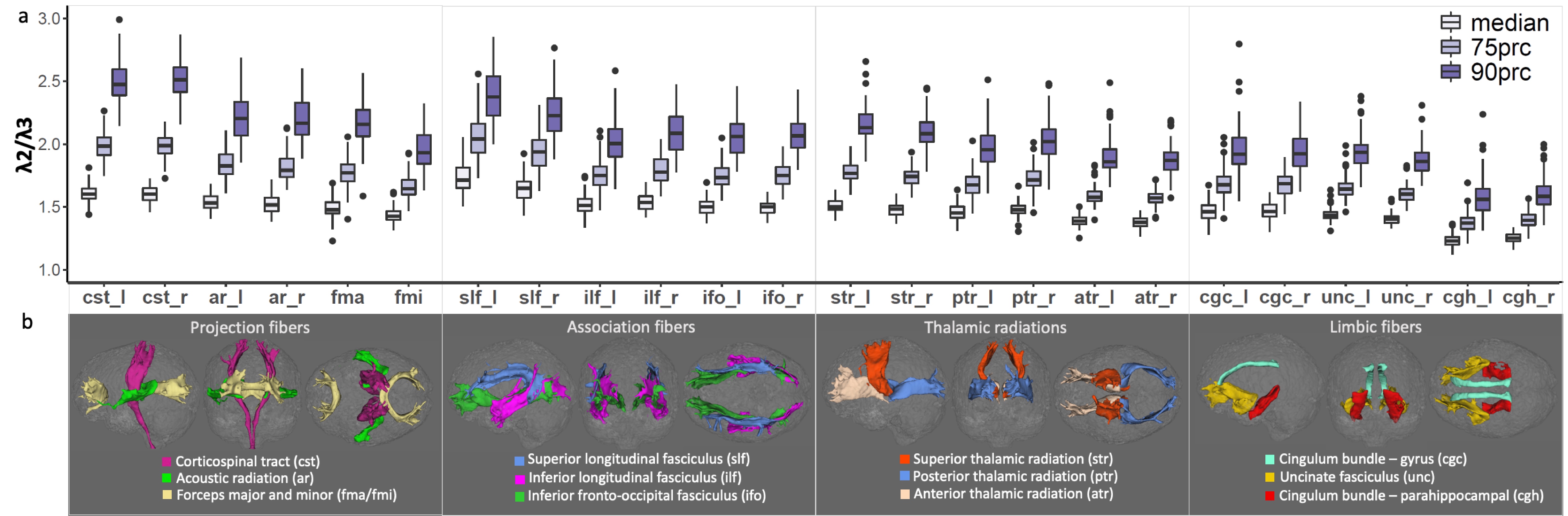
Figure 4: Widespread radial asymmetry in white matter tracts, supporting hypothesis 1. (a) Boxplots of the median, 75th, and 90th percentile distributions of λ2/λ3 for white matter tracts in 78 cognitively normal participants. All tracts exhibit λ2/λ3 ratios well above 1, providing evidence for widespread radial asymmetry. (b) Rendered representations of the 24 tracts displayed in sagittal (left), coronal (center), and axial (right) views, categorized into four groups. Abbreviations: prc, percentile; l, left; r, right.
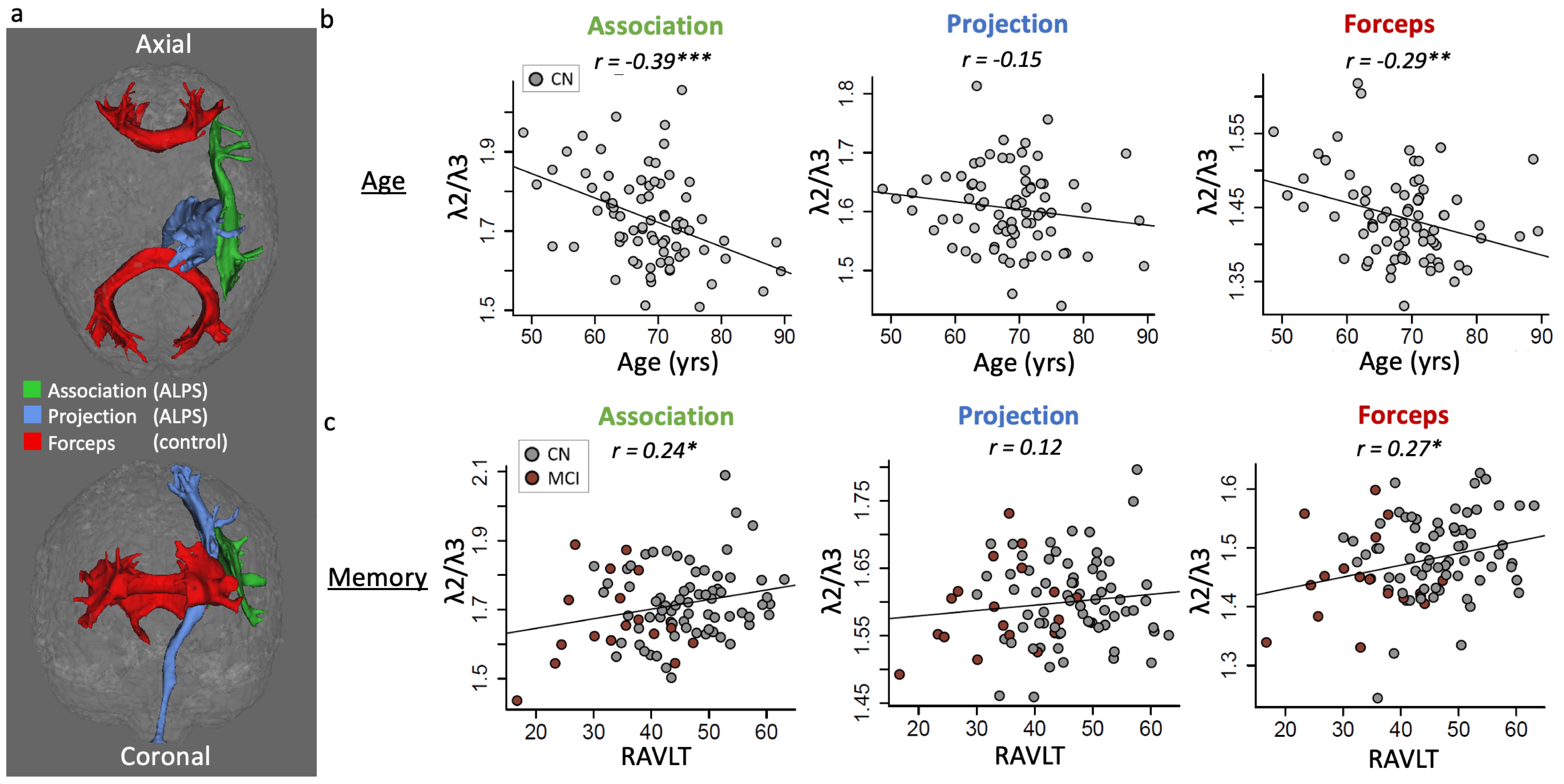
Figure 5: Radial asymmetry decreased with advancing age and decreased memory function, supporting hypothesis 2. (a) Three tracts were analyzed: The association (green) and projection (blue) tracts, which are used to calculate the ALPS-index, and the control tracts represented by the forceps (red). (b) λ2/λ3 decreased with advancing age and (c) was associated with worse memory function in the association and forceps tracts. Significance levels: * p < .05, ** p < .01, and *** p < .001. CN, cognitively normal; MCI, mild cognitive impairment.