0796
Characterizing blood and cerebrospinal fluid flow by D* tensor derived from intravoxel-incoherent-motion-diffusion-tensor-imaging1Department of Radiology and Nuclear Medicine, Maastricht University Medical Center, Maastricht, Netherlands, 2School for Mental Health and Neuroscience, Maastricht University, Maastricht, Netherlands, 3Department of Psychiatry and Neuropsychology, Maastricht University, Maastricht, Netherlands, 4Department of Neurology, Maastricht University Medical Center, Maastricht, Netherlands, 5School for Cardiovascular Disease, Maastricht University, Maastricht, Netherlands, 6Department of Radiology and Nuclear Imaging, Amsterdam UMC location University of Amsterdam, Amsterdam, Netherlands, 7Imaging and Biomarkers, Cancer Center Amsterdam, Amsterdam, Netherlands, 8Department of Electrical Engineering, Eindhoven University of Technology, Eindhoven, Netherlands
Synopsis
Keywords: IVIM, Diffusion/other diffusion imaging techniques
Motivation: A deeper understanding of brain physiology and pathology can be provided with an intravoxel-incoherent-motion-diffusion-tensor-imaging (IVIM-DTI) MR scan, which simultaneously measures blood and cerebrospinal fluid (CSF) flow and flow directions; parenchymal anisotropy; and microvascular perfusion.
Goal(s): To demonstrate the feasibility of IVIM-DTI to provide a proxy for blood and CSF flow.
Approach: A tensor of the pseudo-diffusion component (D*) was derived from IVIM-DTI and related to arterial and ventricular physiology.
Results: D* ellipsoids align well with arterial blood and CSF flow. D*’s magnitude and anisotropy correspond to the expected flow in arteries and ventricles, indicating the technique's ability of characterizing flow dynamics.
Impact: Assessing blood and cerebrospinal fluid flow with intravoxel-incoherent-motion-diffusion-tensor-imaging (IVIM-DTI) alongside traditional IVIM and DTI measures can provide comprehensive pathophysiological insights into neurological conditions. The finding that these physiological processes contribute to IVIM-derived f and D* is important for their interpretation.
Introduction
Cerebral intravoxel incoherent motion (IVIM) imaging is a diffusion-weighted technique that is sensitive to slow water diffusion within the parenchyma and fast-moving water within randomly-oriented capillaries (i.e., the pseudo-diffusion component D*).1 However, pseudorandom flow due to a distribution of unidirectional flow velocities was recently shown to cause anisotropic signal attenuation in low diffusion-sensitive b-values (<200 s/mm2), thereby affecting D*.By acquiring multiple (low) b-values in multiple diffusion-sensitizing directions, the anisotropy of the D* tensor can be estimated.2,3 We hypothesize that the D* tensor corresponds to blood and cerebrospinal fluid (CSF) flow velocities and direction. Thereby, IVIM-diffusion-tensor-imaging (DTI) has the potential to simultaneously measure the parenchymal anisotropy, the microvascular perfusion, and proxies for CSF and blood flow.
However, due to the lack of signal attributed to the D* effect, retrieving directional information with conventional fitting techniques has not been very successful. Therefore, we explore the use of deep-learning-guided parameters estimation to demonstrate the feasibility of IVIM-DTI to provide a proxy for blood and CSF flow.
Methods
MRI acquisition: Eleven healthy volunteers (age range: 22-59y, 4 males) underwent whole-brain imaging on 3T MRI (Philips, Achieva TX). Diffusion MRI was acquired with 15 b-values (0-1000 s/mm2) in 32 noncollinear gradient directions (single-shot spin-echo echo-planar-imaging (EPI), TR/TE=2279/79ms, multi-band factor=3, acquisition time=17min, voxel size=2.4x2.4x2.4mm). A time-of-flight was acquired to visualize arteries and a T1-weighted image for anatomical reference.
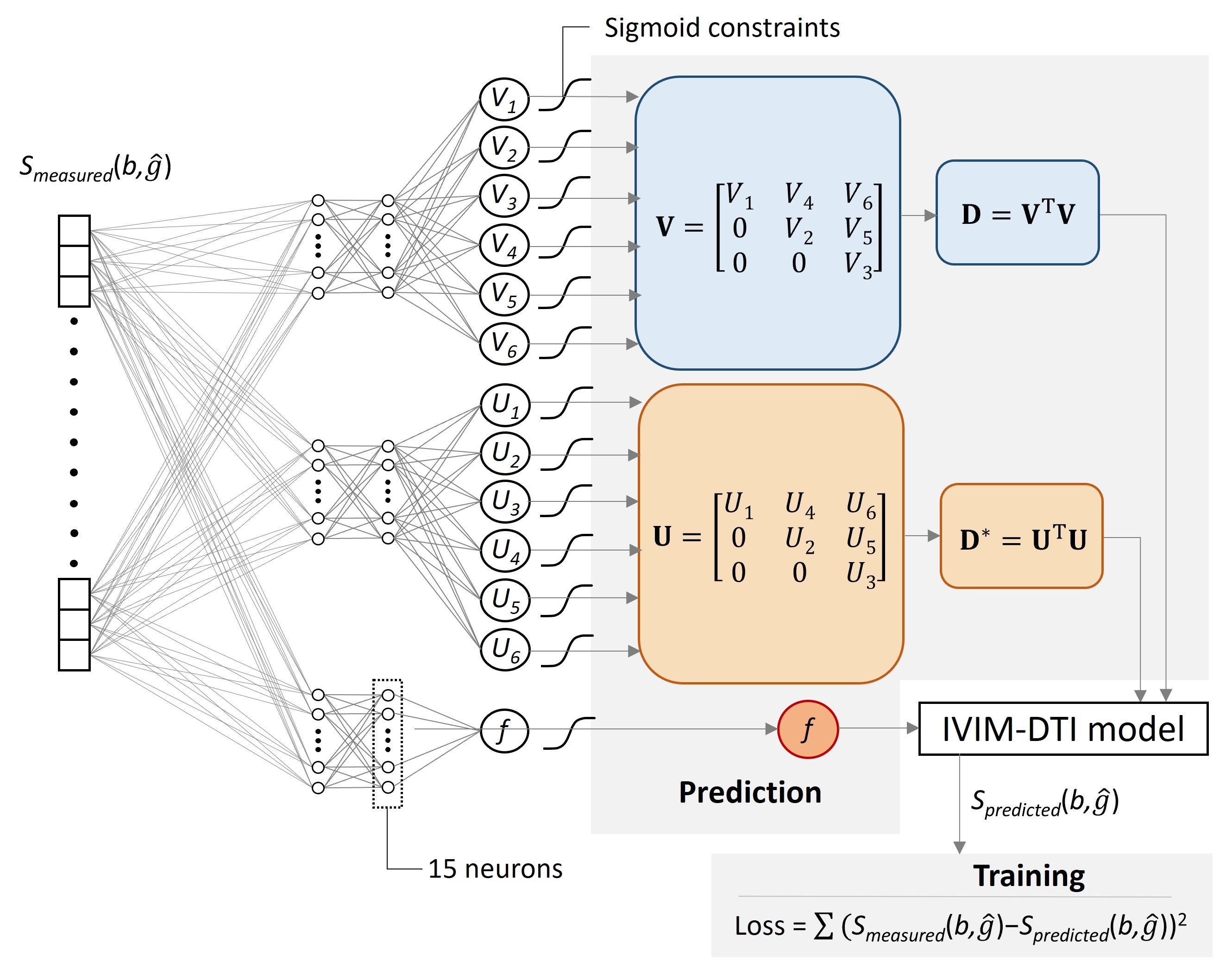
Image analysis: The diffusion images were corrected for geometric EPI distortions (topup, FSL4) and head displacements (ExploreDTI5). Subsequently, the IVIM-DTI model2 was fitted to the diffusion data in a voxel-wise manner using a physics-informed neural network (PI-NN) (Figure 1). Previously, PI-NNs were shown to outperform traditional fitting methods.6,7 Our unsupervised network learns the IVIM-DTI model by incorporating the following model into the loss function:
$$$S(b,\hat{g})=S_{0}[fe^{-b\hat{g}^T\mathbf{D^*}\hat{g}}+(1-f)e^{-b\hat{g}^T\mathbf{D}\hat{g}}]$$$
Here, $$$\hat{g}$$$ is the diffusion-sensitizing direction, $$$f$$$ is a scalar representing the signal fraction of the pseudo-diffusion component, and D* and D are the pseudo-diffusion and parenchymal diffusion tensors, respectively.
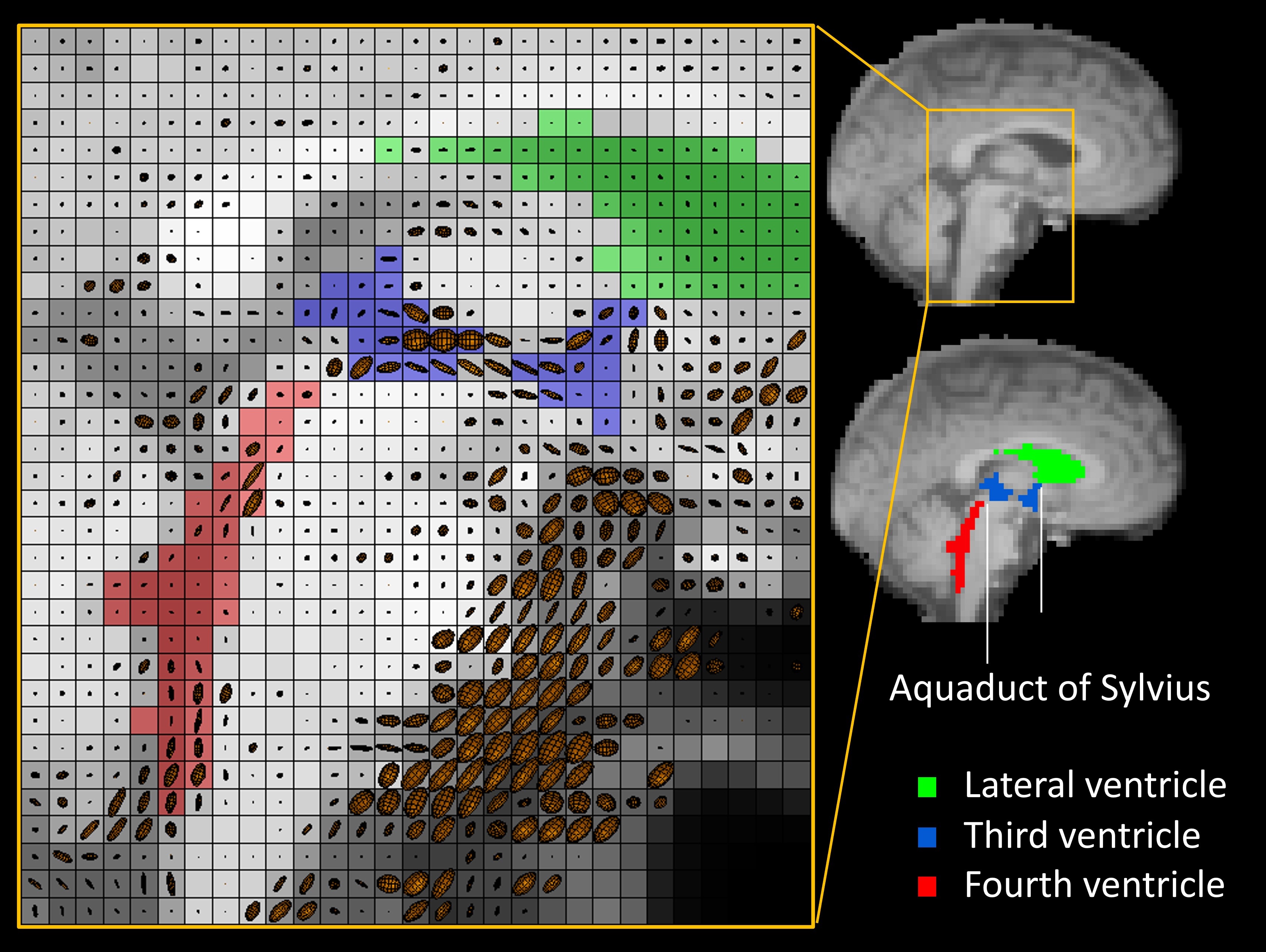
Eigenvectors and eigenvalues were derived from D* and D, from which the fractional anisotropy (FA) and mean diffusivity (MD) were calculated.8 Furthermore, the eigenvalues and eigenvectors of D* were visualized using ellipsoids (fanDTasia9).
The white matter (WM) and lateral, third, and fourth ventricles were segmented using the T1-weighted image (samseg10), while arteries were segmented from the time-of-flight using a signal intensity threshold (top 0.1% of full image). Small arteries (e.g., pericallosal artery, ±2mm diameter) were separated from large arteries (e.g., internal carotid artery, ±5mm diameter) using a morphological opening operation (erosion followed by dilatation).
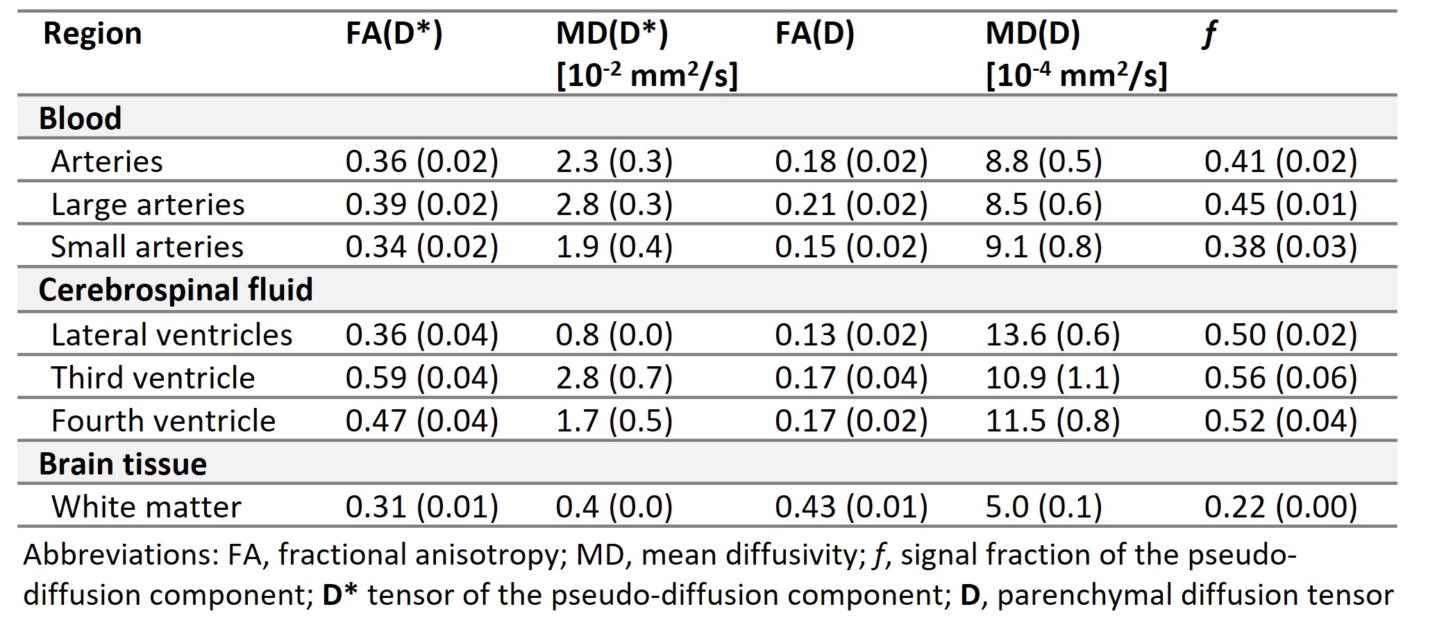
Statistical analysis: Using paired t-tests, we compared FA(D*) and MD(D*) between 1) small and large arteries, expecting a higher variation in flow velocity (i.e., higher FA(D*) and MD(D*)) in large arteries11, 2) the fourth and lateral ventricles, and 3) the fourth and third ventricle, as the variation in CSF flow velocity is highest in the third ventricle, moderate in the fourth ventricle, and lowest in the lateral ventricles.12,13
Results
Figure 2 shows example ellipsoidal representations of D* and their spatial overlap with the arteries. Likewise, Figure 3 visualizes example D* ellipsoids within the ventricles. As can be observed in these figures, the D* ellipsoids align well with the arterial blood flow and CSF flow. Furthermore, an example of the D tensor is shown in Figure 4.The IVIM-DTI parameters per region-of-interest are reported in Table 1. The large arteries had higher FA(D*) and MD(D*) compared to the small arteries (p<0.001). FA(D*) and MD(D*) in the third ventricle were higher than in the fourth ventricle (p<0.001 and p=0.001), and FA(D*) and MD(D*) in the fourth ventricle were higher than in the lateral ventricles (p<0.001).
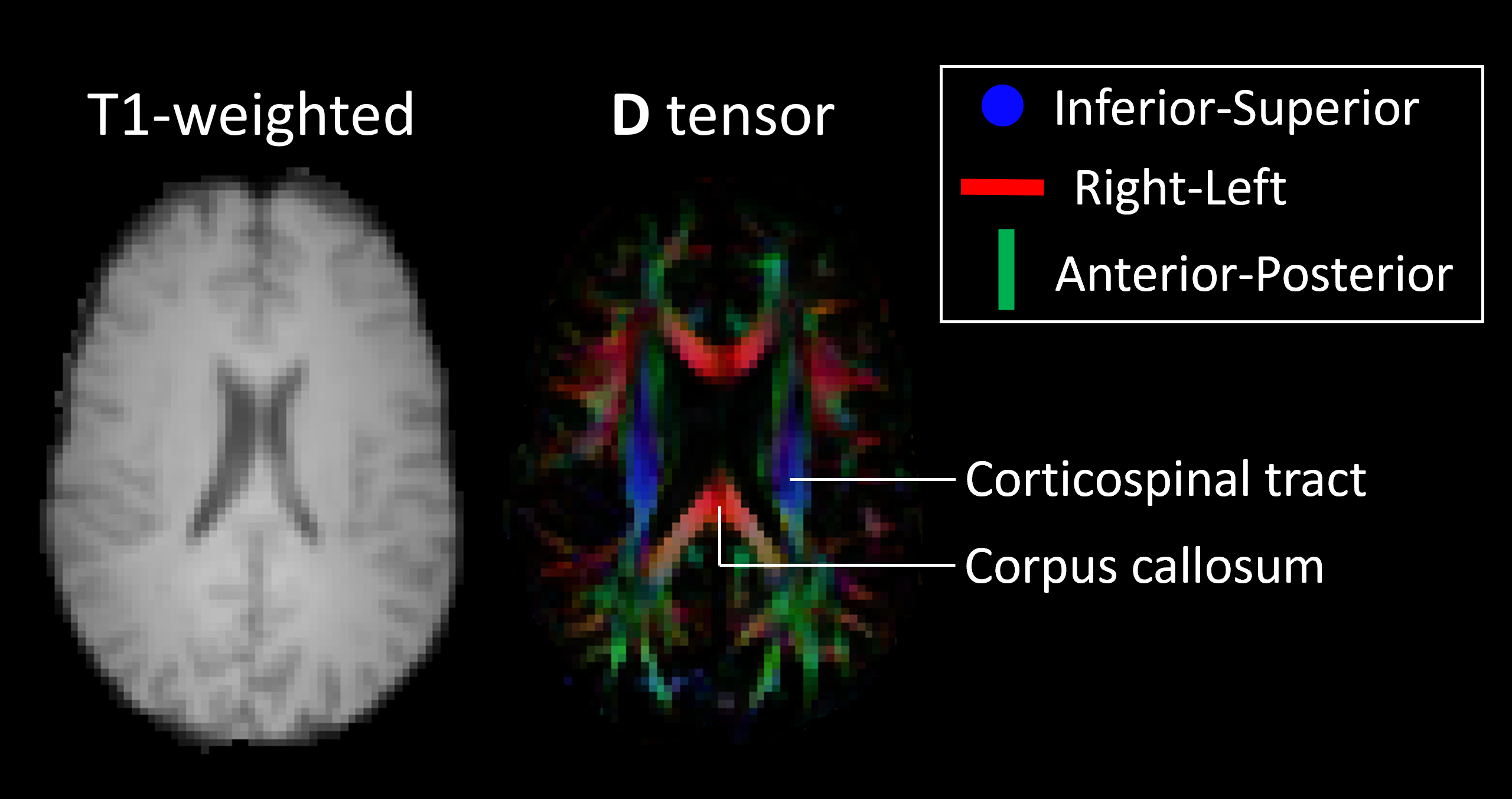
Discussion
This study highlights the potential of IVIM-DTI in characterizing directional blood and CSF flow by showing the alignment of D* ellipsoids with the direction of arteries and CSF cavities. Furthermore, the observed differences in FA(D*) and MD(D*) between large and small arteries, as well as between different ventricles, demonstrate IVIM-DTI’s ability to characterize variation in flow velocities11,13.Moreover, IVIM-DTI can accurately measure the traditional parenchymal diffusion along the WM fibre tracts, as demonstrated by the observed color-coded D tensor and the correspondence of FA(D) in WM with literature14. Future study directions include the evaluation of D* in veins, and the ability of D* to provide a proxy for microvascular architecture.15
Conclusion
IVIM-DTI allows for simultaneous investigation of cerebral microstructural and (micro)vascular alterations, together with information on fluid dynamics, suggesting significant future clinical potential, particularly in conditions such as hydrocephalus and cerebral small vessel disease.12,15Acknowledgements
This work was supported by the European Union’s Horizon 2020 project ‘CRUCIAL’ (grant number 848109).References
1. Le Bihan D. What can we see with IVIM MRI? Neuroimage. 2019;187:56-67.
2. Mozumder M, Beltrachini L, Collier Q, et al. Simultaneous magnetic resonance diffusion and pseudo‐diffusion tensor imaging. Magn Reson Med. 2018;79(4):2367-78.
3. Bito Y, Harada K, Ochi H, Kudo K. Low b-value diffusion tensor imaging for measuring pseudorandom flow of cerebrospinal fluid. Magn Reson Med. 2021;86(3):1369-82.
4. Andersson JL, Skare S, Ashburner J. How to correct susceptibility distortions in spin-echo echo-planar images: application to diffusion tensor imaging. Neuroimage. 2003;20(2):870-88.
5. Leemans A, Jeurissen B, Sijbers J, et al. ExploreDTI: a graphical toolbox for processing, analyzing, and visualizing diffusion MR data. Proc Intl Soc Mag Reson Med; 2009.
6. Kaandorp MP, Barbieri S, Klaassen R, et al. Improved unsupervised physics‐informed deep learning for intravoxel incoherent motion modeling and evaluation in pancreatic cancer patients. Magn Reson Med. 2021;86(4):2250-65.
7. Voorter PH, Backes WH, Gurney-Champion OJ, et al. Improving microstructural integrity, interstitial fluid, and blood microcirculation images from multi-b-value diffusion MRI using physics-informed neural networks in cerebrovascular disease. Magn Reson Med. 2023.
8. Basser PJ, Pierpaoli C. Microstructural and physiological features of tissues elucidated by quantitative-diffusion-tensor MRI. J Magn Reson B. 1996;111(3):209-19.
9. Barmpoutis A, Vemuri BC, Shepherd TM, Forder JR. Tensor splines for interpolation and approximation of DT-MRI with applications to segmentation of isolated rat hippocampi. IEEE Trans Med Imaging. 2007;26(11):1537-46.
10. Puonti O, Iglesias JE, Van Leemput K. Fast and sequence-adaptive whole-brain segmentation using parametric Bayesian modeling. Neuroimage. 2016;143:235-49.
11. Zarrinkoob L, Ambarki K, Wåhlin A, et al. Blood flow distribution in cerebral arteries. J Cereb Blood Flow Metab. 2015;35(4):648-54.
12. Bito Y, Ochi H, Shirase R, et al. Low b-value Diffusion Tensor Imaging to Analyze the Dynamics of Cerebrospinal Fluid: Resolving Intravoxel Pseudorandom Motion into Ordered and Disordered Motions. Magn Reson Med Sci. 2023.
13. Takizawa K, Matsumae M, Hayashi N, et al. The choroid plexus of the lateral ventricle as the origin of CSF pulsation is questionable. Neurol Med Chir (Tokyo). 2018;58(1):23-31.
14. Fox R, Sakaie K, Lee J-C, et al. A validation study of multicenter diffusion tensor imaging: reliability of fractional anisotropy and diffusivity values. Am J Neuroradiol. 2012;33(4):695-700.
15. Dietrich O, Cai M, Tuladhar AM, et al. Integrated intravoxel incoherent motion tensor and diffusion tensor brain MRI in a single fast acquisition. NMR Biomed. 2023:e4905.
Figures