0795
Deep Learning Based Local SAR Prediction for Head imaging at 7T: applicability and accuracy for unknown head coils1Department of Radiology, University Medical Center Utrecht, Utrecht, Netherlands, 2Computational Imaging Group for MR diagnostics & therapy, Center for Image Sciences, University Medical Center Utrecht, Utrecht, Netherlands, 3Tesla Dynamic Coils BV, Zaltbommel, Netherlands, 4Department of Radiotherapy, University Medical Center Utrecht, Utrecht, Netherlands, 5Biomedical Image Analysis, Dept. Biomedical Engineering, Eindhoven University of Technology, Utrecht, Netherlands
Synopsis
Keywords: Safety, Safety, specific absorption rate; deep learning; parallel transmit; convolutional neural network; subject-specific SAR assessment; ultra-high field MRI
Motivation: The methods presented for on-line local SAR evaluation require access to geometric design details of the transmit coil which are not always available.
Goal(s): Evaluate the generalization capabilities of deep learning-based methods when they are used to assess the local SAR distribution for coils not included in the training data.
Approach: We built a diverse synthetic dataset four different coils and trained a neural network: using only samples from each coil, and using samples from all coils except one.
Results: Including a reasonably wide variety of coils in the training process enables local SAR assessment without knowing the design details of the coil.
Impact: The lack of access to design details of the coil makes it challenging to transition the more advanced local SAR assessment methods into clinical practice. Training with a diverse set of coils could enable local SAR assessment without coil information.
PURPOSE
Local SAR evaluation for MRI examinations is an essential part of RF safety assessment for ultrahigh field imaging. Since it cannot be measured it is usually evaluated by off-line numerical simulations. Software tools to perform on-line simulations1,2 and deep learning-based methods3,4 are being developed.Recently a new deep-learning approach was presented for local SAR assessment in brain5.
The brain is indeed the region of greatest clinical interest for ultra-high field MRI. However, all methods presented require access to geometrical design details of the transmit coil. However, these technical specifications and proprietary details are sometimes not shared with users making (on-line) subject-specific simulations impossible. Also deep learning-based approaches will suffer from unknown coil geometries as the coil array intended for use will not be present in the training data set. However, these methods rely on the local relationship between B1+-maps and local SAR and generalizability to other anatomies has been proven to be quite accurate3,4.
In this study, we intend to further test the generalization capabilities of deep learning-based local SAR assessment methods for various coils. Specifically, we will evaluate the local SAR prediction accuracy in the brain for a given coil array, while the network was trained with local SAR distributions from different coil arrays.
METHODS
Intuitively, a broad and diverse training dataset enhances the generalizability of deep learning-based methods. To assess the feasibility of local SAR prediction for out-of-training coil, we built a diverse synthetic dataset (Sim4Life, ZMT, Zürich, Switzerland) using 20 subject-specific head models6-8 and four different head coils for brain imaging at 7T: 1) High-pass birdcage coil with 16 rungs (Diameter:310mm–Length:170mm); 2) 8-fractionated dipole array9,10 3) 4-dipole and 4-loop array; 4) 8-rectangular loop array.For each subject and coil array, we generate complex B1+-maps and corresponding SAR10g distributions3,4 for 1000 random phase-amplitude settings, which results in a total of 80000 (20×4×1000) data samples.
The distributions generated using the models M17, M18, M19 and M20 are used for testing; the other distributions are used for training.
We train a convolutional neural network (U-Net11) to map the relation between complex B1+-maps and the corresponding local SAR distribution by minimizing the RMSE between the predicted and ground-truth local SAR distribution.
To determine whether training with a broader distribution indeed improves predictions beyond the training distribution, we trained the network eight times: four times using only samples from each array, and four times using samples generated from all arrays except one.
Then, for each of the eight training configurations, we assess the local SAR prediction accuracy of the trained networks for unseen coil arrays. Additionally, we explore local SAR prediction using these networks for a 8Tx32Rx head coil (Nova Medical, USA) of which the design details are unknown to us.
RESULTS AND DISCUSSION
Figure 2 shows the scatterplots of predicted versus ground-truth peak local SAR for the networks trained using samples from a single transmit array. A very good correlation can only be observed in scatterplots along the diagonal (same array for training and test).The scatterplots of predicted versus ground-truth peak local SAR for the networks trained using samples from all transmit arrays except one are reported in Figure 3. In this case the scatterplots along the diagonal refer to the tests with the transmit array excluded from the training. However, also in these cases a quite good correlation can be observed.
This improvement is highlighted in Figure 4, reporting the overall scatterplots of predicted versus ground-truth peak local SAR.
Figure 5 shows some examples of the predicted local SAR distribution for the in-silico validation and in-vivo application for the Nova 8Tx24Rx coil array. A reasonably good match between the ground-truth and predicted local SAR distributions can be observed for the birdcage coil and the combined dipole and loop array, even when they are excluded from the training. However, a very poor match is observed for the loop array. Reassuringly, all predicted local SAR distributions for the in-vivo validation are clearly similar.
CONCLUSION
The capability of deep learning-based local SAR prediction in the brain, for unknown or out-of-training head coils, was investigated. Training on a single array always results in poor performance for other arrays. However, training on multiple arrays drastically improves the generalizability. In general, the performance is very good although predicted local SAR distributions still deviate considerably for some transmit arrays (e.g., loop array). Considering these results, we believe that by including a slightly wider variety of transmit arrays in the training process, it will be possible to assess local SAR even without knowing the geometry and design details of the transmit coil.Acknowledgements
This publication is part of the project ‘Finding the hotspot: AI unravels tissue heating in MRI’ with project number 19995 of the Open Technology Program which is (partly) financed by the Dutch Research Council (NWO).References
[1] Villena JF, Polimeridis AG, Eryaman Y, et al. Fast Electromagnetic Analysis of MRI Transmit RF Coils Based on Accelerated Integral Equation Methods. IEEE Trans Biomed Eng. 2016; 63(11):2250-2261.
[2] Brink WM, Yousefi S,Bhatnagar P, Remis RF, Staring M, Webb AG.Personalized local SAR prediction for paralleltransmit neuroimaging at 7T from a singleT1-weighted dataset.Magn Reson Med.2022;88:464-475. doi: 10.1002/mrm.29215
[3] Meliadò EF, Raaijmakers AJE, Sbrizzi A, et al. A deep learning method for image‐based subject‐specific local SAR assessment. Magn Reson Med. 2019;00:1–17.
[4] Meliadò EF, Raaijmakers AJE, Maspero M, et al. Subject-specific Local SAR Assessment with corresponding estimated uncertainty based on Bayesian Deep Learning. Proceedings of the ISMRM 29th Annual Meeting, 8-14 August 2020. p. 4195.
[5] Gokyar S, Zhao C, Ma SJ,Wang DJJ. Deep learning-based local SARprediction usingB1maps and structural MRI of thehead for parallel transmission at 7T.Magn ResonMed. 2023;90:2524-2538. doi: 10.1002/mrm.29797
[6] B. Aubert-Broche, D.L. Collins, A.C. Evans: "A new improved version of the realistic digital brain phantom" NeuroImage, in review - 2006.
[7] B. Aubert-Broche, M. Griffin, G.B. Pike, A.C. Evans and D.L. Collins: "20 new digital brain phantoms for creation of validation image data bases"
[8] Christ A, Kainz W, Hahn EG, et al. The Virtual Family—development of surface‐based anatomical models of two adults and two children for dosimetric simulations. Phys Med Biol. 2010;55:N23–N38.
[9] Steensma BR, Luttje M, Voogt IJ, et al. Comparing Signal-to-Noise Ratio for Prostate Imaging at 7T and 3T. J Magn Reson Imaging. 2019;49(5):1446-1455.
[10] Raaijmakers AJE, Italiaander M, Voogt IJ, Luijten PR, Hoogduin JM, et al. The fractionated dipole antenna: A new antenna for body imaging at 7 Tesla. Magn Reson Med. 2016;75:1366–1374.
[11] O. Ronneberger, P. Fischer, T. Brox, U-Net: Convolutional Networks for Biomedical Image Segmentation, Medical Image Computing and Computer-Assisted Intervention – MICCAI 2015 pp 234-241.
Figures
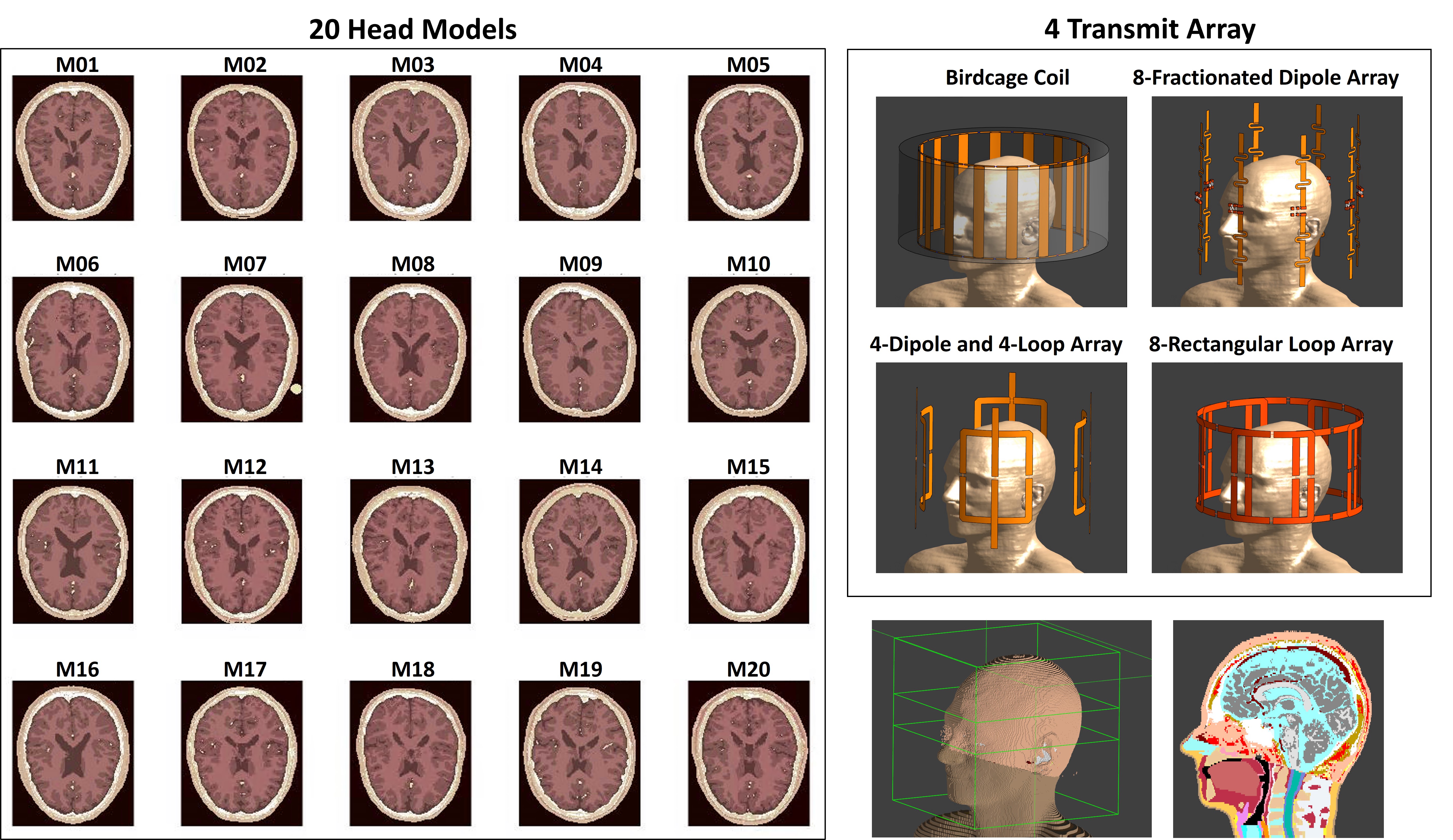
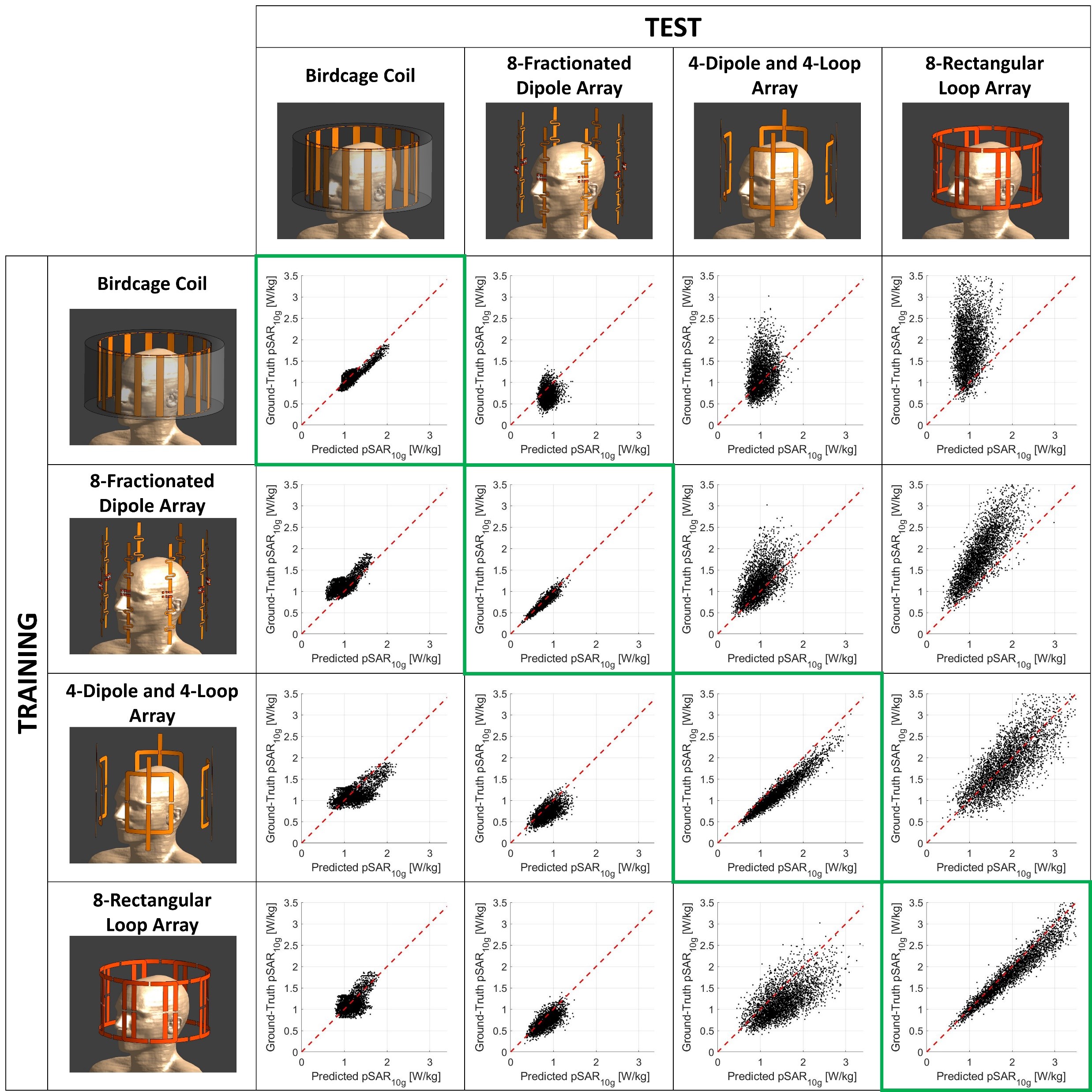
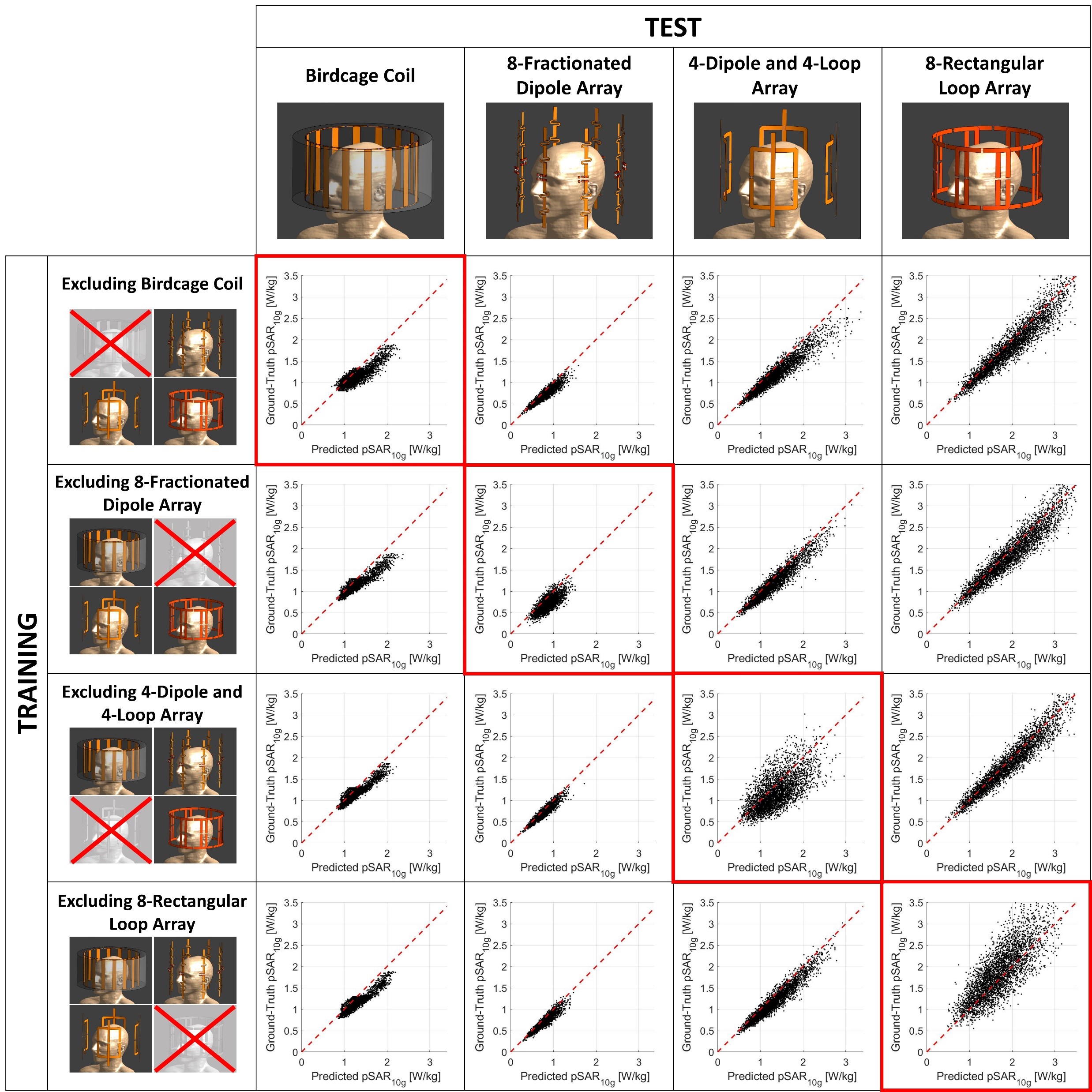
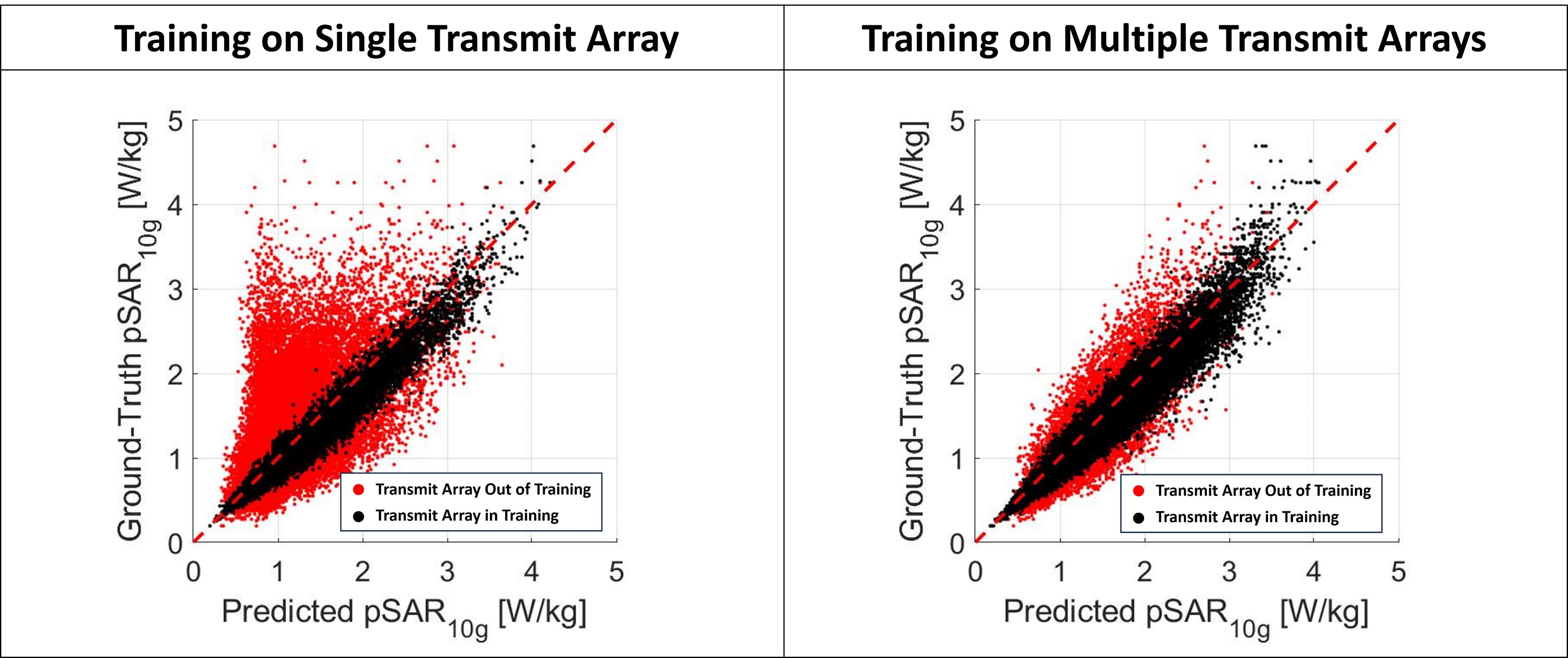
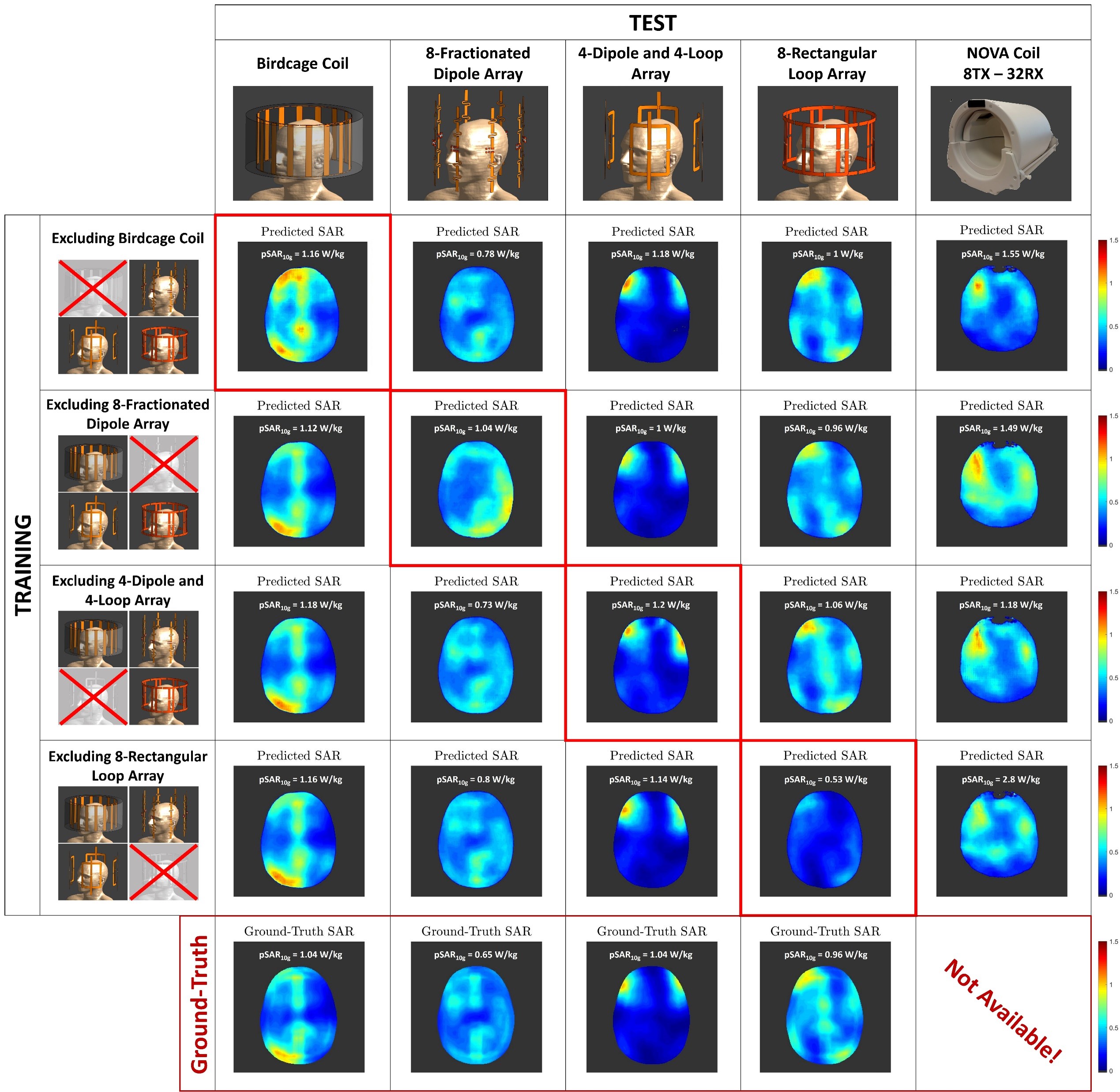
Figure 5: Some examples of the predicted local SAR distribution for the in-silico validation and in-vivo application for the Nova 8Tx24Rx coil array (network trained using samples generated from all arrays except one.).
Written informed consent was obtained according to local IRB regulations.