0770
Sigma-1 receptor changes in chronic knee pain using PET/MRI: Preliminary results of fifteen patients1University of Wisconsin-Madison, Madison, WI, United States, 2Radiology, Stanford University School of Medicine, Stanford, CA, United States
Synopsis
Keywords: Whole Joint, Molecular Imaging, Knee, Pain
Motivation: Diagnosis of chronic knee pain remains a challenge with conventional diagnostic methods leading to unsatisfactory treatment in a large group of patients.
Goal(s): To investigate the use of sigma-1 receptor (S1R) radioligand, [18F] FTC-146 in conjunction with positron emission tomography/magnetic resonance imaging (PET/MRI) for identifying the pain generator in chronic knee pain.
Approach: Comparison of [18F] FTC-146 PET-MRI imaging findings in patients with unresolved chronic knee pain to healthy volunteers.
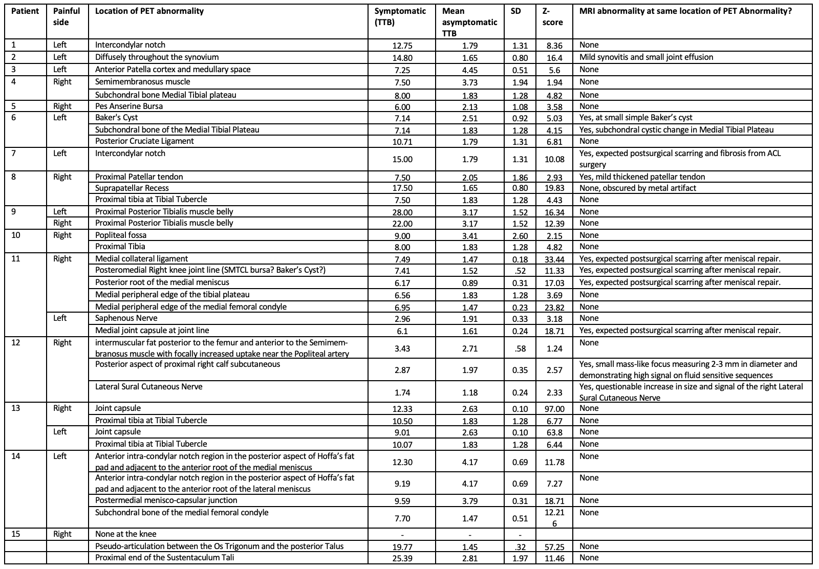
Results: All 15 patients showed statistically significant increased uptake of S1R compared to healthy control subjects in a variety of locations. At sites of abnormal PET uptake, MRI often did not demonstrate abnormalities.
Impact: Future clinical implementation of S1R-PET/MR can potentially help reveal previously unidentified pain generator in patients with chronic knee pain that have exhausted standard clinical care leading to better-targeted treatment.
Introduction
Chronic knee pain is a common medical condition affecting approximately 25% of adults.1 Annually, knee pain costs $635 billion dollars in the U.S.2 The main cause for extended suffering of patients with pain is our inability to identify the pain generator using conventional imaging methods.3 A new method, utilizing the sigma-1 receptor (S1R) radioligand, [18F] FTC-146, has emerged as a promising way to image pain generators. S1R receptor expression is increased in painful and inflamed tissues and modulates a variety of ion channels involved in nociception.4 Tracking this receptor through positron emission tomography (PET) imaging thus allows for identification of pain generators.5,6 S1R PET/MRI has been shown to correlate with injury and pain in the sciatic nerve.7The purpose of this study was to investigate the use of S1R PET/MRI for diagnosing the pain generator in patients with chronic knee pain using an asymptomatic group as control.
Methods
This prospective, single-center, observational study was approved by the IRB and all subjects signed a written informed consent. Patients with chronic knee pain duration >6-month and pain level >4/10 who had failed standard management and asymptomatic controls were included.Whole body PET and MRI scans were acquired simultaneously using a hybrid 3.0T PET/MRI scanner (SIGNA PET/MR; GE Healthcare) in 8-10 bed positions starting 30 minutes after injection of 10 ± 1 mCi of [18F] FTC-146. The following MRI sequences were performed within each bed position: 3D axial fast spoiled gradient-echo, 2D axial T2-weighted fast-spin-echo with triple-echo Dixon. Additional dedicated knee imaging was acquired using multi-planar T1 and T2 weighted sequences with and without fat suppression in addition to 3-dimensional axial double-echo in steady state imaging. PET data was reconstructed using a fully 3-dimensional iterative ordered-subsets expectation maximization algorithm that corrected for attenuation, scatter, dead time and decay.
MR images were first reviewed followed by the PET images in consensus by two musculoskeletal radiologists with more than 15-year experience of interpreting knee MRIs and PET images. Maximum standard uptake value (SUVmax) measurements were performed in Horos (v3.3.2). The SUVmax of corresponding anatomical areas were separately measured in the asymptomatic controls. The SUVmax of the subcutaneous fat of the medial aspect of the knee was measured as background value in all participants. Reference values were established by calculating the asymptomatic control group mean SUVmax and standard deviation for each location. SUVmax to target-to-background (TTB) ratios were calculated in patients and mean SUVmax to TTB ratios in the asymptomatic controls. Single sample Z-score test was used to compare individual patient TTB-ratio against the reference mean TTB and standard deviation. Significance was > 1.96 (95% confidence level). The patient outcomes were followed via self-reported surveys and their electronic medical record.
Results
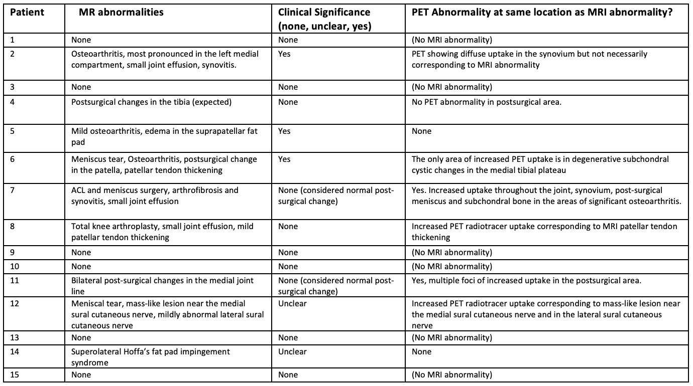
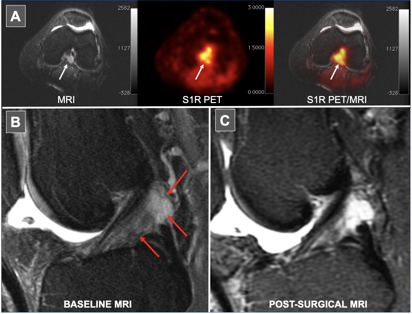
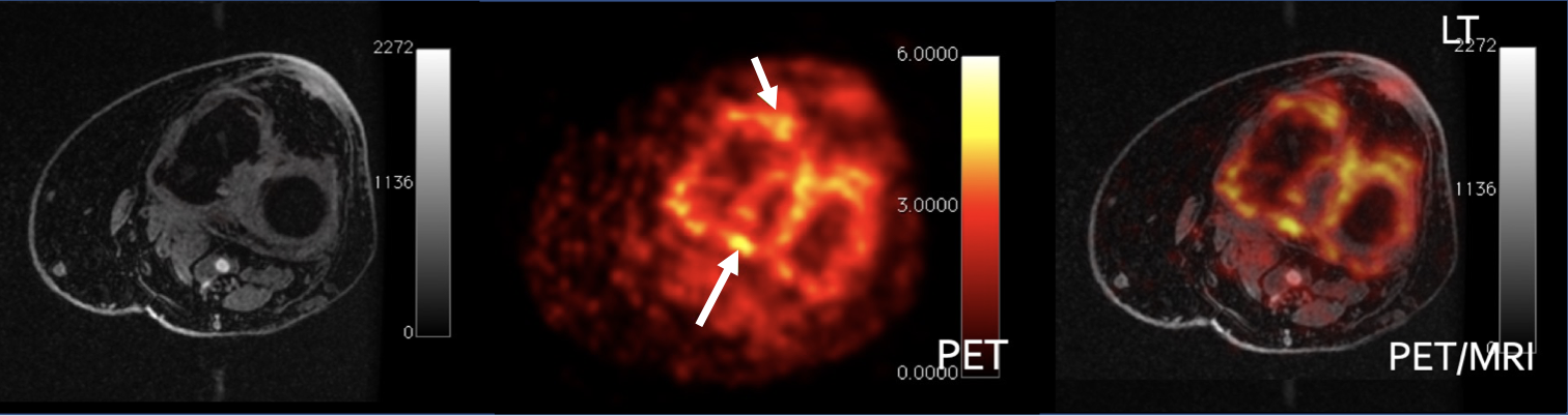
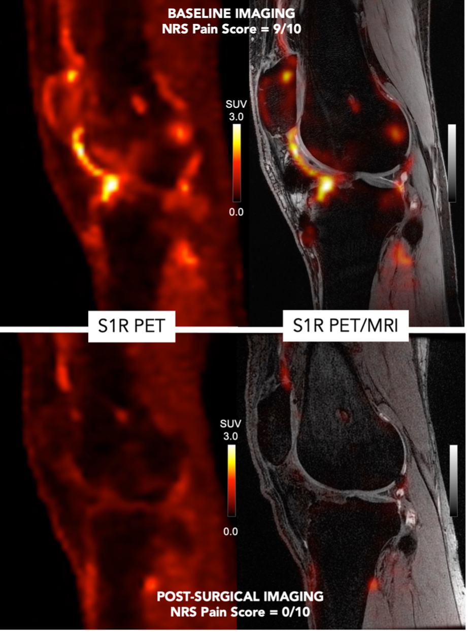
15 patients with chronic knee pain (age 23-78 years, 10 male and 5 female) and 7 asymptomatic controls were included. S1R PET showed statistically significant, abnormally increased S1R uptake in all 15 patients compared to the controls in a variety of locations (Figure 1). In contrast, MRIs showed 6 patients with no MR abnormalities, 4 with expected post-surgical changes that may or may not have been related to the patients’ pain and 5 with findings consistent with joint effusion/synovitis, osteoarthritis and/or meniscal tear (Figure 2). Among the 9 patients that had MRI findings, 4 patients showed abnormal increase S1R uptake at the site of PET abnormality. Clinical follow-up showed a subset of patients benefiting from the PET/MRI insights (Figures 3-5). At least 3 patients, who had been suffering for no less than 2 years of chronic debilitating knee pain, benefited from surgical intervention as a result of the S1R PET/MRI findings. Similarly, at least 3 other patients were deferred surgery since the S1R PET/MRI findings were not supportive of a surgical approach.Discussion and limitations
In this preliminary study, S1R PET/MR imaging appears to have influenced a significant positive outcome in a subset of patients. These patients had achieved complete pain relief when previous standard-of-care measures had failed them. Clinical follow-up was limited in some as they were lost to follow up. Additionally, the referring physician, for a variety of reasons, would or could not act upon the PET/MRI findings in some cases as the findings were novel and could not be easily clarified. Future prospective, longitudinal studies will help determine whether the integration of S1R PET/MR imaging will improve the outcomes of chronic knee pain sufferers.Conclusion
S1R PET/MR can potentially help inform clinical decision making by revealing abnormalities in the areas that are not apparent by MR imaging alone, leading to favorable outcomes in patients with chronic knee pain.Acknowledgements
We would like to acknowledge Sunyoung Park, Kim Halbert, Harsh Gandhi, Dawn Holley, Medhi Khaligi and Peter Cipriano for their technical and administrative assistance and Chris McCurdy PhD for chemistry support. Financial support was provided in part by GE Healthcare. We wish to acknowledge support from Bracco Diagnostics who provide research support to the University of Wisconsin.References
1 Bunt CW, Jonas CE, Chang JG. Knee Pain in Adults and Adolescents: The Initial Evaluation. Am Fam Physician. 2018 Nov 1;98(9):576-585.
2 Institute of Medicine (US) Committee on Advancing Pain Research, Care, and Education. Relieving Pain in America: A Blueprint for Transforming Prevention, Care, Education, and Research. Washington (DC): National Academies Press (US); 2011.
3 Culvenor AG, Øiestad BE, Hart HF, Stefanik JJ, Guermazi A, Crossley KM. Prevalence of knee osteoarthritis features on magnetic resonance imaging in asymptomatic uninjured adults: a systematic review and meta-analysis. Br J Sports Med. 2019 Oct;53(20):1268-1278.
4 National Center for Biotechnology Information. PubChem Database. CID=58511957, https://pubchem.ncbi.nlm.nih.gov/compound/58511957
5 Shen B, Park JH, Hjørnevik T, Cipriano PW, Yoon D, Gulaka PK, Holly D, Behera D, Avery BA, Gambhir SS, McCurdy CR, Biswal S, Chin FT. Radiosynthesis and First-In-Human PET/MRI Evaluation with Clinical-Grade [18F]FTC-146. Mol Imaging Biol. 2017 Oct;19(5):779-786.
6 Tung KW, Behera D, Biswal S. Neuropathic pain mechanisms and imaging. Semin Musculoskelet Radiol. 2015 Apr;19(2):103-11.
7 Shen B, Behera D, James ML, Reyes ST, Andrews L, Cipriano PW, Klukinov M, Lutz AB, Mavlyutov T, Rosenberg J, Ruoho AE, McCurdy CR, Gambhir SS, Yeomans DC, Biswal S, Chin FT. Visualizing Nerve Injury in a Neuropathic Pain Model with [18F]FTC-146 PET/MRI. Theranostics. 2017 Jul 8;7(11):2794-2805.
Figures