0763
Thalamic nuclei atrophy in autosomal dominant Alzheimer's disease1Radiology, University of Massachusetts Chan Medical School, Worcester, MA, United States, 2MGH, Harvard Medical School, Charlestown, MA, United States, 3Norwich Medical School, University of East Anglia, East Anglia, United Kingdom, 4Neurociencias, Universidad de Antioquia, Medellin, Colombia, 5Psychiatry, MGH, Harvard Medical School, Charlestown, MA, United States
Synopsis
Keywords: Alzheimer's Disease, Alzheimer's Disease, thalamic nuclei segmentation
Motivation: The thalamus has not been investigated properly in autosomal dominant Alzheimer's disease (ADAD) despite evidence of early involvement.
Goal(s): We investigated thalamic nuclei atrophy in asymptomatic presenilin carriers vs. non-carriers in an ADAD cohort using a recently proposed multi-atlas segmentation method.
Approach: Thalamic nuclear atrophy and correlations of nuclei volumes with age, amyloid, and tau burden were
analyzed in mutation carriers vs. non-carriers in an ADAD cohort.
Results: Significant atrophy was seen in mediodorsal, pulvinar, and medial geniculate nuclei in carriers compared to non-carriers. These nuclear volumes correlated significantly with age, amyloid, and tau burden as well.
Impact: The pattern of thalamic nuclear atrophy in ADAD presenilin mutation carriers can help understand mechanisms for disease progression as well as aid in possible treatment targets.
Introduction
The thalamus is an important sensorimotor relay and is also associated with memory processing, sleep, wakefulness, and arousal. Despite its widespread connectivity to almost every structure in the brain, it has received scant attention in neuroimaging studies and usually as a single entity instead of a heterogeneous structure comprised of multiple nuclei. Autosomal dominant Alzheimer's Disease (ADAD) offers a unique opportunity to study disease progression and changes in AD. Despite the early involvement of the thalamus in ADAD and other early onset AD, very few studies have examined the thalamus in this context. Here, using a recently proposed sensitive thalamic nuclei segmentation method, we examined thalamic nuclei volumes from T1 MRI in the largest ADAD cohort (Colombia) with the presenilin PSEN1 mutation. Specifically, we quantified thalamic nuclear atrophy in carriers compared to non-carriers in the same kindred. We also examined the associations of thalamic nuclei volumes with age and brain pathology biomarkers such as PET beta-amyloid and tau burden.Methods
T1 MRI scans from 99 subjects (50 non-carriers, 49 PSEN1 E280A mutation carriers) who were part of the Alzheimer's Prevention Initiative of the Colombia-Boston COLBOS study were analyzed. The carrier and non-carrier cohorts were matched with respect to age (mean: 37 years) and gender balance (roughly equal male vs. female). MRI, amyloid PET using 11C Pittsburgh Compound B (PiB) and tau PET using 18F fluortaucipir were acquired as described previously in [1].We used a recently proposed multi-atlas thalamic nuclei segmentation method called Histogram-based polynomial synthesis (HIPS)-THOMAS [2] which has been shown to be more sensitive than Freesurfer-based segmentation [3]. HIPS leverages the improved contrast of white-matter nulled (WMn) MPRAGE by synthesizing that contrast from conventional T1 MPRAGE images using a simple polynomial fitting approach. The synthesized WMn images are then segmented using THOMAS segmentation.
Analyses were carried out using jamovi (2.3.21). Intracranial volumes (ICV) were computed using Freesurfer (eTIV inside aseg stats). ANCOVA was used to determine nuclear atrophy using appropriate covariates. Partial correlation analysis was used to determine the associations of thalamic nuclear volumes with age, amyloid and tau burden.
Results
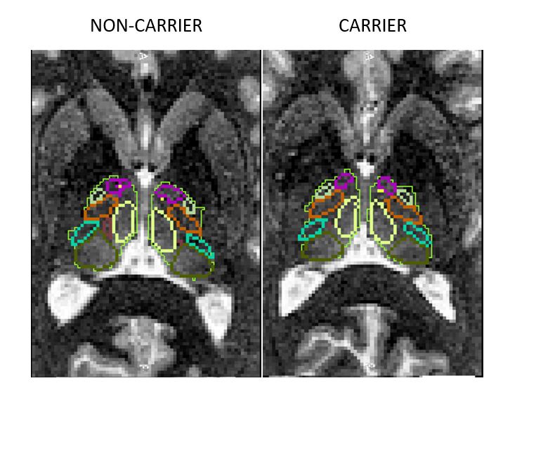
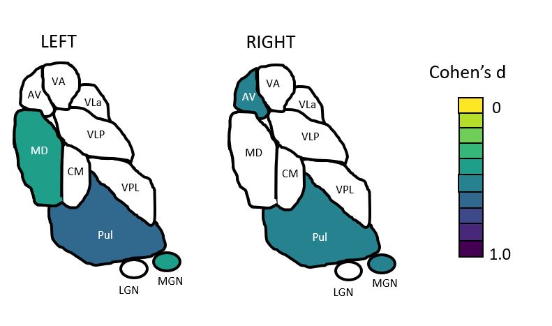
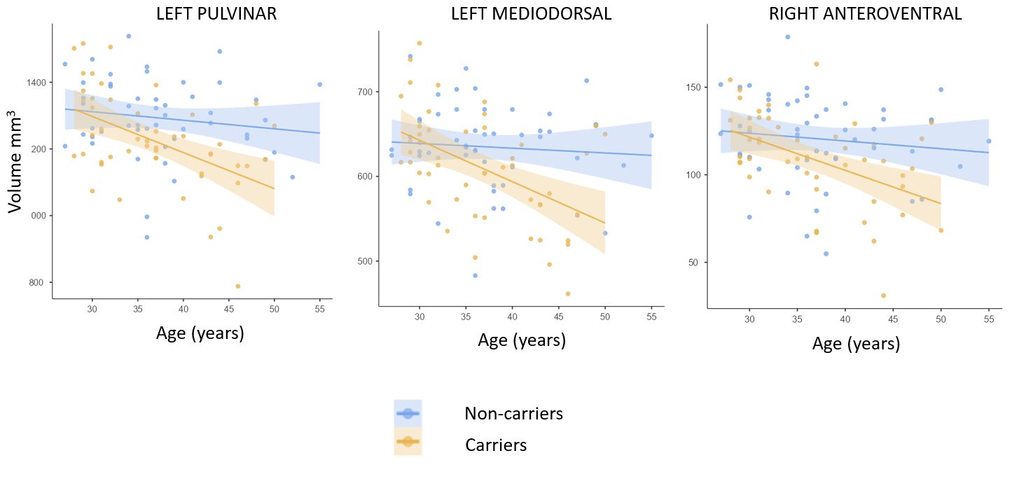
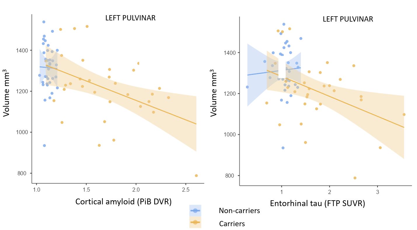
Figure 1 shows HIPS-THOMAS segmentation labels overlaid on WMn images (synthesized from standard T1) from exemplary non-carrier and carrier subjects. THOMAS segments 11 thalamic nuclei per side. In addition to whole left and right thalamus, significant atrophy (effect sizes > 0.5) was observed in mediodorsal, pulvinar and medial geniculate nuclei (MGN) for the left side and in pulvinar, anteroventral, and MGN for the right side in carriers compared to non-carriers after controlling for age, sex, and ICV (Figure 2). Significant age-nuclear volume correlations were observed for all nuclei exhibiting atrophy (Pearson's r -0.28 to -0.42, p < 0.001) after adjusting from sex and ICV. For left medial, left pulvinar, and right anteroventral nuclei, atrophy in carriers deviated significantly from non-carriers as shown in Figure 3 indicating that atrophy is accelerated in these nuclei in carriers on top of age-related atrophy. Lastly, all the nuclei which showed atrophy in carriers also correlated significantly with amyloid and tau burden with left pulvinar showing the highest r (-0.38, p<0.001 for amyloid; -0.34, p <0.003 for tau). The association of left pulvinar volumes with amyloid (left) and tau (right) burden is shown in Fig 4.Conclusions
To our knowledge, this is the first study showing atrophy of specific thalamic nuclei in asymptomatic presenilin carriers in an ADAD cohort. While Padrilla-Delgado [1] also examined thalamic nuclei atrophy in the same cohort, they failed to observe any significant differences between carriers and non-carriers. This could partly be due to the use of Freesurfer which has been shown to be less sensitive to detection of thalamic nuclei atrophy [3] as well as smaller n (67 vs 99 in this study). Their study also used regions comprising of groups of nuclei and combined left and right sides while our study considered individual nuclei from each hemisphere. The atrophy of mediodorsal, pulvinar and MGN in asymptomatic carriers is remarkably similar to that demonstrated in early mild cognitive impairment in an analysis of ADNI data by Bernstein et al [4]. Our correlations with amyloid and tau burden are concordant with [1] who reported medial and posterior regional correlations. In conclusion, the use of a sensitive segmentation method allowed us to delineate specific thalamic nuclear atrophy in asymptomatic mutation carriers. This could be of immense value in studying disease progression mechanisms in AD as well as design of treatment targets.Acknowledgements
Grants: R01AG054671 from the National Institute on Aging (Quiroz), R01EB032674 from the National Institute of Biomedical Imaging and Bioengineering (Saranathan)References
1. Padrilla-Delgado et al. Brain Communications 2021 May 10;3(2):fcab101.
2. Vidal et al.
arXiv:2304.07167 [eess.IV]
3. Williams et al.
arXiv:2309.15053 [eess.IV]
4. Bernstein et al. J. Alzheimer's Disease. 82:361-371 (2021)
Figures