0762
Associations of quantitative susceptibility mapping with cortical atrophy and brain connectome in Alzheimer's disease: a multi-modal study1Department of Radiology, China–Japan Friendship Hospital, Beijing, China, 2China–Japan Friendship Hospital, Beijing, China
Synopsis
Keywords: Alzheimer's Disease, Alzheimer's Disease
Motivation: Associations between abnormal quantitative susceptibility mapping (QSM), brain atrophy, and altered brain connectome in AD remain unclear.
Goal(s): We aim to examine imaging markers from various MRI modalities, with a focus on their spatial correlations, to enhance our understanding of AD pathology.
Approach: By combining multi-contrast MRI techniques, our study provides new insights into the overlapping relationships among brain atrophy, altered regional QSM, and brain connectome.
Results: We observed a remarkable overlap between reduced cortical thickness and abnormal QSM in seven distinct brain regions. In AD patients, we identified specific regional correlations between cortical thickness and network topology from these overlapping brain regions.
Impact: Our
study provides new insights into the complex relationships among iron
accumulation, brain atrophy, and brain connectome in Alzheimer's disease.
INTRODUCTION
Susceptibility ron abnormality and brain network disconnections are observed in Alzheimer's disease (AD), with disrupted iron homeostasis hypothesized to be linked to AD pathology and neuron loss. However, associations between abnormal quantitative susceptibility mapping (QSM), brain atrophy, and altered brain connectome in AD remain unclear.METHODS
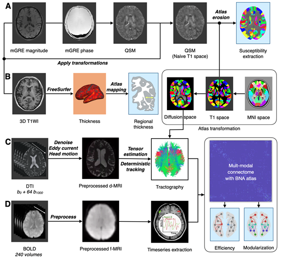
This study utilized multi-modal MRI techniques to explore overlapped regions between cortical atrophy and increased susceptibility and investigate relationships between structural and functional brain connectome, gray matter volumes, and regional QSM on 30 AD patients and 26 healthy controls enrolled at China-Japan Friendship Hospital. We calculated the QSM signal and cortical thickness in 246 brain regions and identified the overlapped regions. The structural and functional connectomes were constructed based on diffusion MRI tractography and functional connectivity, respectively. The network topology was quantified using graph theory analyses.RESULTS
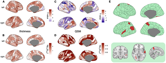
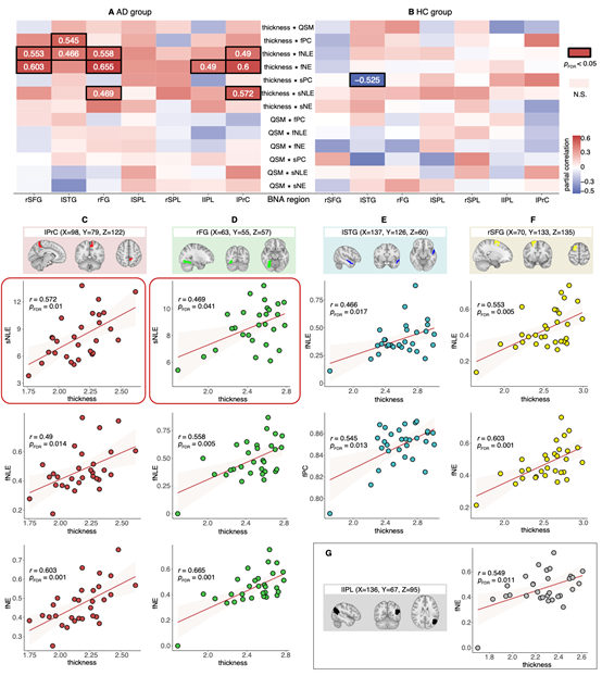
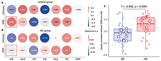
We revealed seven brain regions with both reduced cortical thickness and abnormal QSM, including right superior frontal gyrus, left superior temporal gyrus, right fusiform gyrus, left superior parietal lobule, right superior parietal lobule, left inferior parietal lobule, and left precuneus. Unique correlations between cortical thickness and network topology specific to the AD group were observed in five of these regions, with stronger associations in functional compared to structural topology. Decreased spatial covariance of QSM and global efficiency of the structural network was also found in AD patients.CONCLUSION
Our study provides new insights into the complex relationships among iron accumulation, brain atrophy, and brain connectome in Alzheimer's disease (AD). By combining multi-contrast MRI techniques, we observed distinct coupling patterns between brain connectivity and cortical iron deposition in AD, highlighting the potential role of iron in neurodegeneration. Our findings contribute to a better understanding of the underlying mechanisms involved in AD pathology. Further research is needed to validate and expand upon these findings, with the aim of identifying potential therapeutic targets for AD.Acknowledgements
The authors thank Dr. Lizhi Xie from GE Healthcare for help in solving MR technical problems.References
No reference found.Figures