0759
Alterations in superficial white matter tracts are associated with pathological deposition in early-stage Alzheimer’s disease1The Second Affiliated Hospital of Zhejiang University School of Medicine, Hangzhou, China, 2University of Electronic Science and Technology of China, Chengdu, China, 3Harvard Medical School, Boston, MA, United States
Synopsis
Keywords: Alzheimer's Disease, Brain, diffusion magnetic resonance imaging, superficial white matter
Motivation: Between-cortical connections largely depend on the superficial white matter (SWM) fibers, which are less studied in the AD continuum.
Goal(s): To determine the relationship between superficial white matter (SWM) fiber microstructure and local pathology, and the SWM's impact on cognition.
Approach: We defined cohort groups in the early AD continuum. We quantified the microstructure of SWM fiber tracts (diffusion MRI) and the regional pathological deposition (PET). We analyzed associations between SWM fiber microstructure and regional pathologies in cortical areas connected by the tract.
Results: SWM tract microstructure is affected by pathology in the cortical regions connected by the tract, and this affects memory.
Impact: We localize pathology-affected SWM connections, assess their roles in cognition, and provide new insights into white matter abnormalities in the AD continuum.
Introduction
Alzheimer's disease (AD), the most common type of dementia, is a neurodegenerative disorder characterized by cognitive deterioration that is associated with degeneration in white matter connections1. In AD, alterations in white matter microstructure have been reported to be affected by the accumulation of pathologies2. AD pathology is understood to originate in cortical regions3–5 and to primarily affect, and even spread along, the white matter tracts directly connected to these cortical regions 6–8.Altered white matter microstructure has been associated with the severity of the disease status9,10 and is considered a key factor in cognitive deficits in AD11,12. While associations between the microstructure of long-range deep white matter (DWM) tracts and the pathological deposition in their connecting cortical regions have been found13–20, thus far, the superficial white matter (SWM) is less studied.The SWM is the thin layer of white matter underneath the cortex that includes short-range association connections (u-fibers). Most previous studies have quantified the microstructure of SWM regions at large scales, such as the entire or lobar SWM connections21–23, which precludes the investigation of particular white matter fiber connections within the SWM. The purpose of the current study is to quantify the local microstructure of SWM connections and to determine relationships between alterations in SWM microstructure and cortical pathological deposition across the early stages of the AD continuum.Method
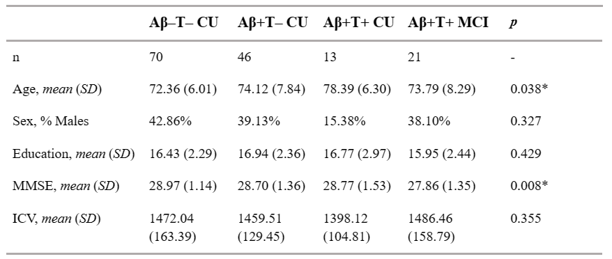
The study included 150 participants from the Alzheimer’s Disease Neuroimaging Initiative (ADNI) with positron emission tomography (PET) data (129 cognitively normal and 21 mild cognitive impairment patients). We defined four pathological staging groups in the AD continuum based on an AD-related biomarker framework: Aβ-T- CU; Aβ+T- CU; Aβ+T+ CU; and Aβ+T+ MCI. We used diffusion magnetic resonance imaging (dMRI) for SWM fiber tractography in the whitematteranalysis (WMA) software package, provided via SlicerDMRI24-25. The method is based on a well-established fiber clustering pipeline26-27 in conjunction with an anatomical white matter tract atlas28, and quantified the microstructure of SWM fiber clusters using a multi-tensor diffusion model29, including free water (FW) and FW-corrected fractional anisotropy (FAt). We quantified the regional pathological deposition of Aβ and tau using PET. We first compared between-group differences in SWM fiber cluster microstructure by constructing generalized linear regression models for each of the 396 fiber clusters. Statistically significant fiber clusters were selected as the clusters of interest (COIs) for subsequent analyses. Furthermore, we constructed linear regression models to analyze the correlation between the diffusion measures of COIs and the regional pathology deposition in two areas connected by COIs. Then, we constructed linear regression models between the microstructure of each COI and cognitive scores for five cognitive domains. Finally, we constructed mediation models to investigate the mediating role of the microstructure of specific SWM fiber clusters between regional pathology and cognition.Results
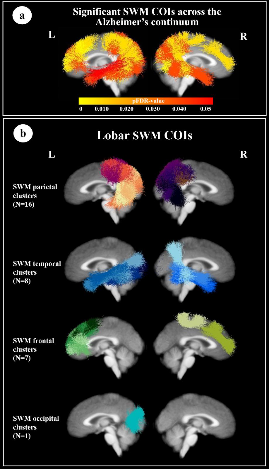
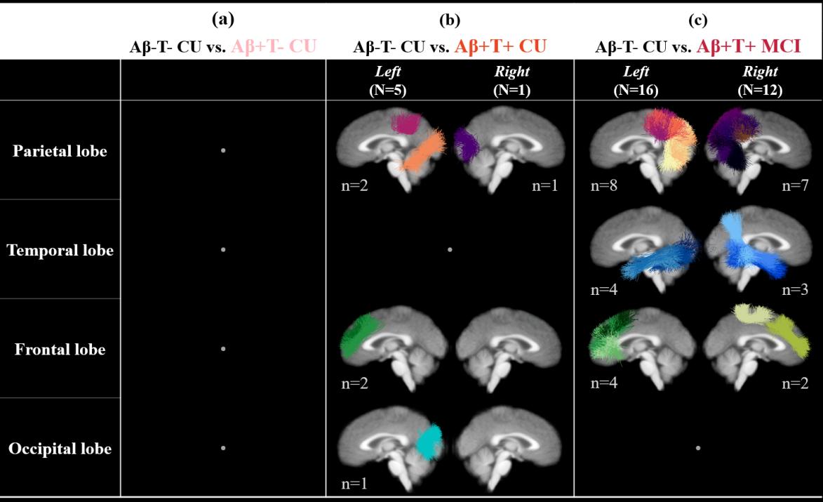
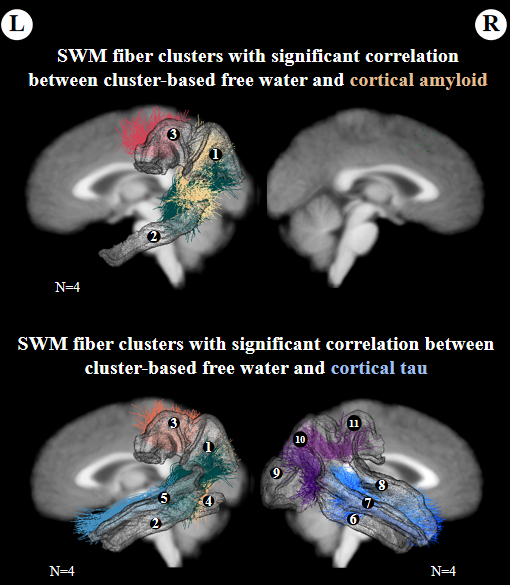
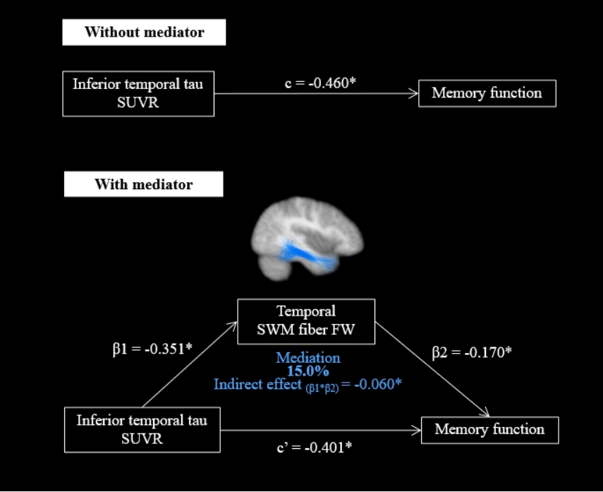
Alterations in SWM fiber cluster microstructure were observed in the early clinical stages of AD, manifesting as a significant increase in FW, mainly distributed in the SWM of the temporal and parietal lobes. The microstructure of the SWM COIs was significantly correlated with Aβ and/or tau deposition in the anatomically connected cortical regions. The SWM COI microstructure in the temporal lobe was significantly correlated with memory function. Mediation analysis showed that the impact of temporal tau pathology deposition on memory function was mediated by the microstructure of an SWM COI in the temporal lobe.Discussion
This study applied dMRI tractography to assess SWM fiber tract microstructure across the early stages of the AD continuum, and investigated the association between SWM cluster microstructure and pathologies in the cluster-connected cortical regions, as well as the impact of SWM on cognition The main findings can be summarized as follows: (i) Between stages of the AD continuum, the number of SWM clusters with significant between-group differences in microstructure increases with the increase of pathological deposition, especially in the parieto-occipito-temporal cortical region; (ii) FW in specific SWM clusters is associated with Aβ and tau deposition in the cluster-specific/connected cortical regions, and these pathology-affected SWM clusters are mostly located in the posterior lobes; (iii) FW in SWM COIs is significantly associated with memory function and, furthermore, FW in a particular SWM COI mediates the association between tau in the cluster-connected cortical regions and memory function.Conclusion
Overall, we performed a detailed investigation of individual SWM connections in the early stages of AD and provided new insights into white matter abnormalities between stages of the AD continuum. In addition, this study localized pathology-affected SWM connections and demonstrated their significance for cognition.Acknowledgements
The authors gratefully acknowledge the following funding grants: the National Natural Science Foundation of China(Grant Nos. 82302138, 62371107), R01MH119222, R01MH132610, R01MH125860, R01NS125781.References
1. Dubois B, Hampel H, Feldman HH, Scheltens P, Aisen P, Andrieu S, et al. (2016): Preclinical Alzheimer’s disease: Definition, natural history, and diagnostic criteria. Alzheimers Dement 12: 292–323.
2. Collij LE, Ingala S, Top H, Wottschel V, Stickney KE, Tomassen J, et al. (2021): White matter microstructure disruption in early stage amyloid pathology. Alzheimers Dement 13: e12124.
3. Mattsson N, Palmqvist S, Stomrud E, Vogel J, Hansson O (2019): Staging β-Amyloid Pathology With Amyloid Positron Emission Tomography. JAMA Neurol 76: 1319–1329.
4. Schöll M, Lockhart SN, Schonhaut DR, O’Neil JP, Janabi M, Ossenkoppele R, et al. (2016): PET Imaging of Tau Deposition in the Aging Human Brain. Neuron 89: 971–982.
5. Braak H, Braak E (1995): Staging of Alzheimer’s disease-related neurofibrillary changes. Neurobiol Aging 16: 271–8; discussion 278–84.
6. Kantarci K, Murray ME, Schwarz CG, Reid RI, Przybelski SA, Lesnick T, et al. (2017): White-matter integrity on DTI and the pathologic staging of Alzheimer’s disease. Neurobiol Aging 56: 172–179.
7. Vogel JW, Iturria-Medina Y, Strandberg OT, Smith R, Levitis E, Evans AC, et al. (2020): Spread of pathological tau proteins through communicating neurons in human Alzheimer’s disease. Nat Commun 11: 2612.
8. Araque Caballero MÁ, Suárez-Calvet M, Duering M, Franzmeier N, Benzinger T, Fagan AM, et al. (2018): White matter diffusion alterations precede symptom onset in autosomal dominant Alzheimer’s disease. Brain 141: 3065–3080.
10. Sachdev PS, Zhuang L, Braidy N, Wen W (2013): Is Alzheimer’s a disease of the white matter? Curr Opin Psychiatry 26: 244–251.
11. Li K, Wang S, Luo X, Zeng Q, Jiaerken Y, Xu X, et al. (2020): Progressive Memory Circuit Impairments along with Alzheimer’s Disease Neuropathology Spread: Evidence from in vivo Neuroimaging. Cereb Cortex 30: 5863–5873.
12. Pereira JB, Janelidze S, Ossenkoppele R, Kvartsberg H, Brinkmalm A, Mattsson-Carlgren N, et al. (2021): Untangling the association of amyloid-β and tau with synaptic and axonal loss in Alzheimer’s disease. Brain 144: 310–324.
13. Strain JF, Smith RX, Beaumont H, Roe CM, Gordon BA, Mishra S, et al. (2018): Loss of white matter integrity reflects tau accumulation in Alzheimer disease defined regions. Neurology 91: e313–e318.
14. Teipel SJ, Kuper-Smith JO, Bartels C, Brosseron F, Buchmann M, Buerger K, et al. (2019): Multicenter tract-based analysis of microstructural lesions within the Alzheimer’s disease spectrum: Association with amyloid pathology and diagnostic usefulness. J Alzheimers Dis 72: 455–465.
15. Sánchez SM, Duarte-Abritta B, Abulafia C, De Pino G, Bocaccio H, Castro MN, et al. (2020): White matter fiber density abnormalities in cognitively normal adults at risk for late-onset Alzheimer’s disease. J Psychiatr Res 122: 79–87.
16. Wen Q, Mustafi SM, Li J, Risacher SL, Tallman E, Brown SA, et al. (2019): White matter alterations in early-stage Alzheimer’s disease: A tract-specific study. Alzheimers Dement 11: 576–587.
17. Lee S-H, Coutu J-P, Wilkens P, Yendiki A, Rosas HD, Salat DH, Alzheimer’s disease Neuroimaging Initiative (ADNI) (2015): Tract-based analysis of white matter degeneration in Alzheimer’s disease. Neuroscience 301: 79–89.
18. Raghavan S, Przybelski SA, Reid RI, Lesnick TG, Ramanan VK, Botha H, et al. (2022): White matter damage due to vascular, tau, and TDP-43 pathologies and its relevance to cognition. Acta Neuropathol Commun 10: 16.
19. Pichet Binette A, Theaud G, Rheault F, Roy M, Collins DL, Levin J, et al. (2021): Bundle-specific associations between white matter microstructure and Aβ and tau pathology in preclinical Alzheimer’s disease. Elife 10. https://doi.org/10.7554/eLife.62929
20. Archer DB, Moore EE, Shashikumar N, Dumitrescu L, Pechman KR, Landman BA, et al. (2020): Free-water metrics in medial temporal lobe white matter tract projections relate to longitudinal cognitive decline. Neurobiol Aging 94: 15–23.
21. Bigham B, Zamanpour SA, Zare H (2022): Features of the superficial white matter as biomarkers for the detection of Alzheimer’s disease and mild cognitive impairment: A diffusion tensor imaging study. Heliyon 8: e08725.
22. Bigham B, Zamanpour SA, Zemorshidi F, Boroumand F, Zare H, Alzheimer’s Disease Neuroimaging Initiative (2020): Identification of Superficial White Matter Abnormalities in Alzheimer’s Disease and Mild Cognitive Impairment Using Diffusion Tensor Imaging. J Alzheimers Dis Rep 4: 49–59.
23. Phillips OR, Joshi SH, Piras F, Orfei MD, Iorio M, Narr KL, et al. (2016): The superficial white matter in Alzheimer’s disease. Hum Brain Mapp 37: 1321–1334.
24. Norton I, Essayed WI, Zhang F, Pujol S, Yarmarkovich A, Golby AJ, et al. (2017): SlicerDMRI: Open Source Diffusion MRI Software for Brain Cancer Research. Cancer Res 77: e101–e103.
25. Zhang F, Noh T, Juvekar P, Frisken SF, Rigolo L, Norton I, et al. (2020): SlicerDMRI: Diffusion MRI and Tractography Research Software for Brain Cancer Surgery Planning and Visualization. JCO Clin Cancer Inform 4: 299–309.
26. O’Donnell LJ, Wells WM 3rd, Golby AJ, Westin C-F (2012): Unbiased groupwise registration of white matter tractography. Med Image Comput Comput Assist Interv 15: 123–130.
27. O’Donnell LJ, Westin C-F (2007): Automatic tractography segmentation using a high-dimensional white matter atlas. IEEE Trans Med Imaging 26: 1562–1575.
28. Zhang F, Wu Y, Norton I, Rigolo L, Rathi Y, Makris N, O’Donnell LJ (2018): An anatomically curated fiber clustering white matter atlas for consistent white matter tract parcellation across the lifespan. Neuroimage 179: 429–447.
29. Pasternak O, Sochen N, Gur Y, Intrator N, Assaf Y (2009): Free water elimination and mapping from diffusion MRI. Magn Reson Med 62: 717–730.
Figures