0755
Respiratory Triggered MRI using an NMR-on-a-chip Sensor1Internal Medicine II, Ulm University Medical Center, Ulm, Germany, 2Institute of Smart Sensors, University of Stuttgart, Stuttgart, Germany
Synopsis
Keywords: Motion Correction, Motion Correction
Motivation: We propose an NMR-on-a-chip sensor as contactless und hysteresis-free alternative for conventional respiratory belts in respiratory-triggered MRI.
Goal(s): The objective of this work was to demonstrate the feasibility of a local field probe for monitoring respiratory motion induced magnetic field changes as respiratory motion surrogate.
Approach: Respiratory belt and field probe signal were recorded simultaneously clearly showing the accurate identification of the respiratory stage by the field probe.
Results: The field probe signal was analysed and fed back to the MR scanner in real-time for proofing its applicability for triggered lung MRI, yielding a sharp lung-liver interface compared with the non-triggered version.
Impact: The feasibility of NMR-on-a-chip sensors for monitoring physiologically-induced magnetic field variations is shown. They enable contactless, hysteresis-free and easy-to-use monitoring of physiologically-induced field variations, which can be fed back to the scanner for real-time respiratory motion monitoring and triggering.
Motivation
Cardiac and lung MRI usually require motion synchronization to achieve sufficient image quality. This can be either done retrospectively e.g. by self-gating1 or prospectively using ECG/respiratory belts2. Bellows-gating has been already investigated extensively3,4 indicating that a bellows has a higher gating efficiency compared to other compensation strategies, is independent of any preparation pulses as may be needed for e.g. navigator-based gating3 and provides a continuous signal. As the breathing belt suffers from hysteresis effects and does not work contactless, we suggest replacing it with an NMR-on-a-chip sensor5. Field probes have previously been shown to monitor physiological signals6, but not been applied for respiratory motion synchronization of an MRI acquisition.Methods
The setup for using the NMR-on-a-chip sensor is sketched in fig.1. Commercially available hardware from National Instruments (NI, Austin, USA) is used for data acquisition and digitalization. A custom-made backend preprocesses the acquired signals and serves as interface for the rest of the hardware. The NMR probes are made of 0.8mm capillaries filled with CuSO4-doped water. High-Q capacitors and 0.3mm thick enameled copper wire is used for building the resonance circuit.To monitor physiological variations of the magnetic field, the sensor was mounted to the examination table. With a temporal resolution of 50ms an FID was acquired and the respective current local resonance frequency determined by linear fitting of the phase. Validation of the resulting respiratory signal was done by correlation with a breathing belt.
Real-time analysis of the acquired respiratory signal was implemented with the NI LabView Software. Identification of the current respiratory stage is done based on the analysis of the acquired frequency shifts. It relies on a convolutional approach in combination with a derivative to detect zero crossings for identification of local minima. The desired respiratory motion state is defined relative to the local minima and once detected, the acquisition of the MRI is triggered using a TTL signal.
The approach was validated in a simple Cartesian FLASH technique with acquisition parameters as: TE = 0.58ms, TR = 100ms, a TFE factor of 4, a slice thickness of 12mm, a FOV of 400mm by 400mm and a resolution of about 3.5mm.
All experiments were carried out on a Philips Ingenia 3.0T CX MRI with a 32-channel cardiac receive phased-array coil (Philips Healthcare, Best, NL).
Results
Fig.2 shows the field-probe recording of a volunteer during free-breathing with intermediate breath hold phase. Please note, the sensor was placed on the chest of the volunteer and respiratory motion as well as cardiac motion induced field shifts can be clearly appreciated as indicated by the two distinct dominant frequencies in the signal.To evaluate the performance of an NMR-on-a-chip probe as contactless sensor, it was positioned on the patient support beneath the volunteer and the signal compared with the simultaneously recorded respiratory belt signal. The acquired signals are shown in fig.3 clearly proofing the field changes being caused by the respiratory motion.
Finally, the sensor was used for synchronizing the MRI measurement of a lung with the measured respiratory signal. Expiratory triggered and free-breathing images are shown in fig.4.
Discussion and Conclusion
The results shown clearly indicate the applicability of the NMR-on-a-chip sensor approach for sensitive monitoring of physiologically-induced field variations. Even though in good correlation with the respiratory belt, as expected the sensor derived respiratory curves show negligible hysteresis.Real-time use of the derived signal for respiratory triggering works well and indicates the potential use of the approach for general respiratory synchronization. Considering the also quite obvious signal modulations by the cardiac motion implies the future potential of the approach especially for general retrospective gating.
Furthermore, using NMR-on-a-chip sensors may also allow monitoring the played-out gradient waveforms during image acquisition simultaneously to the measurement of the physiologically-induced field shifts leading to an improvement of the image quality of the reconstructed data7.
Acknowledgements
This project has received funding from the German Federal Ministry of Education and Research under grant agreement 03ZU1110CA. The authors thank the Ulm University Centre for Translational Imaging MoMAN for its support. Technical support from Philips Healthcare is gratefully acknowledged.References
1. Metze P. et al. Non-Uniform Self-Gating in 2D Lung Imaging. Frontiers in Physics 2022; 10.
2. van Heeswijk RB et al. Motion compensation strategies in magnetic resonance imaging. Crit Rev Biomed Eng. 2012;40(2):99-119.
3. Santelli C et al. Respiratory bellows revisited for motion compensation: preliminary experience for cardiovascular MR. Magn Reson Med. 2011 Apr;65(4):1097-102.
4. McConnell MV et al. Comparison of respiratory suppression methods and navigator locations for MR coronary angiography. AJR Am J Roentgenol. 1997 May;168(5):1369-75.
5. Dreyer F. et al. A 5–780-MHz Transceiver ASIC for Multinuclear NMR Spectroscopy in 0.13-μm BiCMOS. IEEE Transactions on Circuits and Systems I: Regular Papers, vol. 70, no. 9, pp. 3484-3496, Sept. 2023.
6. Gross et al. Physiology recording with magnetic field probes for fMRI denoising. NeuroImage. 2017; 154:106-114.
7. Wilm BJ et al. Higher order reconstruction for MRI in the presence of spatiotemporal field perturbations. Magn Reson Med. 2011 Jun;65(6):1690-701.
Figures
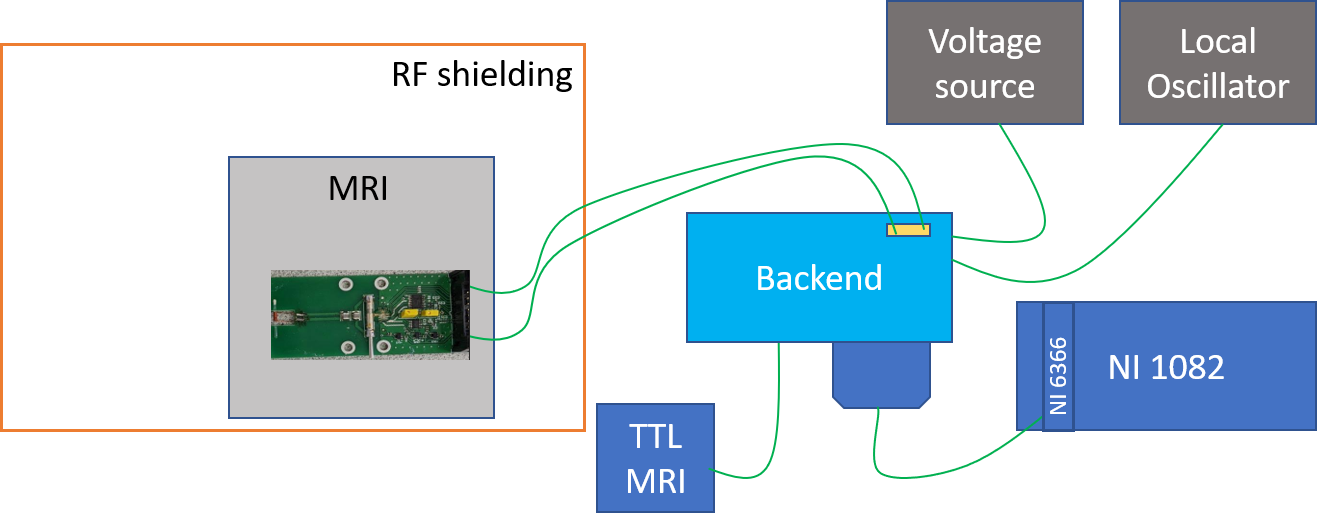
Figure 1: Setup for measurements with the NMR-on-a-chip sensor. Commercially available NI hardware is used for acquisition and digitalization together with a custom designed backend for preprocessing and as interface for the rest of the hardware.
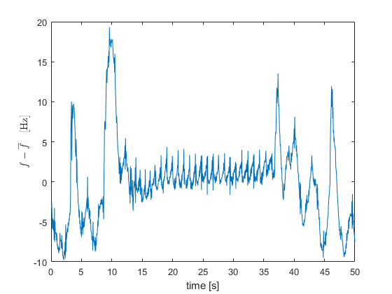
Figure 2: Measured frequency drift with the NMR-on-a-chip sensor on the breast of a volunteer. The signal was acquired with a time resolution of 50ms. In the middle of the measurement, the volunteer was asked to perform a breath hold to allow separation of respiratory and cardiac-induced magnetic field variations.
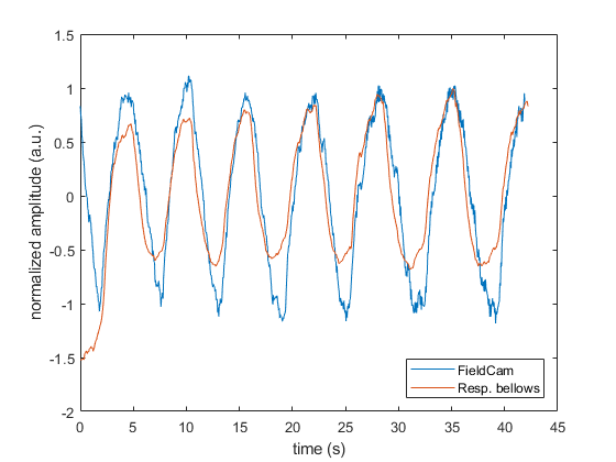
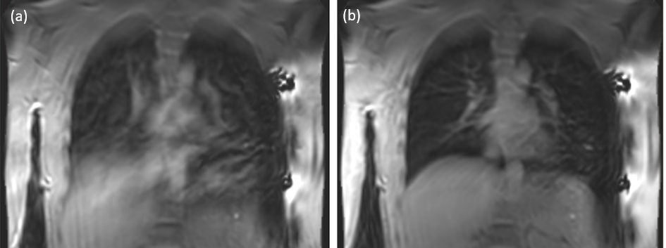
Figure 4: 2D cartesian TSE of a lung. Comparison of free breathing (a) with the synchronized measurement (b) using the NMR-on-a-chip sensor to monitor the respiratory motion state. In (a), the lung-liver interface shows a severe blur compared to (b).