0749
Dynamic Mode Decomposition (DMD) Cardiac Phase Estimation for adult and fetal real-time MRI1Electrical and Computer Engineering, University of Southern California, Los Angeles, CA, United States
Synopsis
Keywords: Motion Correction, Fetus, retrospective-gating
Motivation: Cardiac synchronization in adult and fetal imaging requires external devices (electrocardiogram, Doppler-ultrasound), which may compromise image quality and increase scan time. Self-gating with real-time imaging can mitigate this but may be less reliable for irregular motions and limited in fetal applications.
Goal(s): To develop a fast image-based cardiac phase estimation method with no assumption on the heart rate and minimal user input.
Approach: Dynamic Mode Decomposition is used to estimate cardiac motion signal for retrospective-gating.
Results: DMD cardiac phase estimation captures cardiac motion despite the irregularities and other bulk motions, as demonstrated in real-time adult and fetal cardiac imaging, including a twin gestation.
Impact: The proposed technique, Dynamic Mode Decomposition cardiac phase estimation, constructs cardiac signal with no assumption on periodicity, no iterations, and only minimal user input. This may be valuable in fetal cardiac imaging, where the cardiac signal is not readily available.
Introduction
MRI is an important tool for the noninvasive assessment of adult, pediatric, and fetal cardiac function, anatomy, and pathology [1,2]. For cardiac synchronization, electrocardiogram (ECG) gating is typically used in adults but can result in poor image quality, when there is insufficient gating signal. In the fetus, Doppler ultrasound (DUS) is commonly used but is sensitive to fetal/maternal movement [3,4]. Real-time imaging overcomes these, as no gating is required, and data can be combined retrospectively using self-gating approaches [5,6]. These may require specific imaging trajectories, are less reliable for irregular motions, and are more limited for fetal applications. One state-of-the-art self-gating method for fetal cardiac imaging is Metric Optimized Gating (MOG), which constructs a synthetic waveform by optimizing the image quality [7,8]. Although it is a very powerful method, an iterative optimization is needed.Dynamic mode decomposition (DMD) is a data-driven method that originated in the fluid mechanics community and has since been applied in various fields, including medical imaging [9-12]. Here, we propose using DMD for cardiac phase estimation with no assumption on the periodicity, no iterations, and only minimal user input. We evaluate this approach for real-time adult and fetal cardiac imaging, including a particularly challenging case, twin gestation.
Methods
Figure 1a shows the input-output relation of the DMD algorithm; given a dynamic input, DMD returns the eigendecomposition of the dynamic system, where eigenvectors $$$\phi$$$ show the spatial correlations, and eigenvalues $$$\lambda$$$ show the growth/decay and the frequency. Figures 1b-1c display DMD for real-time adult and fetal cardiac imaging. Selected outputs show the fundamental harmonics of the respiratory and cardiac motion with the corresponding frequency.Figure 2 shows the flowchart of the proposed DMD cardiac phase estimation. Given an image series, the only manual input from the user is the region-of-interest (ROI). DMD modes are ordered based on the signal energy contained in the ROI, which is used to find an automatic threshold. A time domain signal S is estimated from the eigenvalues of the selected modes. The peaks/valleys of this waveform represent systole/diastole and can be used for retrospective gating.
All experiments were performed on a whole-body 0.55T system (prototype MAGNETOM Aera, Siemens Healthineers, Erlangen, Germany) equipped with high-performance gradients (45 mT/m amplitude, 200 T/m/s slew rate) [13]. Real-time imaging was performed using the RTHawk system (Vista.ai, Palo Alto, California) [14]. A pseudo-golden-angle spiral bSSFP acquisition was used with TE/TR = 0.72/5.35ms, FA = 100° [15,16]. Two volunteers (1F, age 24±4) for adult cardiac and four pregnant females (gestational age 29-36w) for fetal imaging were scanned after providing written informed consent under protocols approved by our Institutional Review Board. All images were reconstructed with spatiotemporally constrained reconstruction (STCR) [17] and temporal resolution of 45ms.
Results
Figure 3 contains representative in-vivo adult cardiac images. Figure 3a shows a volunteer experiencing premature-ventricular-contraction. DMD cardiac phase estimation captures the cardiac motion despite the irregularities and achieves stable diastole/systole separation. Figure 3b shows a breath-hold scan. Input is a heavily undersampled image (2 arms/frame, gridded); however, the cardiac motion can still be estimated and used for retrospective binning.Figure 4 contains in-vivo results from two fetal scans acquired in short-axis and 4-chamber orientation, respectively. The proposed method can detect extreme motion states to identify diastole/systole regardless of the scan plane. Notice that the separation is reliable even with bulk motion due to maternal breathing.
Figure 5 contains in-vivo results from a particularly challenging case of twin gestation. ROI selection, in combination with DMD cardiac phase estimation, enables flexibility. The cardiac phases of both fetuses with different heartbeats can be independently extracted by selecting appropriate ROIs.
Discussion
This approach can identify the extreme motion states and seems robust to bulk motion. The peaks/valleys of the estimated waveform correspond to extreme states; however, it is ambiguous which is diastole/systole.The same approach can be applied for respiratory motion by placing the ROI over the chest or liver; however, for a reliable motion signal that can be used for binning, more analysis is needed. Our preliminary work manually placed the ROI; however, this could be automated to cover recognizable organs [18,19].
SNR of the image can significantly impact the accuracy of the estimated waveform. Even though challenging scenarios are presented in this work, more tests need to be performed.
Conclusion
We propose a novel way of extracting cardiac signal, Dynamic Mode Decomposition cardiac phase estimation with no assumption on the periodicity, no iterations, and only minimal user input. We believe this method is especially useful in fetal cardiac imaging, where the cardiac signal is not readily available.Acknowledgements
We acknowledge grant support from the National Institutes of Health (U01-HL167613, R21-HL159533) and National Science Foundation (Award #1828736) and research support from Siemens Healthineers.
References
1. Rajiah PS, François CJ, Leiner T. Cardiac MRI: State of the Art. Radiology. 2023;307(3):e223008. doi:10.1148/radiol.223008
2. Marini D, van Amerom J, Saini BS, Sun L, Seed M. MR imaging of the fetal heart. Journal of Magnetic Resonance Imaging. 2020;51(4):1030-1044. doi:10.1002/jmri.26815
3. Kording F, Yamamura J, De Sousa MT, et al. Dynamic fetal cardiovascular magnetic resonance imaging using Doppler ultrasound gating. J Cardiovasc Magn Reson. 2018;20(1):17. doi:10.1186/s12968-018-0440-4
4. Vollbrecht TM, Hart C, Zhang S, et al. Fetal Cardiac Cine MRI with Doppler US Gating in Complex Congenital Heart Disease. Radiology: Cardiothoracic Imaging. 2023;5(1):e220129. doi:10.1148/ryct.220129
5. Xue H, Kellman P, LaRocca G, Arai AE, Hansen MS. High spatial and temporal resolution retrospective cine cardiovascular magnetic resonance from shortened free breathing real-time acquisitions. J Cardiovasc Magn Reson. 2013;15(1):102. doi:10.1186/1532-429X-15-102
6. Nayak KS, Lim Y, Campbell-Washburn AE, Steeden J. Real-Time Magnetic Resonance Imaging. Journal of Magnetic Resonance Imaging. 2022;55(1):81-99. doi:10.1002/jmri.27411
7. Jansz MS, Seed M, van Amerom JFP, et al. Metric optimized gating for fetal cardiac MRI. Magnetic Resonance in Medicine. 2010;64(5):1304-1314. doi:10.1002/mrm.22542
8. Roy CW, Seed M, van Amerom JFP, et al. Dynamic imaging of the fetal heart using metric optimized gating. Magnetic Resonance in Medicine. 2013;70(6):1598-1607. doi:10.1002/mrm.24614
9. Schmid PJ. Dynamic mode decomposition of numerical and experimental data. Journal of Fluid Mechanics. 2010;656:5-28. doi:10.1017/S0022112010001217
10. Jovanović MR, Schmid PJ, Nichols JW. Sparsity-promoting dynamic mode decomposition. Physics of Fluids. 2014;26(2):024103. doi:10.1063/1.4863670
11. Kuntz, J.N. et al. (2016) Dynamic Mode Decomposition: Data Driven Modeling of Complex Systems. Siam.
12. Ilicak E, Ozdemir S, Zapp J, Schad LR, Zöllner FG. Dynamic mode decomposition of dynamic MRI for assessment of pulmonary ventilation and perfusion. Magnetic Resonance in Medicine. 2023;90(2):761-769. doi:10.1002/mrm.29656
13. Campbell-Washburn AE, Ramasawmy R, Restivo MC, et al. Opportunities in Interventional and Diagnostic Imaging by Using High-Performance Low-Field-Strength MRI. Radiology. 2019;293(2):384-393. doi:10.1148/radiol.2019190452
14. Santos JM, Wright GA, Pauly JM. Flexible real-time magnetic resonance imaging framework. Conf Proc IEEE Eng Med BiolSoc. 2004;2004:1048-1051.
15. Tian Y, Cui SX, Lim Y, Lee NG, Zhao Z, Nayak KS. Contrast-optimal simultaneous multi-slice bSSFP cine cardiac imaging at 0.55 Tesla. Magnetic Resonance in Medicine.
16. Yagiz E, Garg P, Nayak KS, Tian Y. Simultaneous multi-slice real-time cardiac MRI at 0.55T. Proc. ISMRM 2023.
17. Tian Y, Mendes J, Pedgaonkar A, Ibrahim M, Jensen L, Schroeder JD, Wilson B, DiBella EVR, Adluru G. Feasibility of multiple-view myocardial perfusion MRI using radial simultaneous multi-slice acquisitions. PLoS One 2019;14(2): e0211738.
18. Budai A, Suhai FI, Csorba K, et al. Fully automatic segmentation of right and left ventricle on short-axis cardiac MRI images. Computerized Medical Imaging and Graphics. 2020;85:101786. doi:10.1016/j.compmedimag.2020.101786
19. Hu H, Gao Z, Liu L, et al. Automatic Segmentation of the Left Ventricle in Cardiac MRI Using Local Binary Fitting Model and Dynamic Programming Techniques. Han W, ed. PLoS ONE. 2014;9(12):e114760. doi:10.1371/journal.pone.0114760
Figures
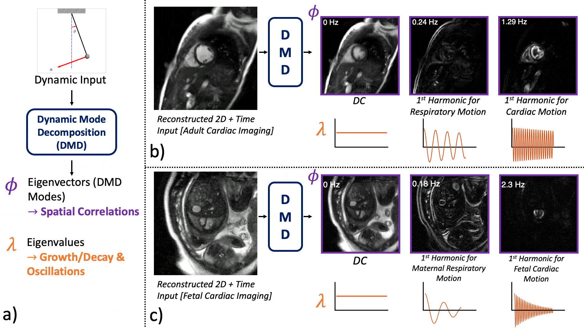
Figure 1: Overview of the DMD algorithm showing the input-output relation. a) Given measurements from a dynamic system, DMD extracts the eigenvectors (or DMD modes, Φ), spatial correlations, and the eigenvalues (λ), growth/decay, and oscillations of the underlying dynamics. Input is STCR [17] reconstructed real-time b) adult cardiac images, c) fetal cardiac images. The DMD Modes correspond to DC, and the first harmonics due to b) respiratory and cardiac c) maternal respiratory and fetal cardiac motion are selected and shown together with the temporal frequency and the eigenvalues.
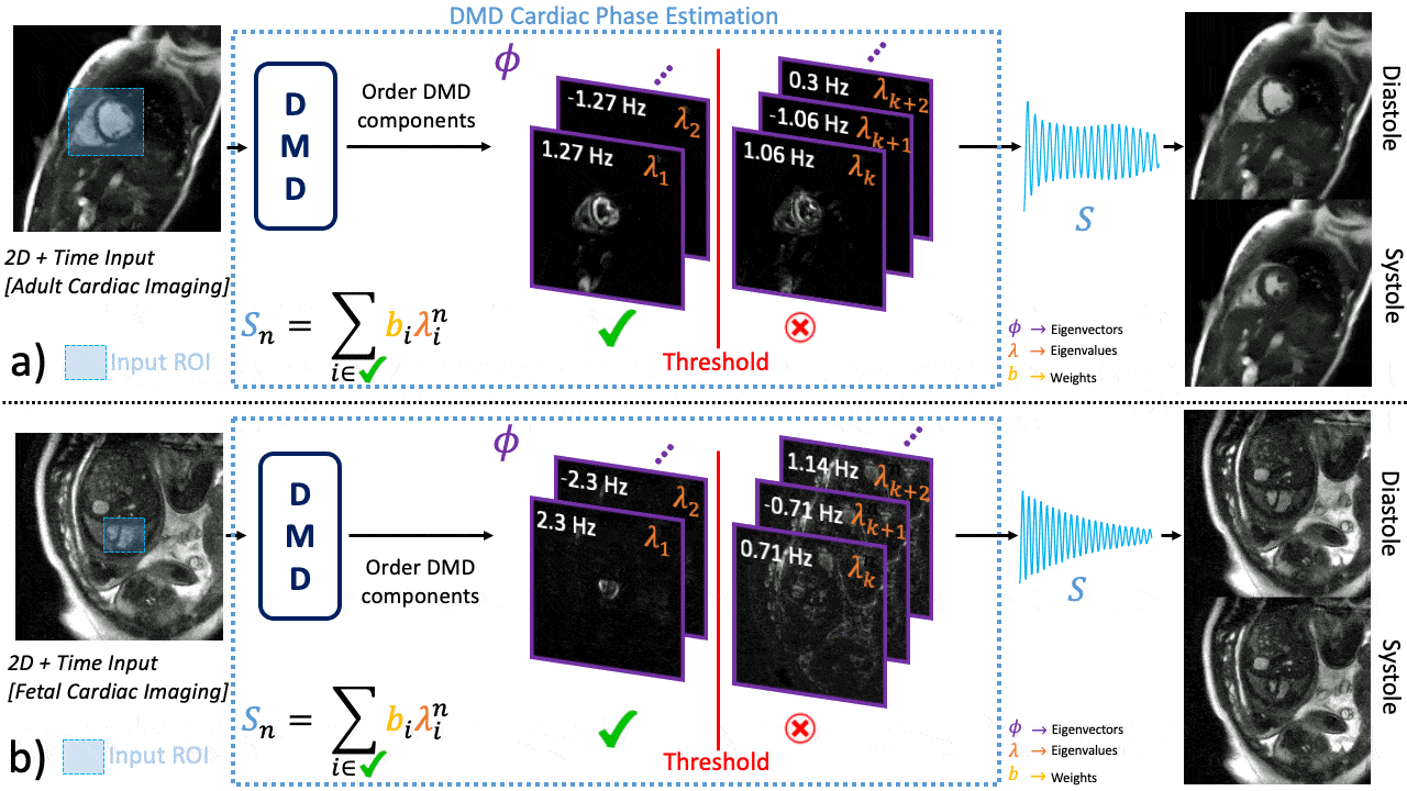
Figure 2: Flowchart of Dynamic Mode Decomposition (DMD) cardiac phase estimation. Given an image series, the user inputs a single region of interest (ROI) over the heart. Then, DMD modes are ordered based on the signal energy contained in the ROI, which is then used to find a threshold. A time domain signal Sn is estimated by weighted summation of the eigenvalues of the selected modes. The peaks/valleys of result Sn represents the systole/diastole and can be used for retrospective gating.
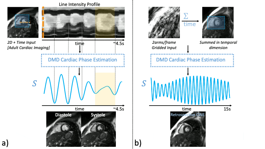
Figure 3: In-vivo results from adult real-time cardiac images. a) a volunteer experiencing premature ventricular contraction. Irregular beats are highlighted in the line intensity profile and in the estimated cardiac signal S (yellow shading). DMD-based estimation can capture the cardiac motion despite the irregularities and achieve stable diastole/systole separation. b) a breath-hold scan. Input is a heavily undersampled image (2 arms/frame, gridded). DMD cardiac phase estimation can still identify the motion, and this can be used for retrospective binning for a cine image.
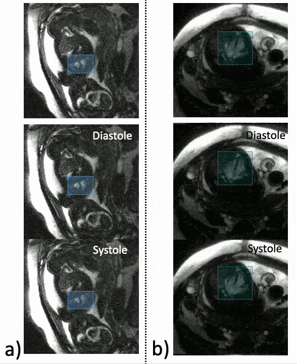
Figure 4: In-vivo results from two fetal real-time cardiac images. a) Real-time images acquired in short-axis orientation. Despite the bulk motion due to maternal breathing, diastole/systole can be extracted. b) Real-time images acquired in 4-chamber orientation. The proposed method doesn’t assume a scan plane and can detect the extreme motion states within the acquired data to separate diastole/systole.
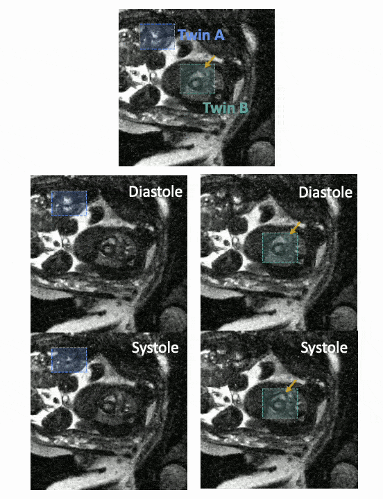
Figure 5: In-vivo results from a twin gestation. This result shows the flexibility of ROI selection in combination with DMD cardiac phase estimation. By selecting appropriate ROIs, the cardiac phases of both fetuses with different heartbeats can independently be extracted. Twin A shows a good diastole/systole separation. Even though the LV motion is minimal for Twin B, RV motion can still be captured (indicated by arrows).