0748
Harmonizing 2D and 3D FLAIR MRIs in white matter hyperintensity quantification1Neurology, Washington University School of Medicine, St. Louis, MO, United States, 2Mallinckrodt Institute of Radiology, Washington University School of Medicine, St. Louis, MO, United States
Synopsis
Keywords: White Matter, White Matter, FLAIR, harmonization, white matter hyperintensity
Motivation: Harmonizing neural imaging datasets respectively acquired with 2D and 3D FLAIR MRI.
Goal(s): Converting 2D FLAIRs to high-resolution 3D FLAIRs.
Approach: We employed a ResUNet-based deep learning approach to learn the complex transformation from 2D to 3D FLAIR.
Results: The converted 3D FLAIRs bear a high resemblance to the acquired 3D FLAIR in terms of image similarity measures and white matter hyperintensity segmentation.
Impact: With this proposed approach, we can harmonize the 2D FLAIRs from the ADNI study with the 3D FLAIRs in the UK Biobank study.
Introduction
FLAIR sequence can suppress the CSF signal in the brain and provide superb contrast for detecting white matter abnormalities known as white matter hyperintensities (WMHs). WMHs are commonly observed in patients with cerebral small vessel disease or Alzheimer’s Disease. Current neuroimaging studies use either 2D or 3D FLAIR protocols, resulting in different FLAIR image contrasts and slice thickness. We have developed a deep learning-based approach to harmonize the FLAIR images by converting 2D to 3D FLAIR MRI.Methods
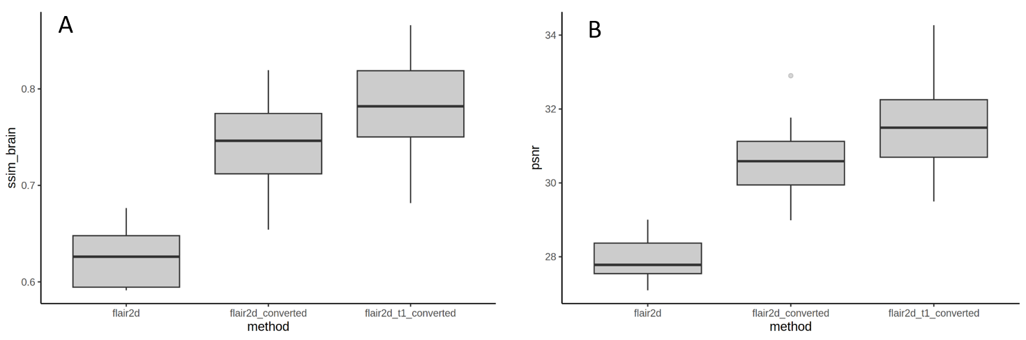
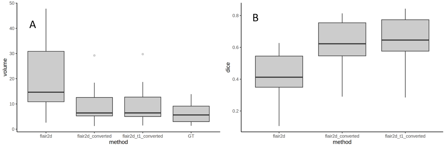
This is an IRB-approved study. We have acquired both 2D and 3D FLAIRs and T1-MPRAGE from 65 subjects (mean age: 69.26; 58% Female). 2D FLAIRs have an in-plane resolution of ~1mm with a through-plain resolution of 3mm. 3D FLAIRs were acquired with an isotropic 1mm resolution. Skull-stripping and inhomogeneity correction were performed with the FSL toolkit. 2D FLAIR and T1-MPRAGE were registered to the 3D FLAIR acquired from the same subject. We have employed a previously developed 3D patch-based ResUNet [1] to learn this complex transformation from 2D to 3D FLAIR. The patch size used is 64x64x64, and the L1 loss between the estimated and the ground-truth 3D FLAIRs is minimized during the training process. The 65 subjects were randomly divided into training (47), validation (5), and testing (13) sets. We trained two models using 1) 2D FLAIR only and 2) 2D FLAIR with T1-MPRAGE as inputs. The image qualities of the converted 3D FLAIRs were measured with structural similarity index (SSIM) and peak signal-to-noise ratio (PSNR) using the acquired 3D FLAIR as the reference. In addition, using a pre-trained 3D FLAIR WMH segmentation UNet, we have compared the segmentation results from the registered 2D and the converted 3D FLAIRs to the ground-truth segmentation obtained from the acquired 3D FLAIRs. In all the comparisons, Tukey’s Honest Significant Difference test was used to identify the significant differences among the registered 2D and converted FLAIRs.Results
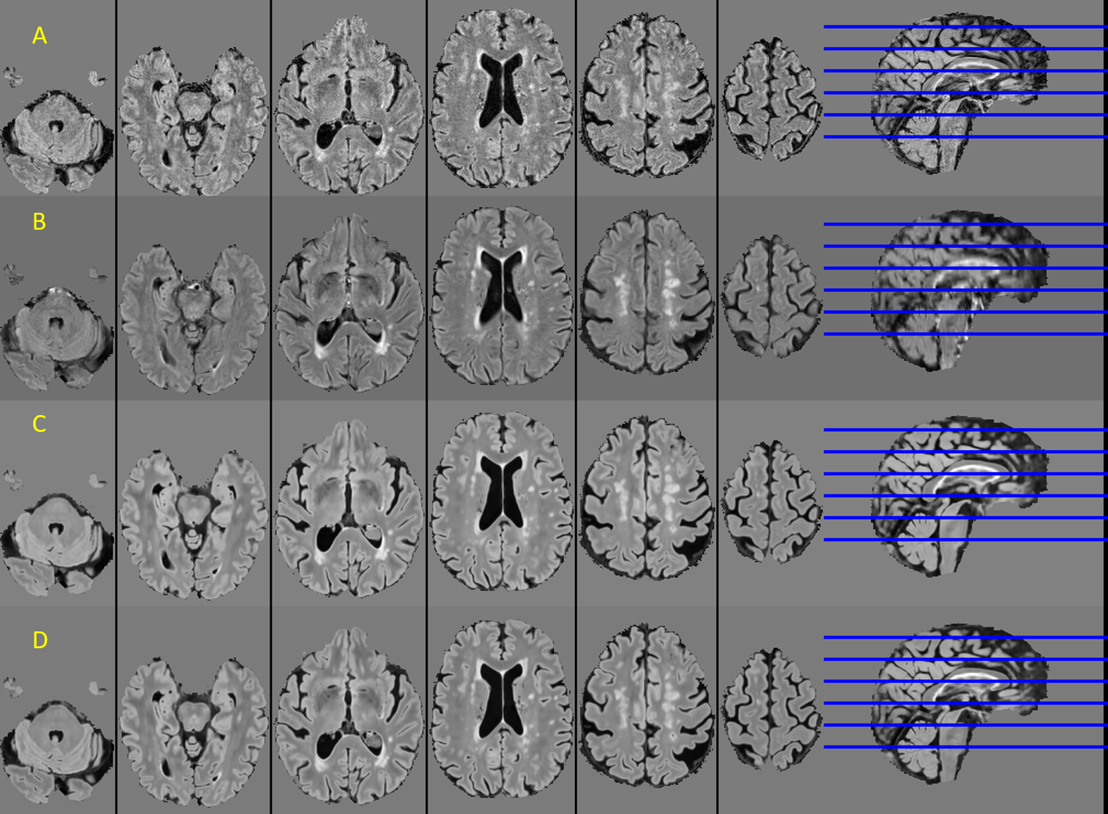
One representative example of the acquired 3D, registered 2D, and the converted FLAIRs is given in Fig. 1. WMHs appear brighter and larger in the registered 2D than the acquired 3D FLAIR, and the converted FLAIRs bear higher resemblance to the acquired 3D FLAIR than the registered 2D FLAIR. The converted 3D FLAIRs also demonstrate significantly improved SSIM (p<10-9) and PSNR (p<10-9) compared to the registered 2D FLAIRs (Fig. 2). The converted 3D FLAIR using both 2D FLAIR and T1-MPRAGE demonstrated marginal improvement in these similarity measures comparing to the ones converted with 2D FLAIR alone. WMH volume from the registered 2D FLAIR was significantly higher than both the converted and the acquired 3D FLAIRs (p<10-2), and no significant difference was found between the WMH volumes from the converted FLAIRs with ground-truth segmentation (Fig. 3A). Similarly, the converted FLAIRs also have significantly higher DICE ratios in WMH overlapping with the ground-truth segmentation than the registered 2D FLAIRs (p<10-3, Fig. 3B). The converted 3D FLAIR using both 2D FLAIR and T1 demonstrated marginal improvement than using 2DD FLAIR alone in WMH segmentation (Fig. 3B).Conclusion
To the best of our knowledge, this study may be the first in 1) harmonizing the 2D to 3D FLAIR MRI; and 2) systematically comparing the segmentation between the 2D and 3D FLAIRs from the same subjects. One previous study found pitfalls in using 3D FLAIR to replace 2D FLAIR [2]. We demonstrated that the brighter and larger appearance of the WMHs in 2D FLAIR leads to a significantly larger volume in WMH segmentation. As a result, there will be a large discrepancy in WMH quantifications if 2D and 3D FLAIR images are mixed in a neuroimaging study. The proposed approach dramatically reduces this discrepancy by producing similar WMH segmentation results between 2D and 3D FLAIR MRIs. In addition, our results also demonstrated improved image quality through conversion. Potentially, the proposed method may harmonize the WMH quantifications in large multi-center datasets, for example, the ADNI and the UK Biobank studies.Acknowledgements
This study was supported by NIH grants RF1 NS116565 and 2R01HL129241.References
[1] Yasheng Chen, Chunwei Ying, Michael M Binkley, Meher R Juttukonda, Shaney Flores, Richard Laforest, Tammie L S Benzinger, Hongyu An. Deep learning-based T1-enhanced selection of linear attenuation coefficients (DL-TESLA) for PET/MR attenuation correction in dementia neuroimaging. MRM 86(1):499-512 (2021)
[2] Shingo Kakeda, Yukunori Korogi, Yasuhiro Hiai, Norihiro Ohnari, Toru Sato, Toshinori Hirai. Pitfalls of 3D FLAIR Brain Imaging: A Prospective Comparison with 2D FLAIR. Academic Radiology, Vol. 19, Issue 10: 1225-1232 (2012).
Figures