0745
Neuroimaging signatures of severe depression in people with HIV1Department of Global Health and Infection, Brighton & Sussex Medical School, University of Sussex, Brighton, United Kingdom, 2Edinburgh Neuroscience, The University of Edinburgh, Edinburgh, United Kingdom, 3Clinical Imaging Sciences Centre, Brighton & Sussex Medical School, University of Sussex, Brighton, United Kingdom
Synopsis
Keywords: Infectious Disease, Diffusion/other diffusion imaging techniques, HIV
Motivation: People with HIV and co-morbid severe depression are rarely included in research, despite a critical need for identifying biomarkers for early detection of depression in this group.
Goal(s): We explored whether neuroimaging biomarkers may distinguish between people with HIV experiencing severe or mild depressive symptoms.
Approach: We recruited 11 participants with HIV and severe or mild depressive symptoms, who underwent standard and diffusion-weighted MR spectroscopy and dynamic contrast-enhanced MRI.
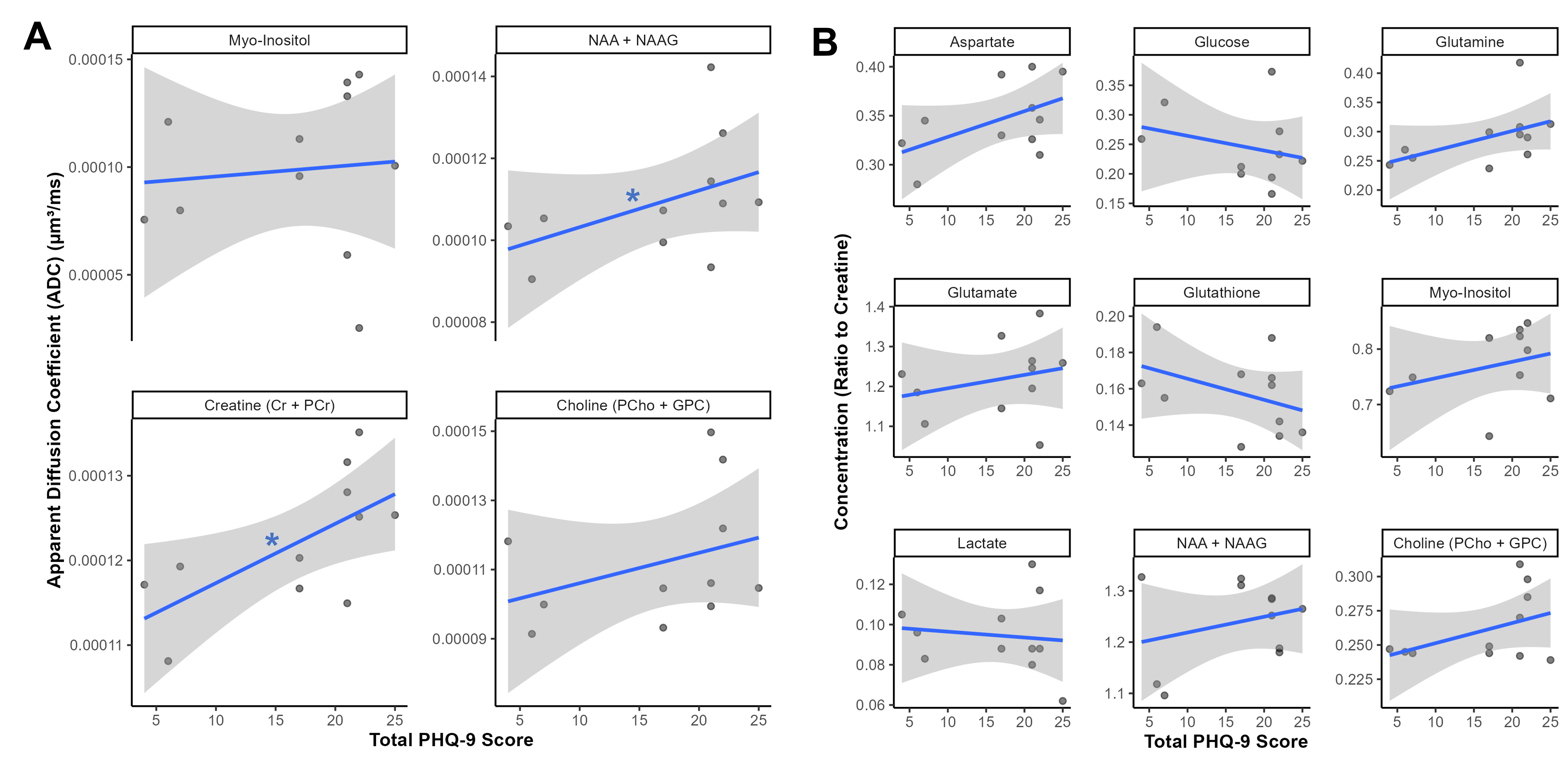
Results: We found no significant group differences, but observed correlations of depressive symptom severity with creatine (ρ = 0.66) and NAA (ρ = 0.64) diffusion, though these findings did not survive multiple comparisons correction.
Impact: This is the first study to successfully quantify neurometabolite diffusion in people with HIV and depression. Intracellular diffusion of neurometabolites may be associated with depressive symptom severity in this community, and well-powered studies are needed to resolve this relationship.
Introduction
People with HIV exhibit neuroinflammation and blood-brain barrier (BBB) dysfunction, which persists despite successful antiretroviral therapy1,2 and may contribute to the increased risk for depressive symptoms in this community.3,4 Severe, treatment-resistant depression is associated with increased inflammation in the general population.5 However, people with HIV and co-morbid severe depression are rarely included in research studies, despite a critical need for improving detection and treatment strategies for depression in this group. Diffusion-weighted magnetic resonance spectroscopy (DW-MRS) was recently shown to be sensitive to experimentally-induced neuroinflammation in humans, and may thus represent a useful non-invasive tool to aid in detection of inflammatory depression.6 Here, we explored whether neuroimaging biomarkers of inflammation can distinguish between people with HIV experiencing severe depressive symptoms and those with mild depressive symptoms.Methods
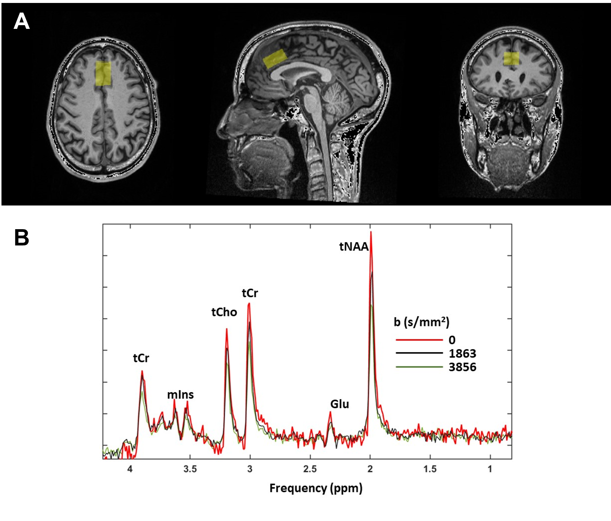
For this cross-sectional pilot study, we recruited adults with HIV in Brighton, UK. Participants completed the Patient Health Questionnaire (PHQ-9), using which they were categorised into a high (“HD”, PHQ-9 ≥ 15) or low (“LD”, PHQ-9 ≤ 7) depressive symptom severity group.Neuroimaging was performed on a 3T Siemens Prisma MR scanner (Erlangen, DE) and 64 channel array receiver coil. Following a scout image and a T1 weighted anatomical image, a 2cm (RL) x 1.5cm (FH) x 3cm (AP) volume of interest (VOI) was positioned on the anterior cingulate cortex (ACC) (Figure 1). MRS data were acquired using a semi LASER sequence (TE/TR = 30/3000 ms, 64 averages, GOIA-WURST pulses). DW-MRS data were acquired with the same sequence, equipped with a bipolar diffusion weighting scheme7 with three mutually orthogonal DW directions and three b values (0, 1863 and 3856 s/mm2), 32 averages per DW condition and TE/TR = 80ms/3 heart beats (cardiac synchronization with pulse oximeter unit). From these data, we quantified apparent diffusion coefficients (ADCs) for four metabolites, and concentrations for nine metabolites relative to total creatine and total water, correcting for T2 and water content differences between gray matter, white matter and CSF. Only metabolites for which CRLB <20% for all participants and all DW conditions were considered.
Whole-brain dynamic contrast enhanced MRI (DCE-MRI) was acquired with a T1-weighted gradient echo pulse sequence: 100 volumes, time resolution 9.34s, FA=7deg, voxel size=2.5mm3, total acquisition time=15.5mins. Dotarem (gadoterate meglumine; 0.1mMol/kg) was injected after 90s of baseline scanning. Volume leakage constant (Ktrans, a proxy for BBB permeability) was extracted by fitting the Patlak model to the data.
Statistical analyses were performed in R v.4.2.1. We compared log-transformed biomarker values between HD and LD groups using Wilcoxon’s rank-sum tests. In sensitivity analyses, we assessed partial Spearman’s correlations between log-transformed biomarker values and PHQ-9 score as a continuous variable, adjusted for fractional white matter volumes. All p-values were corrected for multiple comparisons using the False Discovery Rate method to yield corrected q values.
Results
Participants were N = 11 people with HIV, with median [interquartile range (IQR)] age of 54 [46.5, 54] years, of whom 82% self-identified as men and 82% as White. Participants in the HD (n = 8, median [IQR] PHQ-9 score 21 [20, 22]) and LD (n = 3, median [IQR] PHQ-9 score 6 [5, 6.5]) groups had comparable sociodemographic characteristics. Metabolite concentrations, metabolite ADCs, and Ktrans did not significantly differ between groups (all q > 0.05, Figure 2). Similarly, these biomarkers were not significantly correlated with PHQ-9 score (all q > 0.05, Figure 3), except there were strong correlations of PHQ-9 score with creatine ADC (ρ = 0.66, p = 0.027, q = 0.088) and NAA ADC (ρ = 0.64, p = 0.035, q = 0.088) which did not survive FDR correction.Discussion
To our knowledge, this is the first study to successfully acquire DW-MRS data and quantify neurometabolite diffusion in people with HIV and depression. In our small sample, we observed no statistically significant differences in metabolite diffusion, metabolite concentrations, or BBB permeability in the ACC between participants with severe depression and those with low depressive symptom severity. However, we observed promising trends towards strong correlations of creatine and NAA diffusion with depressive symptoms, which may be resolved with larger sample sizes. Increase in creatine ADC has been associated with increase in metabolic activity8 and reported in multiple sclerosis9 and neuropsychiatric lupus10, both conditions characterised by neuroinflammation. Our findings thus warrant further investigation of creatine ADC as a potential biomarker of inflammatory depression.Conclusion
We have demonstrated the feasibility of using DW-MRS to quantify neurometabolite diffusion in people with HIV and depression. Recruitment for this study is ongoing; with a larger sample size, we may substantiate distinct neuroimaging biosignatures of depression severity in people with HIV.Acknowledgements
We thank Dr. Edward Auerbach, Dr. Malgorzata Marjanska and Dr. Dinesh Deelchand from the Center for Magnetic Resonance Research at the University of Minnesota for making available their MRS and DW-MRS sequences to us.References
1. Vera JH, Guo Q, Cole JH, Boasso A, Greathead L, Kelleher P, Rabiner EA, Kalk N, Bishop C, Gunn RN. Neuroinflammation in treated HIV-positive individuals: a TSPO PET study. Neurology. 2016;86(15):1425-1432.
2. Caligaris G, Trunfio M, Ghisetti V, Cusato J, Nigra M, Atzori C, Imperiale D, Bonora S, Di Perri G, Calcagno A. Blood–brain barrier impairment in patients living with HIV: predictors and associated biomarkers. Diagnostics. 2021;11(5):867.
3. Do AN, Rosenberg ES, Sullivan PS, Beer L, Strine TW, Schulden JD, Fagan JL, Freedman MS, Skarbinski J. Excess burden of depression among HIV-infected persons receiving medical care in the United States: data from the medical monitoring project and the behavioral risk factor surveillance system. PloS one. 2014;9(3):e92842.
4. Mudra Rakshasa-Loots A, Whalley HC, Vera JH, Cox SR. Neuroinflammation in HIV-associated depression: evidence and future perspectives. Molecular Psychiatry. 2022:1-14. doi:10.1038/s41380-022-01619-2
5. Strawbridge R, Hodsoll J, Powell TR, Hotopf M, Hatch SL, Breen G, Cleare AJ. Inflammatory profiles of severe treatment-resistant depression. Journal of affective disorders. 2019;246:42-51.
6. De Marco R, Ronen I, Branzoli F, Amato ML, Asllani I, Colasanti A, Harrison NA, Cercignani M. Diffusion-weighted MR spectroscopy (DW-MRS) is sensitive to LPS-induced changes in human glial morphometry: A preliminary study. Brain, Behavior, and Immunity. 2022;99:256-265.
7. Genovese G, Marjańska M, Auerbach EJ, Cherif LY, Ronen I, Lehéricy S, Branzoli F. In vivo diffusion‐weighted MRS using semi‐LASER in the human brain at 3 T: Methodological aspects and clinical feasibility. NMR in Biomedicine. 2021;34(5):e4206.
8. Branzoli F, Techawiboonwong A, Kan H, Webb A, Ronen I. Functional diffusion‐weighted magnetic resonance spectroscopy of the human primary visual cortex at 7 T. Magnetic resonance in medicine. 2013;69(2):303-309.
9. Bodini B, Branzoli F, Poirion E, García-Lorenzo D, Didier M, Maillart E, Socha J, Bera G, Lubetzki C, Ronen I. Dysregulation of energy metabolism in multiple sclerosis measured in vivo with diffusion-weighted spectroscopy. Multiple Sclerosis Journal. 2018;24(3):313-321.
10. Ercan E, Magro-Checa C, Valabregue R, Branzoli F, Wood ET, Steup-Beekman GM, Webb AG, Huizinga TW, van Buchem MA, Ronen I. Glial and axonal changes in systemic lupus erythematosus measured with diffusion of intracellular metabolites. Brain. 2016;139(5):1447-1457.
Figures
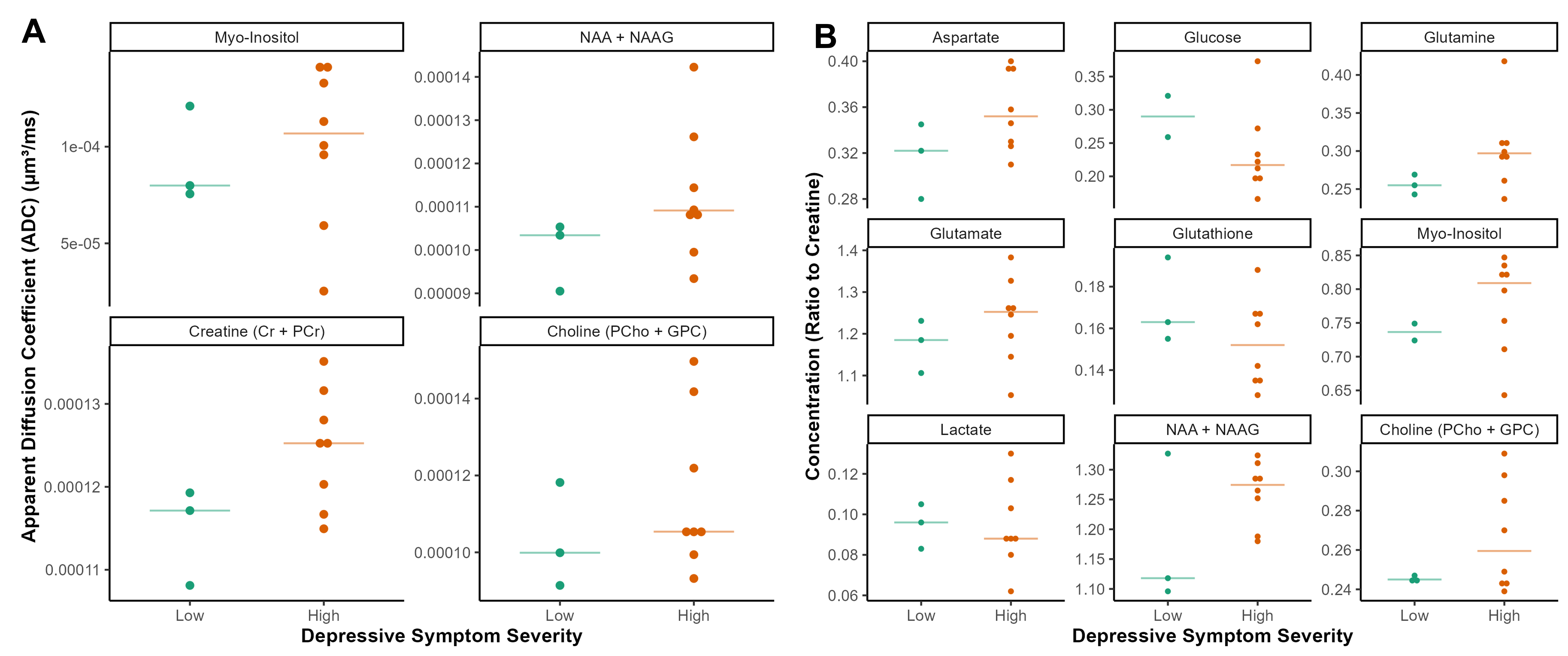
(A) Apparent Diffusion Coefficients (ADCs) for four metabolites and (B) creatine-referenced concentrations for nine metabolites, stratified by depressive symptom severity as measured using the PHQ-9. Horizontal lines indicate median values for each group.