0741
Evaluating metabolic disruption following radiotherapy in the developing brain: a preclinical juvenile model1Biomedical MRI / Dept. Imaging & Pathology, KU Leuven, Leuven, Belgium, 2Department of Oncology, KU Leuven, Leuven, Belgium
Synopsis
Keywords: Neuroinflammation, Radiotherapy, 1H MRS
Motivation: Cognitive performance in paediatrics is severely impacted by current brain tumour treatment plans such as radiotherapy.
Goal(s): Our goal is to decipher the underlying mechanism of this cognitive decline, which remains unknown and is particularly difficult to decipher in paediatric patients due to confounding developmental variables.
Approach: Longitudinal 1H MRS analysis in the brain following radiotherapy in a juvenile rat model provides valuable insight into metabolic disruption.
Results: Myo-inositol levels, linked to other neurodegenerative and cognitive conditions, were elevated in the hippocampus and cerebellum. Deviance in N-acetyl-aspartate levels between irradiated and healthy rats over a developmental period of 12.5 weeks was also revealed.
Impact: 1H MR spectroscopy reveals valuable insight into longitudinal impacts of radiotherapy on the developing brain. A juvenile rat model enables acute and chronic alterations in inflammatory markers, membrane synthesis, bioenergetics and viability to be monitored, distinguishing irradiation and development effects.
Introduction
Brain tumours constitute 85-90% of all primary central nervous system tumours and yearly affect ±300,000 people worldwide from which ~17% are children. Despite improving survival rates, the neuropsychological impact of treatment remains ill understood. The components of brain tumour treatment plans, particularly radiotherapy, are known to negatively impact cognitive performance.1 However, the underlying mechanism of this cognitive decline remains unknown. To unravel the mechanism(s) and longitudinal impact of radiotherapy in the developing brain, paediatric preclinical models are a consequently key first step. Using a longitudinal 1H MR spectroscopy (MRS) assessment of regional alterations in inflammatory markers such as myo-inositol and choline, as well as in membrane synthesis, cellular bioenergetics and compartment-specific viability, we seek to establish the effects of radiotherapy in the developing brains of juvenile rats following a clinically relevant workflow complementary to ongoing clinical studies.Methods
Animal model and radiotherapy: Juvenile Fischer rats (female, n=6) were irradiated at 6 weeks of age using a designated preclinical photon irradiation platform designed to mimic clinic workflow (Small Animal Radiation Research Platform, Xstrahl GmbH).2 Longitudinal analysis compared irradiated rats to healthy, sex/age-matched controls (n=4). Using an integrated CT, reference images were acquired (60 kV, 0.8 mA, 67 sec) for subsequent treatment planning performed in Muriplan. Two subsequent parallel beam lines with a 10x10 mm collimator targeted the full brain excluding the olfactory bulb and cerebellum, while also avoiding the eyes (see Fig.1). Rats received a single fraction of 12.5 Gy (2.6 Gy per minute).MR Acquisitions: Baseline MRI at 5.5 weeks of age with follow-up 3-d, 10-d, 5.5-w and 12.5w post-radiotherapy were acquired for whole brain irradiated (WBI) and healthy control (HC) rats. Data were acquired using the Bruker Biospec 9.4 T/20, 400-MHz horizontal magnet equipped with a designated rat head surface coil and linear volume coil located within the MoSAIC Core Facility. ParaVision 360 V3.0 (Bruker Biospin, Inc.) was used for all acquisitions. 2D T2-weighted anatomical scans with 0.125 mm2 in-plane resolution were acquired via turboRARE (TE 7.5ms, TR 6s, 2 averages). T2-weighted images provided anatomical reference for precise MRS voxel positioning and confirmation of radiotherapy-induced lesions. Metabolite assessment was carried out using a PRESS sequence (TE, TR = 20ms, 2.5s; 256 averages). Voxels were localized within the hippocampus (2x1.5x3 mm) and cerebellum (2.5x2x4 mm) in subsequent acquisitions (Fig.2). Linewidths were 0.02 ± 0.01 ppm and 0.04 ± 0.01 ppm for the hippocampus and cerebellum, respectively. Signal-to-noise was calculated in LCModel3 and corresponded to the maximum peak of each spectra (hippocampus 14.0 ± 3.4 and cerebellum 19.7 ± 3.1).
Analysis: Pre-processing of MRS was completed in MATLAB. Bruker data was first converted to LCModel compatible format with subsequent HLSVD water removal. Spectra were then further processed in LCModel. A metabolite basis set was used in combination with the unsuppressed water signal for estimation of absolute metabolite quantification. Metabolite ratios to choline were also calculated. Although the full metabolite basis set was used to fit each spectrum, including standard lipid and macromolecules, the metabolites of interest were restricted to those with an average Cramer Rao lower bounds (CRLB) less than 15%. This corresponded to 10 of 18 metabolites in both regions undergoing further analysis. Statistical analysis was performed in Prism GraphPad using a mixed-model effects, p<0.05 (all graphs are mean ± SD).
Results
Anatomical MRI confirmed presence of irradiation-induced lesions starting as early as 3 days post-treatment. In the hippocampus (Fig.3), myo-inositol significantly increased from baseline ten days following radiotherapy (p=0.0370). Interestingly, healthy animals exhibited a reduction in NAA over 5.5 weeks (p=0.0085) while radiotherapy treated rats trended to increasing NAA over the full 12.5 week period. In the cerebellum (Fig.4), irradiated rats demonstrated elevated Cre/Cho ratio immediately following radiotherapy (day 3, p=0.0283; day 10, p=0.0011) and significantly increased myo-inositol levels 10 days post-treatment (p=0.0125).Discussion
Single-voxel 1H MRS is a highly sensitive approach to quantify regional metabolite alterations in the developing brain. In particular, elevation of myo-inositol indicates an early, albeit not necessarily acute, neuroinflammatory response in both the hippocampus and cerebellum, two regions susceptible to volume reduction in patients undergoing similar treatment. Furthermore, development-specific metabolic changes are accounted for with age/sex-matched healthy controls. While most other studies focus on limited/late-stage patient data or adult models thus limiting the scope outside of paediatric patients, this is the first study to assess radiotherapy on metabolic activity in the developing brain using a juvenile rat model.Conclusion
Spectroscopic insight into the metabolic deviations immediately following low-dose photon irradiations in juvenile rats provides key insights into the effects radiotherapy has on the developing brain.Acknowledgements
No acknowledgement found.References
1. Feigin, V. L. et al. Lancet Neurol 18:459–480 (2019).
2. M. Riva et al. Neurosurgery 88:E205-E215 (2021).
3. Provencher SW. NMR Biomed 4:260-264 (2001).Figures
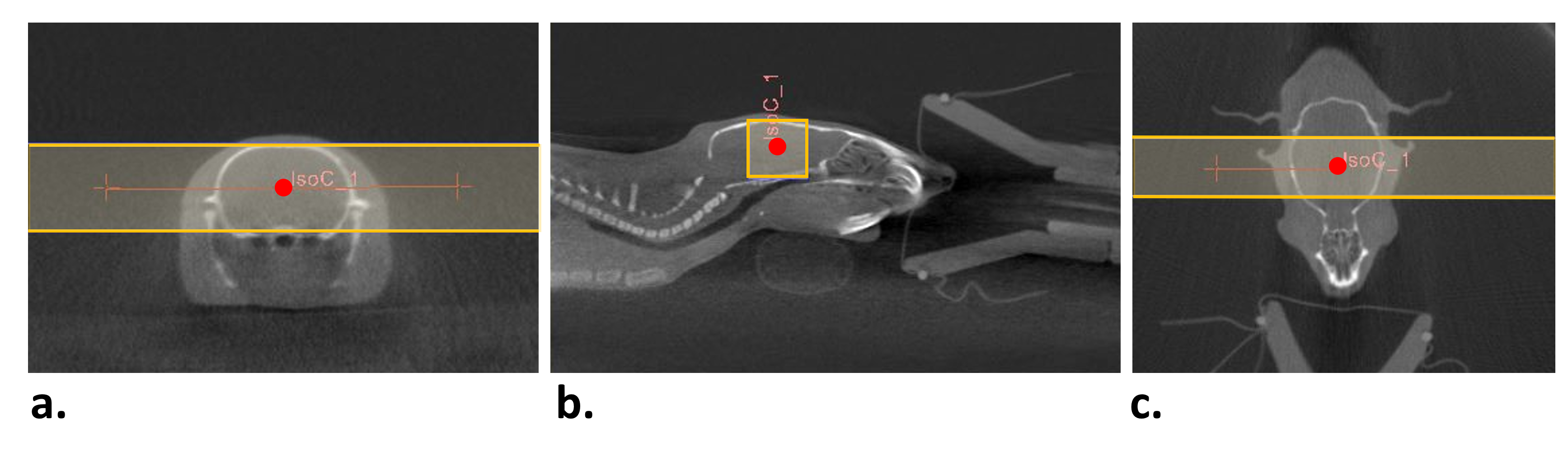
Figure 1. Radiotherapy treatment plan. Representative CT images in all three planes (a-c) with overlapping beam line placement (yellow lines) and iso-centre (red) indicating two parallel photon beams irradiating 1 cm of the brain, avoiding the olfactory bulb and cerebellum.
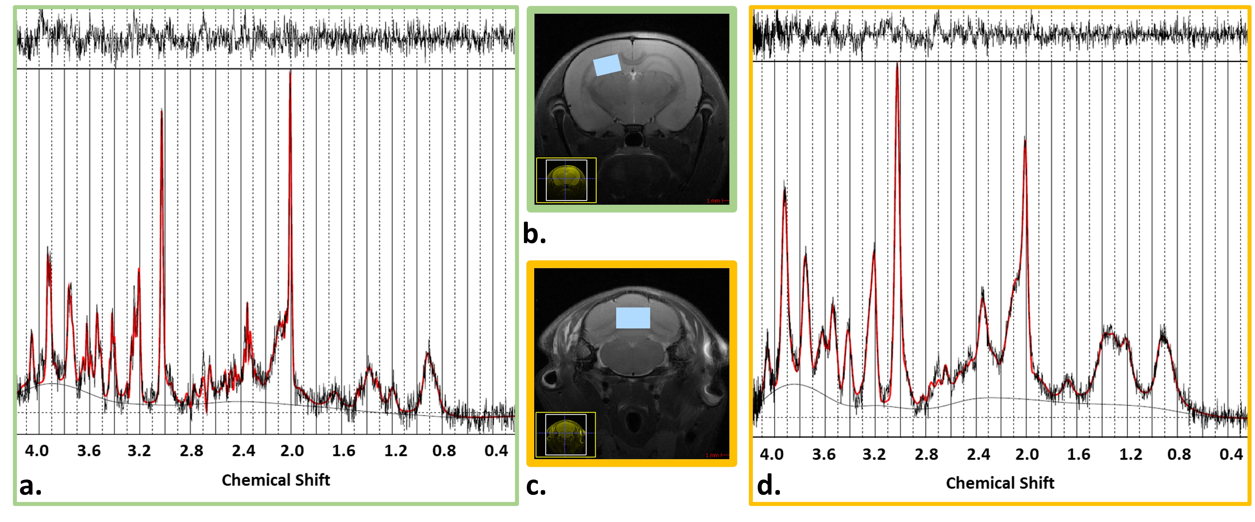
Figure 2. Representative 1H MRS. Baseline spectra and voxel placement in the hippocampus (a,b) and cerebellum (c,d) of a representative 5 week old rat. Spectra was quantified in LCModel. The fitted data, seen in red, is overlayed on the original from 0.2 to 4.2 ppm. The residual data is visualized in the row above each spectrum.
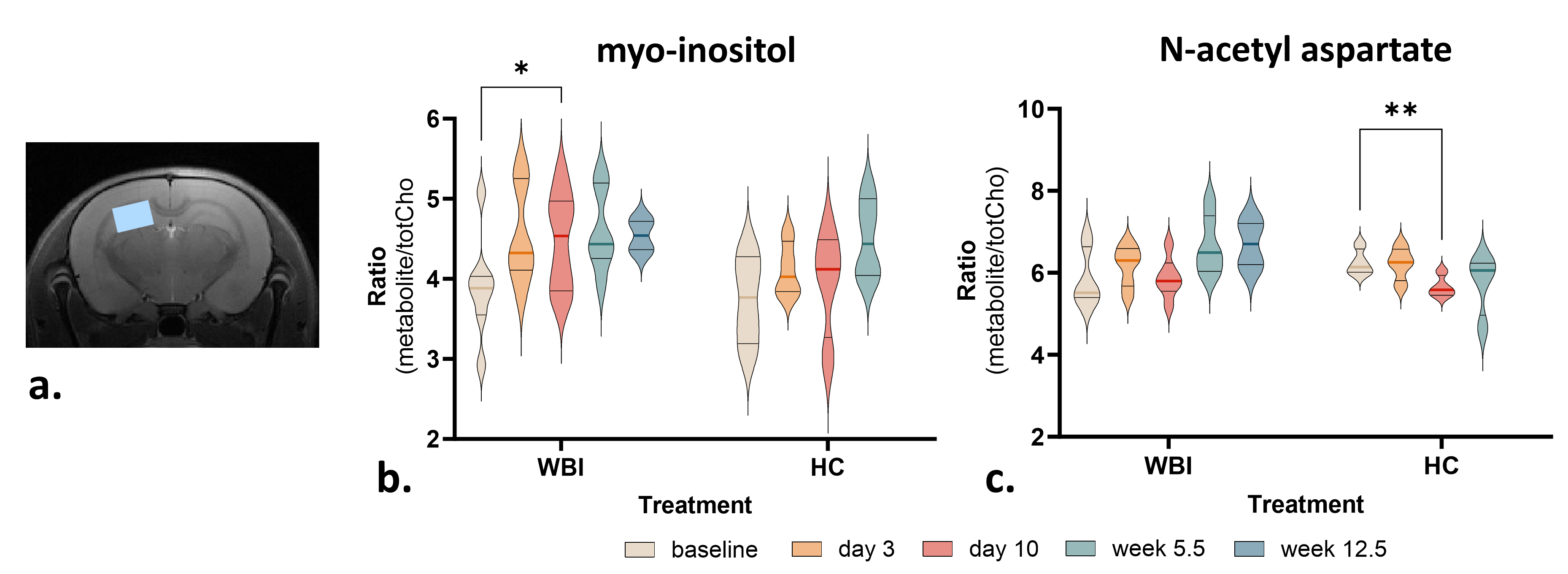
Figure 3. Metabolite changes in the hippocampus for whole brain irradiated (WBI) and healthy controls (HC). a) 1H MRS via PRESS was localized to the hippocampus at five timepoints (baseline, days 3 and 10, weeks 5.5 and 12.5). b) Irradiated rats demonstrated an increase in myo-inositol from baseline to day 10 (p = 0.0370). c) A reduction in NAA was seen in healthy controls only between baseline and day 10 (p = 0.0085).
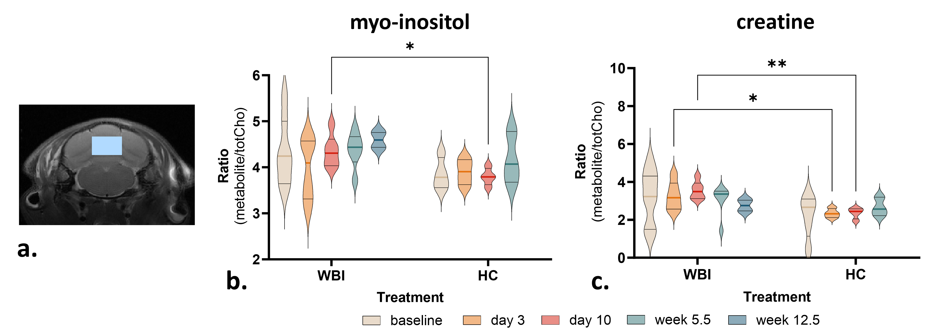
Figure 4. Metabolite changes in the cerebellum for whole brain irradiated (WBI) and healthy controls (HC). a) 1H MRS via PRESS was localized to the cerebellum at five timepoints (baseline, days 3 and 10, weeks 5.5 and 12.5). b) Irradiated rats demonstrated increased myo-inositol on day 10 compared to healthy control (p = 0.0125). c) Irradiated rats also demonstrated elevated creatine immediately following radiotherapy (day 3, p = 0.0283; day 10, p = .0011)