0740
Impaired Glymphatic Clearance Linked to Neuroinflammation and Disability in Multiple Sclerosis1Yale University, New Haven, CT, United States, 2Weill-Cornell Medicine, New York, NY, United States
Synopsis
Keywords: Multiple Sclerosis, Multiple Sclerosis, Brain Lymphatics
Motivation: Multiple sclerosis (MS) involves a proinflammatory state leading to cellular waste accumulation and disability. This study investigates the glymphatic system's waste clearance role, hypothesizing that lymphatic dysfunction contributes to MS-related disability.
Goal(s): Explore the relationship between glymphatic function, meningeal lymphatic vessels (mLV), and MS-related disability.
Approach: The study examined 49 MS patients using MRI to visualize mLVs and evaluate glymphatic stasis via CSF fraction (CSFF), analyzing links between mLV volume, CSFF, and MS outcomes
Results: With age, cortical CSFF increases. mLV volume also increases with CSFF. Higher cortical CSFF is associated with more lesions and disability, suggesting glymphatic dysfunction contributes to MS-related disability.
Impact: Our study suggests that glymphatic dysfunction contributes to lesion burden and disability in multiple sclerosis, highlighting the importance of lymphatic clearance mechanisms in disease progression.
Introduction
In multiple sclerosis (MS), there is a proinflammatory state with ongoing demyelination and neuronal loss, resulting in increased generation of cellular waste1, lesion formation, and accumulation of disability. The glymphatic system and meningeal lymphatic vessels (mLV) play essential roles in clearing cellular waste from the brain and draining cerebrospinal fluid (CSF) into the blood, which are important for optimal PS in healthy individuals. The glymphatic system removes cellular waste from the brain and transfers it to the CSF2, while the mLVs potentially transfer these waste products from the CSF to the blood3. The mLVs also provide a pathway for immune cells to traverse the central nervous system (CNS)3. As such, impaired lymphatic clearance can lead to the accumulation of toxic proteins, disruption of neural function, and accumulation of MS-related disability. To date, no studies have explored the relationship between the glymphatic system, mLV, and clinical disability in MS. We hypothesized that brain lymphatic dysfunction contributes to MS-related disability.Methods
We conducted a cross-sectional study investigating 49 MS patients. All participants underwent high-resolution 3D T2-FLAIR, T1-MPRAGE, and fast acquisition with spiral trajectory and adiabatic T2prep (FAST-T2) sequence on a Siemens 3T MRI scanner. The mLVs along the sagittal sinus were visualized and segmented using a 3D T2-FLAIR sequence. To assist in the segmentation of the mLVs, a template was generated from individual FLAIR images using the ANTs multivariate template construction algorithm4. An inverse transformation was then applied to the template mask, allowing for the conversion of the mLV mask in template space into the native space of each participant. This transformation produced individual mLV masks in native space. During the segmentation process, there was unavoidable contamination from blood and cerebrospinal fluid (CSF) in the venous sinus and subarachnoid space. This contamination was addressed by removing voxels within the mask that was part of a CSF mask obtained from T1-MPRAGE. The CSF fraction (CSFF) was used to assess glymphatic fluid stasis at the voxel level5 CSFF was obtained by fitting a three-water compartment model with multi-echo FAST-T2 data. CSFF is corresponding to the long T2 (T2>1000ms) compartment of the fitting results. The relationship between cerebral cortical CSFF and mLVs was tested. Neuropsychological tests were conducted to assess cognitive domains.Results
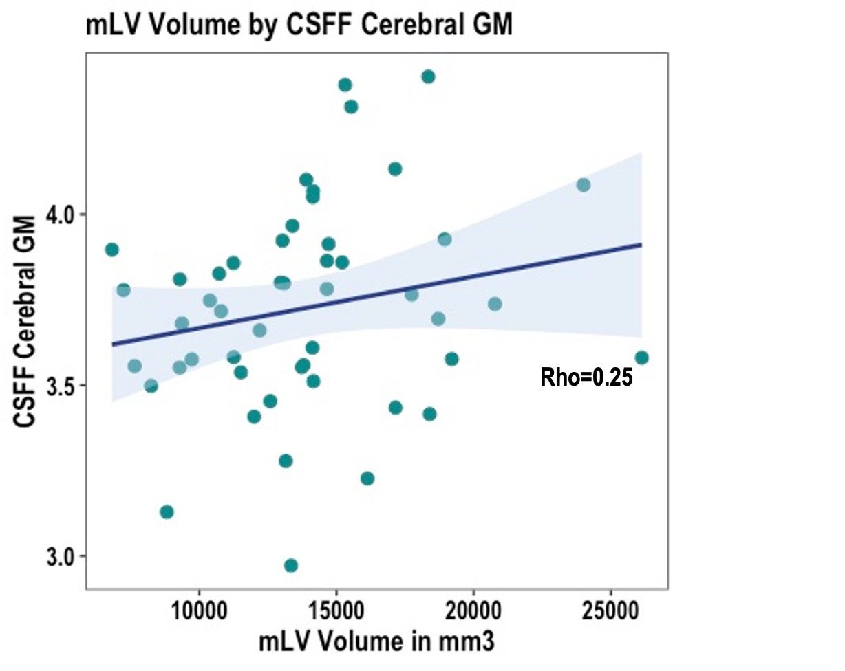
1. Relationship between mLV Volume and Cortical CSFF:Our analysis aimed to understand how glymphatic fluid stasis, as indicated by CSFF, relates to the mLV volume in the cortical grey and white matter. We found that an increase in cortical grey matter CSFF is associated with an enlargement of the mLV volume (rho=0.25, p=0.08, Fig 1). However, no such relationship was found between mLV volume and CSFF in the white matter (rho=0.08, p=0.58).
2. Effect of Age on Brain Lymphatics:
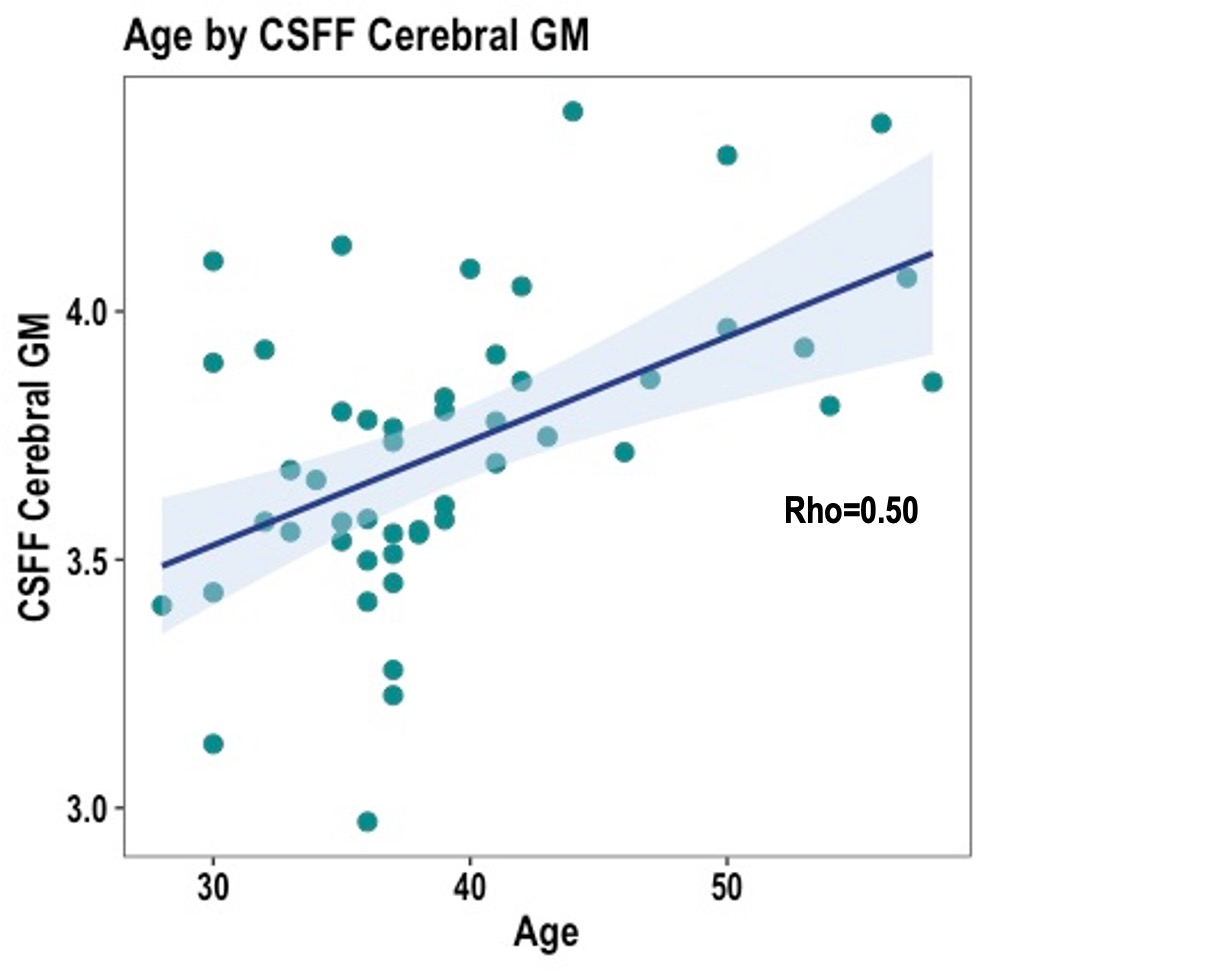
With advancing age, there is a notable rise in cortical grey matter CSFF, suggesting age-related changes in glymphatic circulation (rho=0.50, p=0.0002, Fig 2). While there is an observable trend linking age with increased mLV, this relationship was not statistically significant (rho=0.22, p=0.14).
3. Impact of Brain Lymphatics changes on MS:
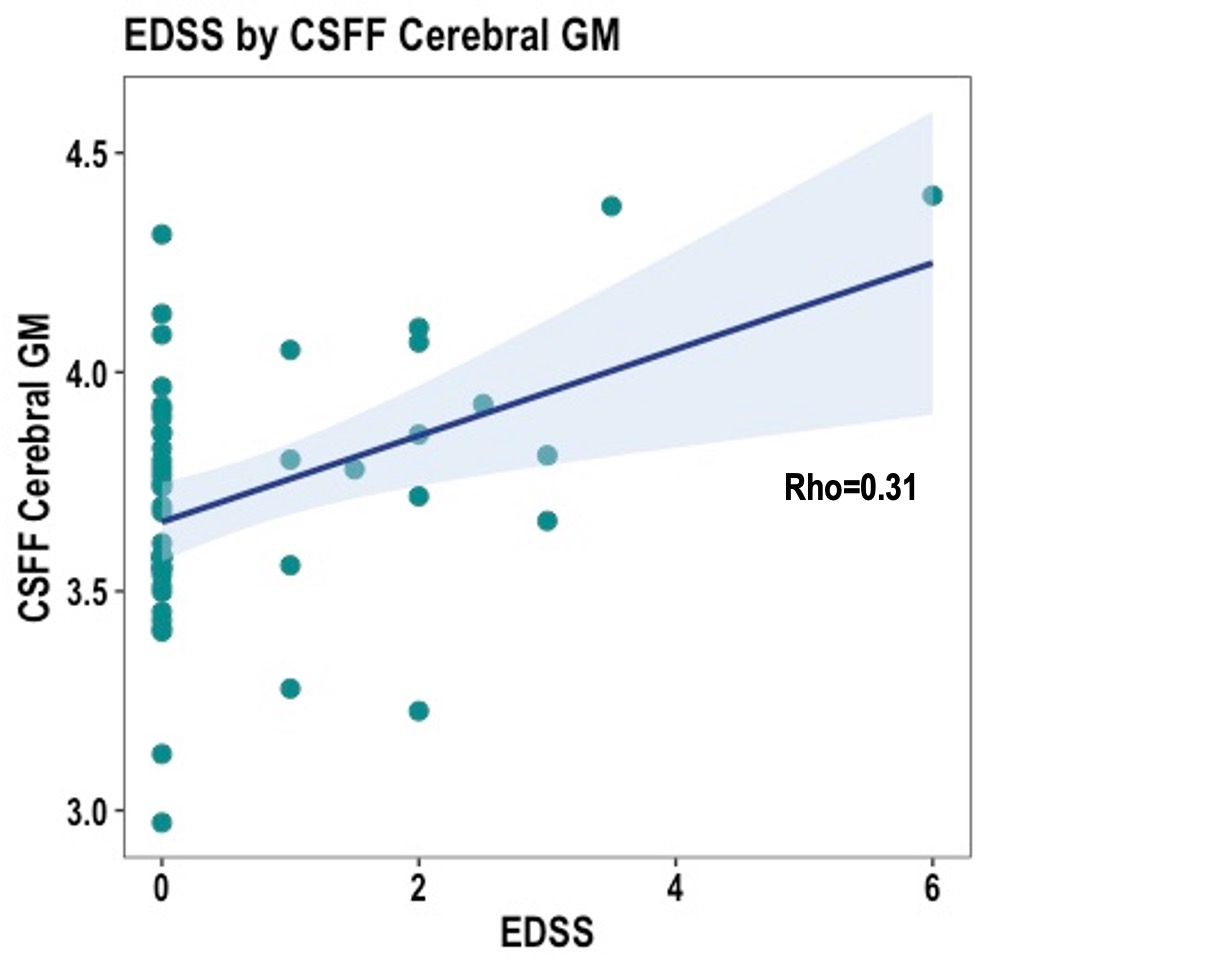
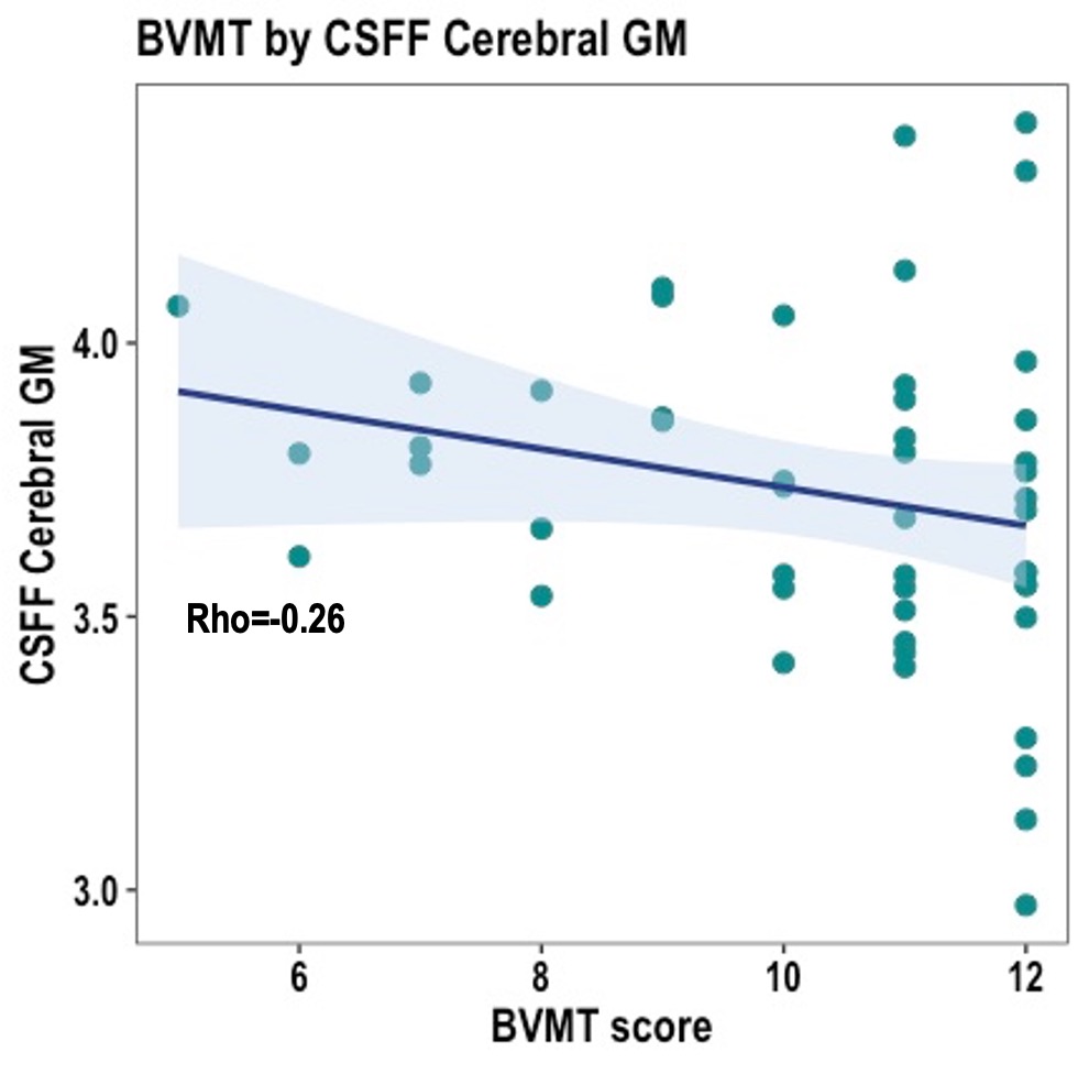
Our results indicate that increase in cortical CSFF is associated with increase in MS-related lesion burden, implying that fluid stasis in the glymphatic system may exacerbate MS lesion formation (rho=0.40, p=0.0046). Furthermore, the presence of rim-positive lesions, which indicate active inflammation, also correlates significantly with higher CSFF levels (rho=0.31, p=0.02), suggesting a potential link between inflammation and impaired glymphatic function. Additionally, increased CSFF was associated with a worsening in the Expanded Disability Status Scale (EDSS; rho=0.31, p=0.03, Fig 3) and lower Brief Visuospatial Memory Test (BVMT; rho=-0.26, p=0.06, Fig 4), suggesting that glymphatic fluid stasis may be related to accumulation of MS disability. There was no relationship between mLV volume and MS-related disease variables.
Discussion
The present results are consistent with emerging evidence that glymphatic system becomes dysfunction in MS. Our results suggest that glymphatic fluid accumulates with age which in turn may result in mLV hypertrophy. In MS, underlying inflammation might impact glymphatic drainage, thereby contributing to an increased lesion burden and disability. Further studies are needed to disentangle the disease-specific consequences of glymphatic alterations from age-specific changes.Acknowledgements
We thank all the patients and volunteers who participated in this research efforts. We thank our lab research interns and coordinators for their help in recruiting patients and volunteering for the study.References
1. Carotenuto, A. et al. Glymphatic system impairment in multiple sclerosis: relation with brain damage and disability. Brain 145, 2785–2795 (2022).
2. Iliff, J. J. et al. A Paravascular Pathway Facilitates CSF Flow Through the Brain Parenchyma and the Clearance of Interstitial Solutes, Including Amyloid β. Science Translational Medicine 4, 147ra111-147ra111 (2012).
3. Louveau, A. et al. Structural and functional features of central nervous system lymphatic vessels. Nature 523, 337–341 (2015).
4. Avants, B. B. et al. A reproducible evaluation of ANTs similarity metric performance in brain image registration. Neuroimage 54, 2033–2044 (2011).
5. Zhou, L. et al. Association of brain tissue cerebrospinal fluid fraction with age in healthy cognitively normal adults. Frontiers in Aging Neuroscience 15, (2023).
Figures