0737
Improving subtle BBB permeability estimation using iterative nonlocal estimation of multispectral magnitudes (iNESMA) filtering1National Institute on Aging, National Institute of Health, Baltimore, MD, United States
Synopsis
Keywords: Data Processing, DSC & DCE Perfusion, Blood Brain Barrier, NESMA filtering, subtle BBB permeability
Motivation: Recently, Dynamic Contrast-Enhanced MRI studies revealed increased Blood-Brain Barrier (BBB) permeability in aging and in Alzheimer’s disease (AD). However, the subtle BBB disruption in aging and in AD yields substantially low contrast extravasation, which results in an intrinsically low signal-to-noise ratio.
Goal(s): An effective filtering method is desirable to suppress noise, while maintaining the spatial variation in contrast dynamics.
Approach: We propose an iterative nonlocal estimation of multispectral magnitudes (iNESMA) filtering approach, which achieves noise-filtering by combining the voxels with similar spectral patterns.
Results: Our results suggest that iNESMA filtering allows accurate and precise determination of kinetic parameters for subtle BBB permeability.
Impact: We propose an effective, yet straightforward, filtering paradigm for improved determination of the kinetic parameters from DCE-MR images. Our proposed iNESMA filtering would allow better characterization of subtle vascular changes in aging and in AD.
Introduction
Increased Blood-Brain Barrier (BBB) permeability, measured using Dynamic Contrast-Enhanced (DCE) MRI, has been recognized as an important physiological phenomenon associated with aging1, 2 and a myriad of neurodegenerative diseases including Alzheimer’s disease (AD)2-4. However, subtle BBB disruption expected in aging and in AD poses great challenges for accurate permeability estimation in the presence of noise. Furthermore, the impact of noise leads to BBB permeability parameter maps exhibiting negative values, which are physiologically not plausible. These voxels were traditionally excluded, reducing the statistical power of the subsequent analyses4. While gaussian filtering has previously been applied to tackle this issue5, the resulting parameter maps exhibited severe blurring due to averaging. Recently, a nonlocal estimation of multispectral magnitudes (NESMA) filtering has been introduced6, 7, and successfully applied to improve myelin water fraction mapping8, cerebral blood flow determination9, and high-dimensional relaxometry-diffusion parameter estimation10. Building on this previous work, we introduce a new iterative version of NESMA (iNESMA) and applied it to detect subtle BBB permeability changes.Methods
DCE-MRI study: We utilized publicly available DCE-MRI dataset from Reference Database to Evaluate Response (RIDER) study11. In this study, a total of 19 patients with recurrent glioblastoma underwent two repeated DCE-MRI studies within 2 days. Each dynamic scan was acquired with 3D FLASH sequence (TE/TR=1.8/3.8ms, voxel size=1×1×5mm). A total of 16 frames was acquired with a temporal resolution of 4.8s. The pre-contrast T1 map was also acquired with multi-flip 3D FLASH sequence, prior to the DCE-MRI sequence.Filtering design: Two different filtering techniques were considered in this study: an iterative 3D-gaussian filtering (iGaussian) and an iterative NESMA filtering (iNESMA). iGaussian was applied with a standard deviation of 1 and iNESMA was applied with a search window of 3 mm in-plane, 1mm-slice dimension, and a similarity threshold of 30%. These filtering schemes were iteratively applied (iteration = 4), in which the previously filtered images were supplied as the input to the next iteration. For both filters, the standard deviation and the threshold, respectively, were progressively reduced by 25% in each iteration.
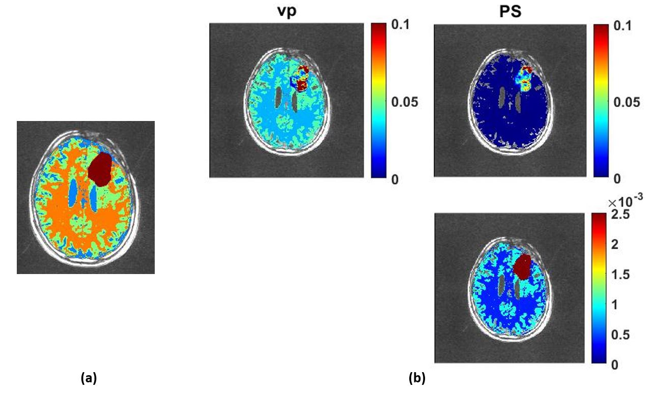
Simulation study: To evaluate the accuracy in BBB permeability estimation using iGaussian and iNESMA, we performed a simulation study. We randomly selected one subject from the RIDER dataset and performed an automatic segmentation using FSL toolbox12 to isolate white matter and gray matter regions (Figure1a). The tumor segmentation was manually performed to preserve spatial heterogeneity. The pharmacokinetic parameter values, adopted from the previous study5, were assigned to both the gray matter and white matter regions, while those of the tumor region were directly imported from the estimation using the in-vivo data (Figure 1b). These kinetic parameter maps were used to simulate noise-free DCE-MRI images using the Patlak model13. The Gaussian noise was then added to the simulated DCE-MRI images. Finally, these images were filtered using iGaussian or iNESMA, followed by the pharmacokinetic model analysis (PKM) for parameter determination. For comparison, a similar analysis was conducted on the unfiltered images.
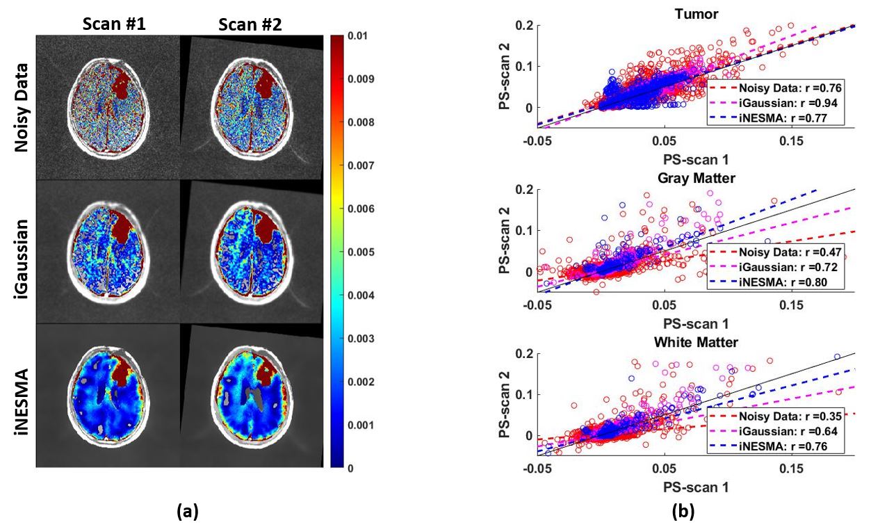
Repeatability test with in-vivo data: Two dynamic scans of the in-vivo data of the same subject were filtered using iGaussian and iNESMA. Then, the PKM analysis was performed on the unfiltered, iGaussian filtered and iNESMA filtered data to assess the BBB permeability (PS). The repeatability of these scans was evaluated using Pearson correlation.
Results & Discussion
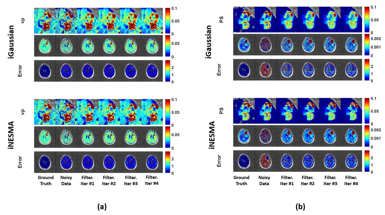
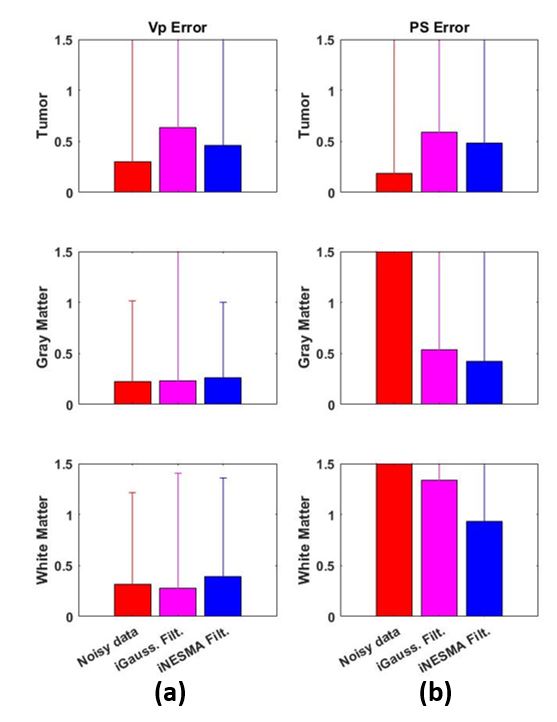
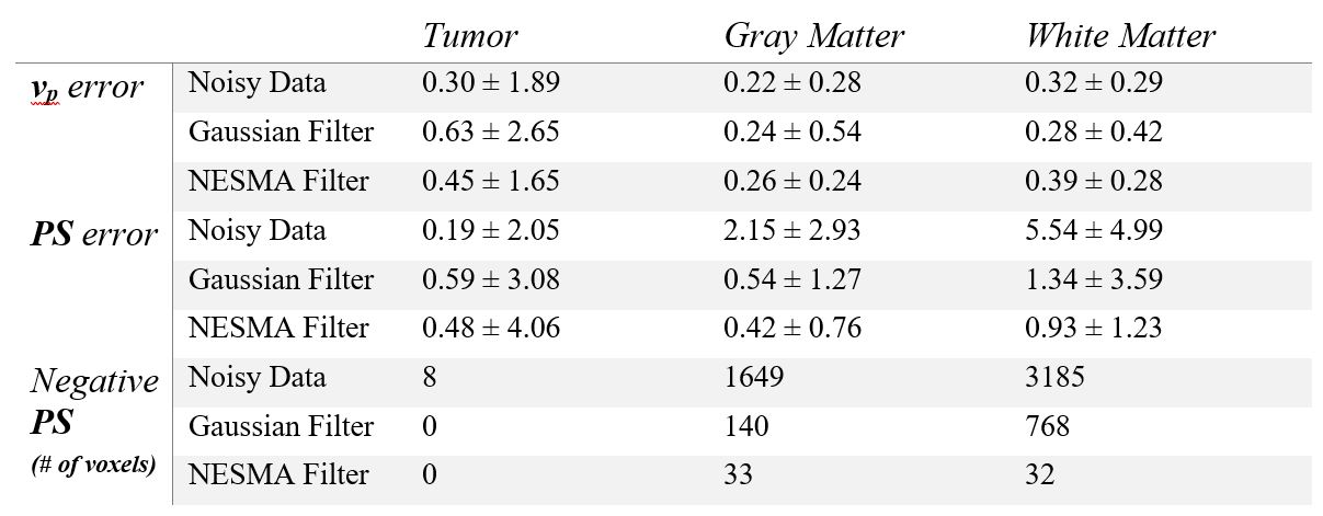
Simulation study: Figure 2 shows the estimated kinetic parameters of each filtering scheme against the assigned ground-truth parameters. As shown in Figure 2b, while both filters exhibit substantial improvement as compared to those derived from the unfiltered images, iNESMA maintained the conspicuity and the heterogeneity of tumor as compared to iGaussian which induces blurring. Moreover, in normal appearing brain regions, iNESMA filtering scheme achieves more clear contrast in PS between the gray matter and the white matter, especially as iteration proceeds, while iGaussian fails to accurately estimate subtle permeability measures. Finally, Figure 3 shows the error in each segmented regions, where iNESMA filtering yields substantially lower errors both in derived regional mean and standard deviation values (Table 1), compared to iGaussian.Repeatability test: Figure 4a shows the estimated PS maps from unfiltered, iGaussian filtered or iNESMA filtered images. Figure 4b demonstrates the correlation between 2 scans for each filtering scheme, where iNESMA outperforms iGaussian, especially in the regions with low permeability.
Conclusion
In this study, we proposed an iterative NESMA filtering method, iNESMA, which can successfully reduce the noise by identifying similar spectral patterns. iNESMA can be easily applied to acquired DCE-MRI to improve determination of subtle BBB permeability.Acknowledgements
This work was supported by the Intramural Research Program of the National Institute on Aging of the National Institutes of Health.References
1. Verheggen, I.C., et al., Increase in blood–brain barrier leakage in healthy, older adults. Geroscience, 2020. 42: p. 1183-1193.
2. Montagne, A., et al., Blood-brain barrier breakdown in the aging human hippocampus. Neuron, 2015. 85(2): p. 296-302.
3. Nation, D.A., et al., Blood–brain barrier breakdown is an early biomarker of human cognitive dysfunction. Nature medicine, 2019. 25(2): p. 270-276.
4. Van De Haar, H.J., et al., Blood-brain barrier leakage in patients with early Alzheimer disease. Radiology, 2016. 281(2): p. 527-535.
5. Bae, J., et al., Improving measurement of blood-brain barrier permeability with reduced scan time using deep-learning-derived capillary input function. NeuroImage, 2023. 278: p. 120284.
6. Bouhrara, M., et al., Noise estimation and reduction in magnetic resonance imaging using a new multispectral nonlocal maximum-likelihood filter. IEEE transactions on medical imaging, 2016. 36(1): p. 181-193.
7. Bouhrara, M., M.C. Maring, and R.G. Spencer, A simple and fast adaptive nonlocal multispectral filtering algorithm for efficient noise reduction in magnetic resonance imaging. Magnetic resonance imaging, 2019. 55: p. 133-139.
8. Bouhrara, M., et al., Use of the NESMA filter to improve myelin water fraction mapping with brain MRI. Journal of neuroimaging, 2018. 28(6): p. 640-649.
9. Alisch, J.S., et al., Sex and age-related differences in cerebral blood flow investigated using pseudo-continuous arterial spin labeling magnetic resonance imaging. Aging (Albany NY), 2021. 13(4): p. 4911.
10. Benjamini, D., et al., Multidimensional MRI for characterization of subtle axonal injury accelerated using an adaptive nonlocal multispectral filter. Frontiers in Physics, 2021. 9: p. 737374.
11. Barboriak, D., Data from RIDER_NEURO_MRI. The Cancer Imaging Archive, 2015. 577.
12. Jenkinson, M., et al., Fsl. Neuroimage, 2012. 62(2): p. 782-790.
13. Patlak, C.S., R.G. Blasberg, and J.D. Fenstermacher, Graphical evaluation of blood-to-brain transfer constants from multiple-time uptake data. Journal of Cerebral Blood Flow & Metabolism, 1983. 3(1): p. 1-7.
Figures