0735
Quantitative Water Permeability Mapping using Biophysical-modeling-based Deep Learning1Cornell University, Ithaca, NY, United States, 2Weill Cornell Medicine, New York, NY, United States
Synopsis
Keywords: Simulation/Validation, Quantitative Imaging, Arterial Spin Labeling, Brain, Vessels
Motivation: In diffusion-weighted arterial spin labeling (DW-ASL) images, quantification of the water exchange rate $$$k_{w}$$$ uses a single-pass approximation (SPA) which introduces systematic error while fitting the non-linear model is difficult.
Goal(s): Our goal was to reduce the blood-brain-barrier (BBB) water exchange rate ($$$k_{w}$$$) quantification errors in DW-ASL images.
Approach: We introduced the biophysical-modeling-based deep learning method (QTMNet) and tested both the simulated and in vivo data.
Results: On simulated data, QTMNet has 90% less normalized root mean square error (NRMSE) compared to the traditional kinetic model.
Impact: The improvement in evaluation accuracy by QTMNet may benefit Alzheimer’s Disease detection where $$$k_{w}$$$ has significant reduction.
Summary of findings
We compared the $$$k_{w}$$$ obtained using conventional means as well as QTMnet in simulated brain diffusion-weighted arterial spin labeling images. The QTMNet method gives 3.7% NRMSE in quantification on $$$k_{w}$$$ compared to 31.2% for the traditional model.Introduction
Blood-brain-barrier (BBB) water exchange rate ($$$k_{w}$$$) is an emerging biomarker of the BBB dysfunction due to the aging or aging-related disorders1. Traditional tracer kinetic model based on single-pass approximation (SPA) ignores the tracer concentration exchange from extravascular extracellular space to the vascular space2. However, ignoring the backflow of the tracer introduces a systematic error and the non-linearity of the SPA makes the inverse problem hard to solve. Here we use QTMNet, a biophysical-modeling-based deep learning method, to quantify $$$k_{w}$$$. We demonstrate that on the simulated brain ASL images, QTMNet achieves a 90% reduction in NRMSE compared to the traditional SPA model.Methods
In QTMnet, a Unet is trained to learn the inverse mapping from diffusion-weighted arterial spin labeled (DW-ASL) to the water exchange rate ($$$k_{w}$$$). The training data for QTMnet consists of simulated data within a cube-shaped tissue of size 32x32x32mm3 obtained as follows. We first extract large vessels from an example MR angiography image and use constrained constructive optimization3-5 to generate synthetic micro-vessel branches. The radius of mother and daughter vessels follows cubic rules: $$$r^3_M=r^3_{d1}+r^3_{d2}$$$4. Meanwhile, unlike the common plug flow assumption6, we applied the quadratic blood velocity profile $$$\textbf{u}(r)$$$7 which is more realistic for vessel flow. The tracer concentration profile is computed by solving the following transport equations:$$\partial_t c_c(r,t)=\nabla\cdot(-\mathbf{u}(r) c_c(r,t))+k_w(r)(c_b(r,t)-c_c(r,t))-R_{1a}c_c(r,t)$$
$$\partial_t c_b(r,t)=-k_w(r)(c_b(r,t)-c_c(r,t))-R_{1b}c_b(r,t)$$
Here $$$c_b (r,t)$$$ and $$$c_c (r,t)$$$ are the tracer concentration inside brain tissue and capillary, respectively, $$$R_{1a}=0.6 s^{-1}$$$ and $$$R_{1b}=0.83 s^{-1}$$$ are the longitudinal relaxation rates of blood and brain tissue, $$$\mathbf{u}(r)$$$ is the velocity field, $$$\nabla$$$ is the gradient operator and $$$\partial_t$$$ is the derivative of time. $$$C(t)=\int_{V_c}c_c(r,t)dV+\int_{V_b}c_b(r,t)dV$$$ is the concentration of the voxel. $$$V_c$$$ and $$$V_b$$$ are the volume of the capillary and brain tissue, respectively. Finally, the tracer concentrations were summed over each voxel to obtain DW-ASL images. In the training, we minimize the L1 norm of the predicted $$$k_w$$$ from QTMNet $$$\Psi$$$ and the ground truth with regularization on $$$\nabla k_w$$$:
$$k_w(r)=argmin_{k_w}\|k_w(r)-\Psi(C(t))\|_1+\lambda\|\nabla k_w(r)\|_1$$
Conventional $$$k_w$$$ mapping was performed by fitting the simulated DW-ASL data to the signal model1:
$$\Delta M_b=\frac{2CBF\epsilon M_0\beta}{\lambda}[\frac{e^{-(R_{1a}-R_{1b})ATT}}{R_{1b}}(e^{-R_{1b}(t-\delta)}-e^{-R_{1b} t})-\frac{e^{-(R_{1a}-\alpha)ATT}}{\alpha}(e^{-\alpha(t-\delta)}-e^{-\alpha t})]$$
$$\Delta M_c=-\frac{2CBF\epsilon M_0}{\lambda\alpha}e^{-(R_{1a}-R_{1b})ATT}(e^{-\alpha(t-\delta)}-e^{-\alpha t})$$
$$\Delta M(b)=\Delta M_b e^{-bD_b}+\Delta M_c e^{-bD_c}$$
Here, $$$\Delta M$$$ is the signal of the acquired ASL image; $$$\Delta M_b$$$ and $$$\Delta M_c$$$ are signals of tissue and capillary compartment, respectively. $$$\lambda = 1$$$ is the tissue/blood partition coefficient for the simulated data, and $$$\epsilon = 1$$$ is the tagging efficiency for simulated data. CBF is the cerebral blood flow, $$$\alpha = k_w+R_{1a}$$$ and $$$\beta = \frac{k_w}{k_w+R_{1a}-R_{1b}}$$$
Given the DW-ASL images acquired with different b-value (0,10,20,50,100 $$$s/mm^2$$$) and different post-labeling delay (PLD=1200,1500,1800,2100 ms), The $$$k_w$$$ could be evaluated by minimizing the following cost function.
$$k_w(r)=argmin_{k_w}\sum_b\sum_{PLD}\|\Delta M(b,PLD)-f(k_w)\|_2^2+\lambda\|k_w(r)\|_2^2$$
Results
Figure 1 shows the simulated DW-ASL image frames at PLD=1200, 1500, 1800, 2100 ms under different b-values with the normalized concentration.Figure 2a-c compares the $$$k_{w}$$$ ground truth of the simulated brain DW-ASL images with the quantitative results from the traditional SPA model and QTMNet, respectively. Figure 2d-e shows that QTMNet reduces the $$$k_{w}$$$ error compared to the traditional SPA models.
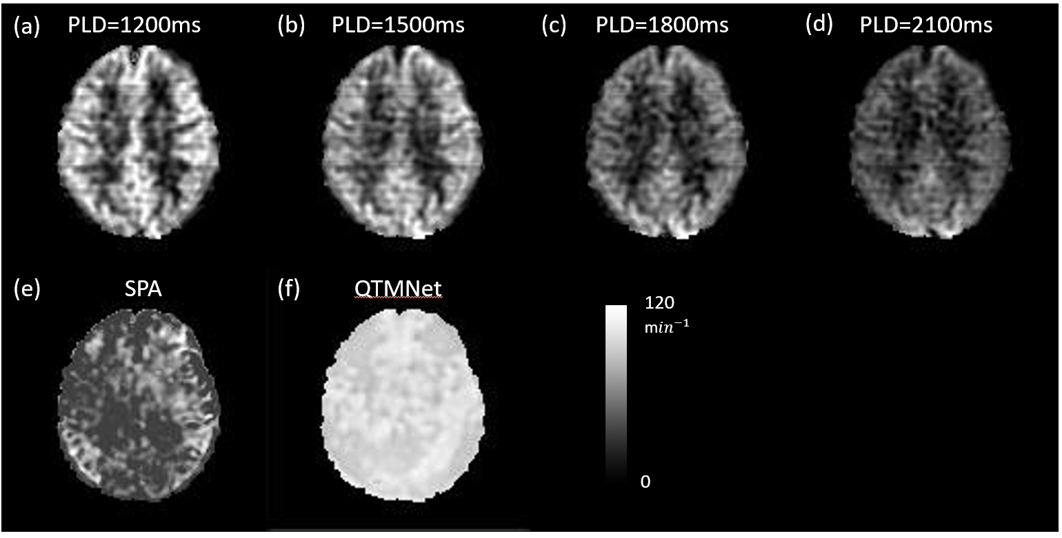
Figure 3a-d show the clinical images with $$$PLD=1200, 1500, 1800, 2100 ms$$$ and $$$b=0$$$. Figure 2e-f show the evaluation of $$$k_{w}$$$ using SPA model and QTMNet, respectively.
Conclusion
We demonstrate the feasibility of QTMNet method in $$$k_{w}$$$ quantification. The QTMNet method surpasses the traditional SPA model on the CFD-simulated DW-ASL data. The use of the biophysical CFD simulation contributes to the accurate $$$k_{w}$$$ mapping. Possible limitations of this work include selecting the $$$k_{w}$$$ values preset in the brain and cube generations. The inconsistency of the reported $$$k_{w}$$$ makes the value selection difficult and might introduce bias into the network.Acknowledgements
We acknowledge the funding from National Institute on Aging (1 R01 AG080011-01A1) and National Institute of Biomedical Imaging & Bioengineering (1 R01 EB034755-01)References
1. Ford JN, Zhang QH, Sweeney EM, Merkler AE, de Leon MJ, Gupta A, Nguyen TD, Ivanidze J. Quantitative Water Permeability Mapping of Blood-Brain-Barrier Dysfunction in Aging. Front Aging Neurosci 2022;14.
2. St Lawrence KS, Frank JA, McLaughlin AC. Effect of restricted water exchange on cerebral blood flow values calculated with arterial spin tagging: A theoretical investigation. Magn Reson Med 2000;44(3):440-449.
3. Karch R, Neumann F, Neumann M, Schreiner W. A three-dimensional model for arterial tree representation, generated by constrained constructive optimization. Computers in Biology and Medicine 1999;29(1):19-38.
4. Murray CD. The physiological principle of minimum work II Oxygen exchange in capillaries. P Natl Acad Sci USA 1926;12:299-304.
5. Neumann F, Schreiner W, Neumann M. Constrained constructive optimization of binary branching arterial tree models. Structural Optimization 1995:181-188.
6. Buxton RB, Frank LR, Wong EC, Siewert B, Warach S, Edelman RR. A general kinetic model for quantitative perfusion imaging with arterial spin labeling. Magn Reson Med 1998;40(3):383-396.
7. Zhou LD, Zhang QH, Spincemaille P, Nguyen TD, Morgan J, Dai WY, Li Y, Gupta A, Prince MR, Wang Y. Quantitative transport mapping (QTM) of the kidney with an approximate microvascular network. Magn Reson Med 2021;85(4):2247-2262.
8. Shao XF, Ma SJ, Casey M, D'Orazio L, Ringman JM, Wang DJJ. Mapping water exchange across the blood-brain barrier using 3D diffusion-prepared arterial spin labeled perfusion MRI. Magn Reson Med 2019;81(5):3065-3079.
Figures
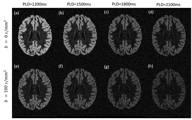
Figure 1. The normalized simulated DW-ASL at four different PLDs under $$$b=0s/mm^2$$$ and $$$b=100s/mm^2$$$ situations.
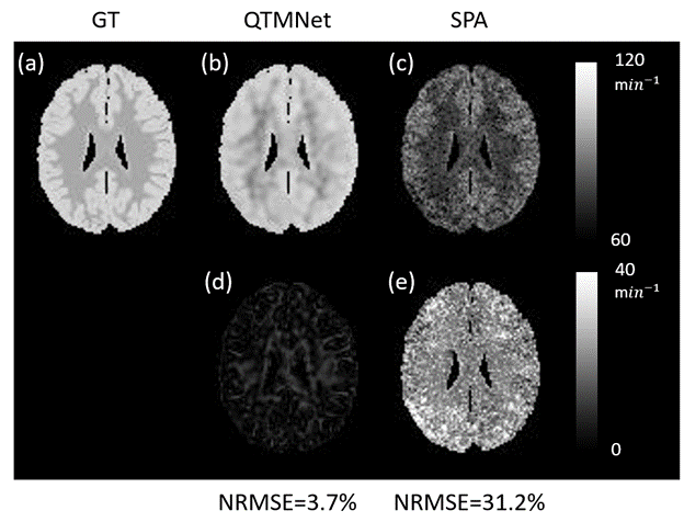
Figure 2. The $$$k_w$$$ quantification of a random slice by QTMNet and traditional SPA model. The first row displays the $$$k_w$$$, and the second row shows the $$$k_w$$$ error compared to the ground truth.