0733
Repeatability of Blood-Brain Barrier Diffusion Weighted Arterial Spin Labeling MRI at Different Post-Label Delays1Biomedical Engineering, University of California, Davis, Davis, CA, United States, 2Neurology, University of California, Davis, Davis, CA, United States
Synopsis
Keywords: Neurofluids, Arterial spin labelling, Blood-brain barrier, Diffusion-weighting
Motivation: Anomalous blood-brain barrier (BBB) water transfer rate (Kw) has the potential to be a novel biomarker for neurological disorders.
Goal(s): However, additional studies are needed to affirm the reliability of MRI sequences that assess Kw.
Approach: This in vivo study sought to determine the intrasession repeatability of the single-delay diffusion-weighted (DW) arterial spin labeling (ASL) MRI sequence at different DW post-label delays (PLDs). [1]
Results: Our findings confirmed that cerebral blood flow (CBF) and Kw were most stable at a DW PLD of 1800ms and that there exists a significant linear correlation between arterial transit time (ATT) and Kw.
Impact: Our findings show that for single-delay diffusion weighted (DW) ASL MRI, properly selecting the DW PLD and consideration of ATT are crucial for robust BBB water permeability measurements. Studies like ours are necessary before Kw imaging in different disease states.
Introduction
Water flows freely, though controlled, across the blood-brain barrier (BBB) through transmembrane aquaporin (AQP) channels whose aberrant expression has been the major explanatory mechanism for decreased water exchange rate (Kw) measured in disease states using the diffusion-weighted (DW) ASL method. [2-4] The theory behind the DW ASL sequence, published by St. Lawrence et al. [5], relies on the different diffusivities of blood and tissue to split the overall ASL signal into an intravascular and extravascular component from which Kw can then be deduced through a set of general kinetic model equations. The sequence itself was pioneered by Shao et al. with initial tests on healthy participants before applications in a cohort with small vessel disease. [1]Thus, the purpose of our work is to assess the intrasession repeatability of this sequence on our scanners at different DW post-label delays (PLDs). We are interested in quantifying the robustness of Kw measurements at different DW PLDs because of prolonged arterial transit times (ATTs) in some patient populations that may necessitate a later DW PLD to compensate for the signal’s later arrival. [6] We also directly investigate if there is any relationship between Kw and ATT.
Methods
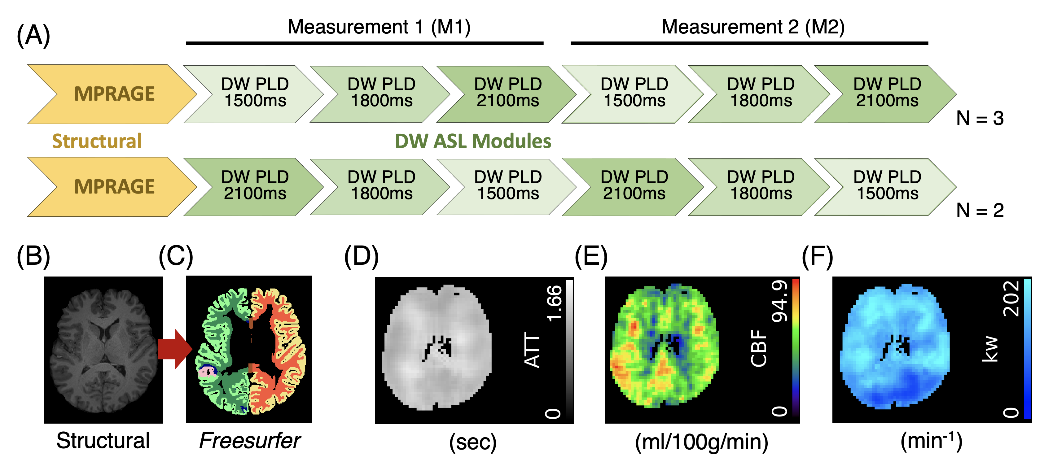
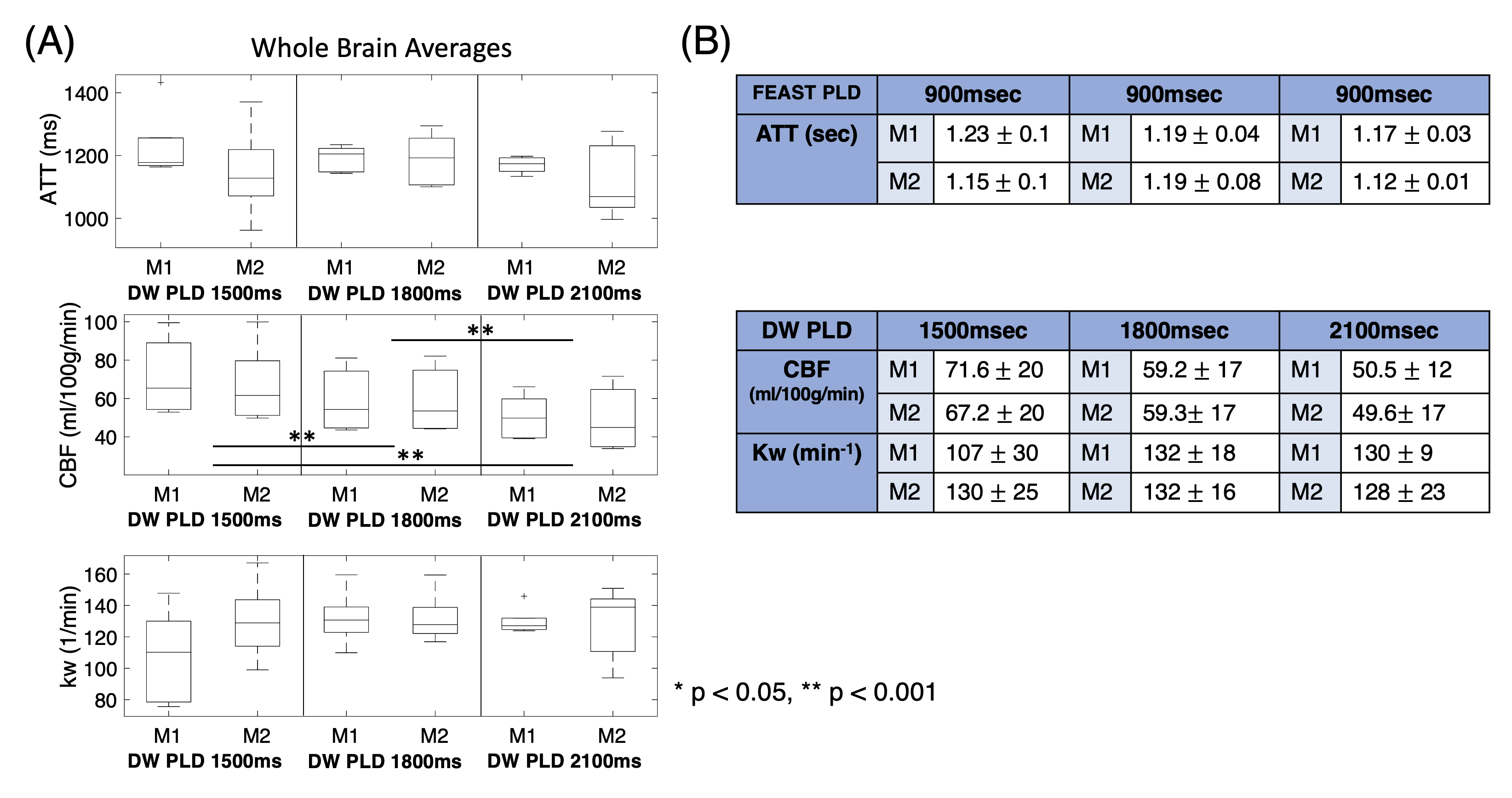
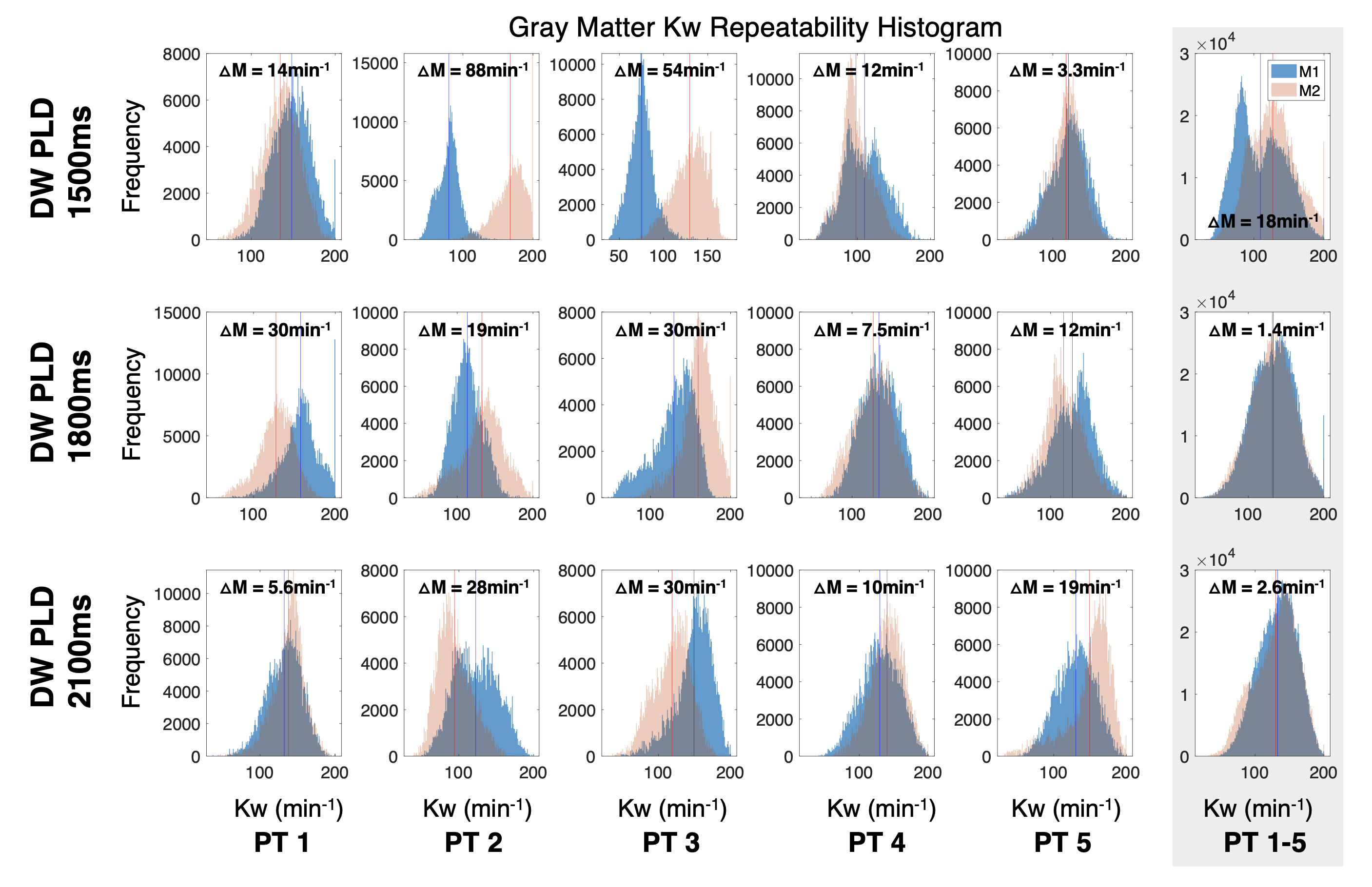
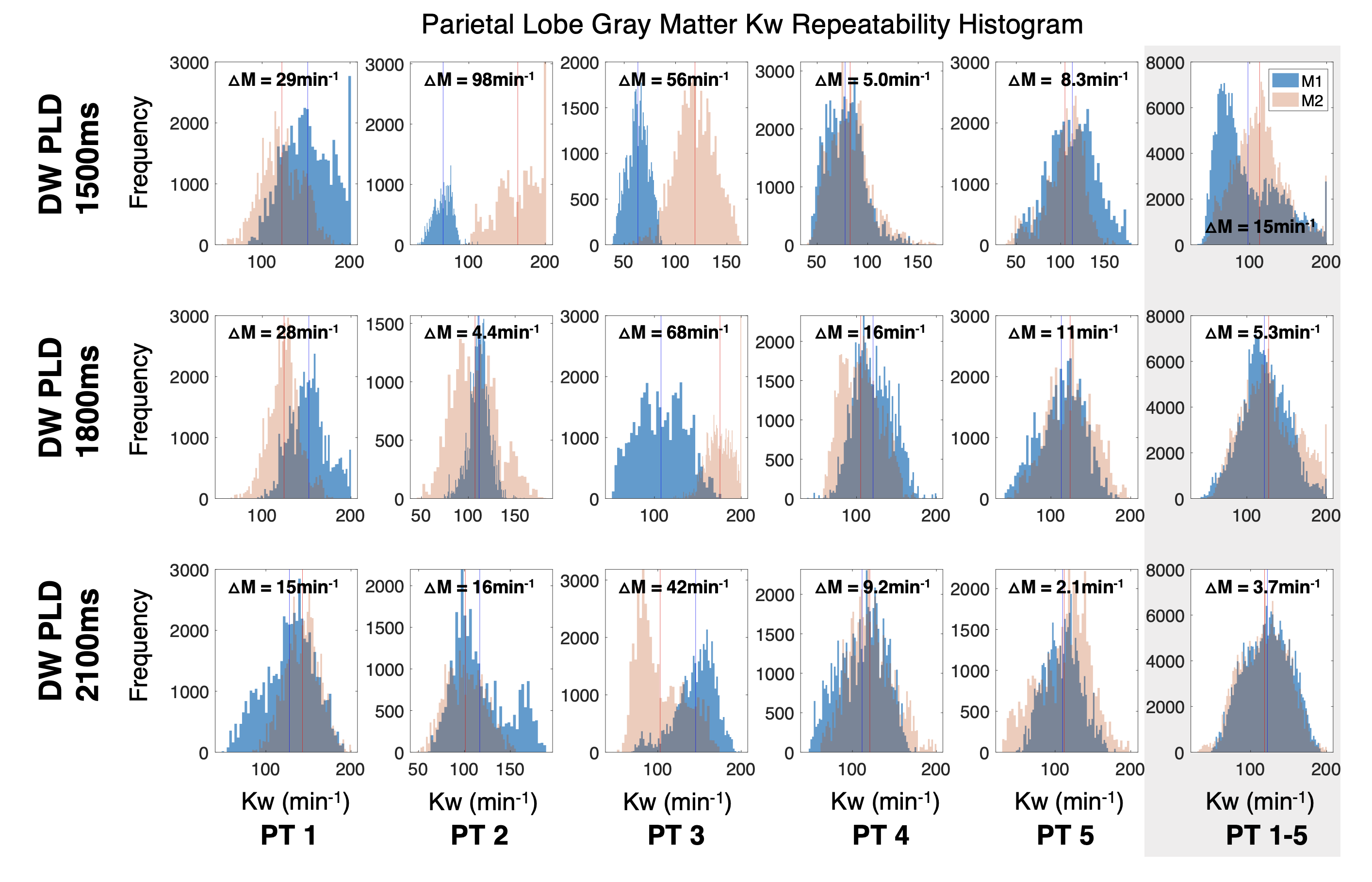
Five healthy participants (2 females, aged 23-29) were scanned on a 3T Siemens Skyra MRI. Structural T1 images were acquired, followed by three DW ASL sequences (M1) to measure Kw at separate individual DW PLDs of 1500ms, 1800ms, and 2100ms, consistent with an optimization experiment in Shao et al. [1] These three modules were then repeated (M2) to assess the intra-session repeatability. In two participants, we reversed the DW PLD order to minimize ordinal bias (Figure 1). The flow encoding arterial spin tagging (FEAST) [7] PLD, used to calculate ATT, was kept at 900ms across all six repetitions.The data was processed with the Water Exchange Quantification (WEQ) Toolbox to calculate whole-brain ATT, cerebral blood flow (CBF), and Kw maps as well as global averages (Figure 2). We used a two-sample t-test to investigate any significant group differences in CBF, ATT, and Kw across our 3 DW PLDs. We then linearly registered the WEQ Toolbox-derived parametric maps to the structural T1 images using FSL FLIRT. [8] We used Freesurfer [9] to segment the structural T1 to isolate the gray matter signal and parietal lobe region of interest (ROI) (Figures 3 & 4). We chose to study the parietal lobe because this region has been shown to exhibit altered BBB permeability in schizophrenia using this DW ASL sequence. [2] The difference between the two distributions’ means was reported as ΔM.
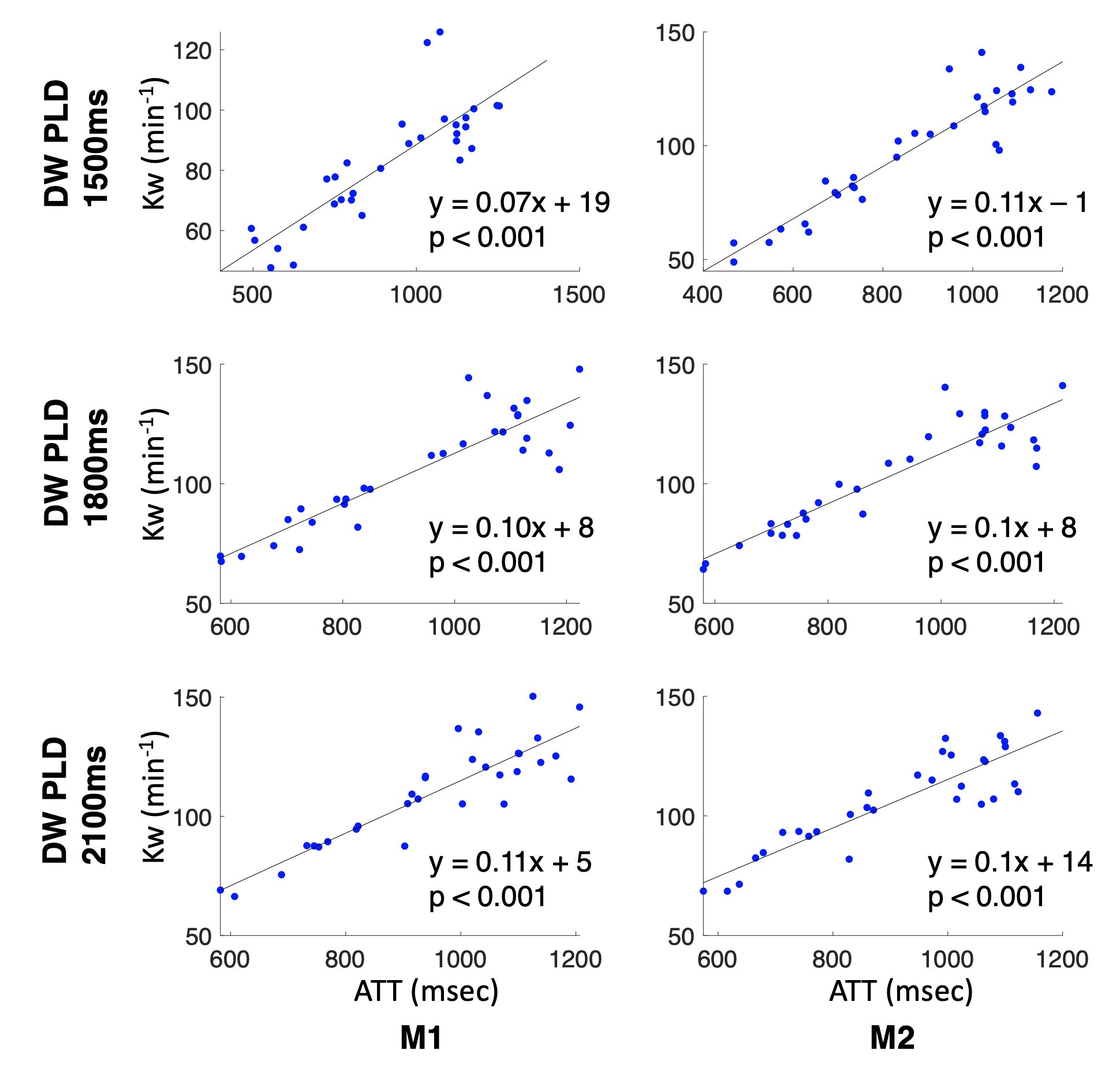
To investigate the correlation between Kw and ATT, we extracted the Kw and ATT averages in 35 Freesurfer ROIs across all five participants and fitted each resulting scatterplot with a simple linear regression model (Figure 5). Four outlier ROIs were excluded.
Results & Discussion
Whole brain averages, calculated via the WEQ Toolbox, showed that CBF was significantly different between the three DW PLDs and that perfusion decreased as the DW PLD increased to 2100ms (Figure 2A). However, whole-brain ATT and Kw did not differ across DW PLD acquisitions. Figure 2B also shows that at a DW PLD of 1800ms, the Kw and CBF group averages are most similar between M1 and M2 in terms of their mean and standard deviation.Gray matter histograms (Figure 3) show that, on a group average level (far right column), the Kw distribution for M1 and M2 have good consistency (low ΔM) for DW PLDs of 1800ms (ΔM=1.4min-1) and 2100ms (ΔM=2.6min-1) compared to shorter DW PLD of 1500ms (ΔM=18min-1). Similar observations were seen on the group average level for the parietal lobe cortical ROI (Figure 4). On an individual level, DW PLDs of 1800ms and 2100ms also showed higher consistency of Kw values across the gray matter than DW PLD of 1500ms, especially in participants 2 and 3. Slightly more variability was observed for parietal lobe regional analysis, in which DW PLD of 2100ms provided the most reliable Kw assessment across individuals.
Correlation analysis between Kw and ATT across Freesurfer ROIs showed a significant and positive association between these two variables at all three DW PLDs (Figure 5). This finding suggests that for patients with longer ATTs (e.g. Alzheimer’s disease or aging), the underlying ATT should be considered when comparing Kw values from single-delay DW ASL scans. Multi-PLD DW ASL, still in development, could provide more robust ATT and Kw metrics. [10] Future work will extend this analysis to different age ranges or patients with a wider biological range of ATT.
Acknowledgements
R01NS128179, UL1 TR001860 (linked award TL1 TR001861). We also thank the LOFT Lab at the University of Southern California (USC) for sharing this sequence.References
1. Shao, X., et al., Mapping water exchange across the blood-brain barrier using 3D diffusion-prepared arterial spin labeled perfusion MRI. Magn Reson Med, 2019. 81(5): p. 3065-3079.
2. Goldwaser, E.L., et al., Evidence of Neurovascular Water Exchange and Endothelial Vascular Dysfunction in Schizophrenia: An Exploratory Study. Schizophr Bull, 2023.
3. Jessen, N.A., et al., The Glymphatic System: A Beginner's Guide. Neurochem Res, 2015. 40(12): p. 2583-99.
4. Palomares, J.A., et al., Water Exchange across the Blood-Brain Barrier in Obstructive Sleep Apnea: An MRI Diffusion-Weighted Pseudo-Continuous Arterial Spin Labeling Study. J Neuroimaging, 2015. 25(6): p. 900-5.
5. St Lawrence, K.S., D. Owen, and D.J. Wang, A two-stage approach for measuring vascular water exchange and arterial transit time by diffusion-weighted perfusion MRI. Magn Reson Med, 2012. 67(5): p. 1275-84.
6. Dai, W., et al., Effects of arterial transit delay on cerebral blood flow quantification using arterial spin labeling in an elderly cohort. J Magn Reson Imaging, 2017. 45(2): p. 472-481.
7. Wang, J., et al., Arterial transit time imaging with flow encoding arterial spin tagging (FEAST). Magn Reson Med, 2003. 50(3): p. 599-607.
8. Jenkinson, M., et al., Fsl. Neuroimage, 2012. 62(2): p. 782-90.
9. Fischl, B., FreeSurfer. Neuroimage, 2012. 62(2): p. 774-81.
10. Shao, X., et al., Quantification of blood-brain barrier water exchange and permeability with multidelay diffusion-weighted pseudo-continuous arterial spin labeling. Magn Reson Med, 2023.
Figures