0732
ASL blood-brain barrier permeability is associated with amyloid and cognitive impairment1Radiology and Nuclear Medicine, Amsterdam UMC locatie VUmc, Amsterdam, Netherlands, 2Department of Neurology, Akershus University Hospital, Oslo, Norway, 3Department of Physics and Computational Radiology, Division of Radiology and Nuclear Medicine, Oslo University Hospital, Oslo, Norway, 4Department of Psychology, Center for Lifespan Changes in Brain and Cognition, University of Oslo, Oslo, Norway, 5Fraunhofer-Institute for Digital Medicine MEVIS, Bremen, Germany, 6Institute of Clinical Medicine, Campus Ahus, University of Oslo, Oslo, Norway, 7Department of Neurology, University Hospital of North Norway, Tromsø, Norway, 8Department of Brain Repair and Rehabilitation, UCL Queen Square Institute of Neurology, University College London, London, United Kingdom, 9Oslo University Hospital, Computational Radiology and Artificial Intelligence, Division of Radiology and Nuclear Medicine, Oslo, Norway, 10Computational Radiology and Artificial Intelligence, Division of Radiology and Nuclear Medicine, Oslo University Hospital, Oslo, Norway, 11Helmholtz-Zentrum Dresden-Rossendorf, Institute of Radiopharmaceutical Cancer Research, Dresden, Germany
Synopsis
Keywords: Alzheimer's Disease, Arterial spin labelling, Blood-brain barrier, Biomarkers
Motivation: Blood-brain barrier (BBB) permeability changes may be implicated in Alzheimer’s Disease (AD) pathophysiology.
Goal(s): To investigate if the exchange time (Tex) of water across the BBB is associated with cognitive and amyloid status.
Approach: We measured Tex with a multi-echo arterial spin labeling MRI sequence in 116 adults older than 50 years and studied its association with cognition (cognitively normal vs mild cognitive impaired) and amyloid (A- vs A+) status.
Results: BBB water permeability is increased in A+ participants and in patients with MCI, compared to healthy controls
Impact: Our results suggest that multi-TE ASL MRI BBB water permeability can be used as a potential early imaging biomarker of AD pathophysiology.
1. Introduction
Introduction: Blood-brain barrier (BBB) dysfunction is potentially one of the earliest microvascular changes in Alzheimer’s disease (AD) and related dementias1. Techniques utilizing gadolinium-based contrast agents (GBCA) to measure BBB permeability are not only invasive and costly but also potentially toxic. Given that GBCAs possess higher molecular weights, they are less sensitive to early, subtle BBB changes, such as disruption of aquaporin-4 channels, which have been linked to aging and AD in both animal models2 and humans3. An emerging technique to image the time of exchange (Tex) of water across the BBB is multi-echo4,5 arterial spin labeling (ASL)4, which obviates the need for exogenous contrast and has been shown to provide reproducible values of BBB integrity in healthy volunteers4. Here, we investigate the associations of Tex values with amyloid positivity and cognitive status. As cerebral blood flow (CBF) can be seen as an established ASL biomarker, we repeated all analyses for CBF for comparison.Methods: Data from 116 participants older than 50 years were selected from the Center for Lifespan Changes in Brain and Cognition (LCBC) and the Dementia Disease Initiation (DDI) cohorts. LCBC comprises a population-based cohort including only cognitively normal (CN) participants (n=77, CNLCBC), while DDI is a clinical outpatient cohort including CN and subjective cognitive decline patients (n=24, merged here into a single cohort CNDDI) and mild cognitive impairment (n=15, MCIDDI)6 patients. Amyloid status was defined as positive (A+) or negative (A-) from the CSF amyloid-beta 42/40 ratio (cut-off ≤ 0.077) or amyloid-PET by visual read, when available. All cohorts were scanned on the same 3T Siemens Prisma scanner with a 32-channel head coil. Two recently developed multi-post-labeling delay (PLD) Hadamard-encoded (HAD) 3D GRASE PCASL sequences were used to estimate Tex and CBF: 1) HAD-8 with a labeling duration (LD) 400 ms, PLD [600:400:3400] ms, and single echo time (TE) 12.5 ms; 2) multi-TE HAD-4 with LD 1000 ms, PLD [1500:1000:3500] ms, and 8 TEs [14.4:28.9:217.2] ms. Data were analyzed with ExploreASL 1.11.0beta7, and gray matter (GM) CBF and Tex were quantified with FSL-FABBER8. Tex and CBF associations with amyloid and cognitive status were assessed using linear regression adjusted for age and sex.
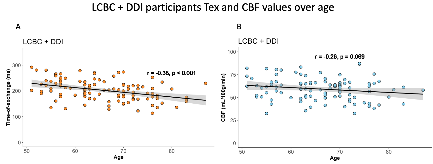
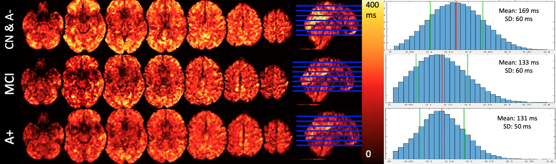
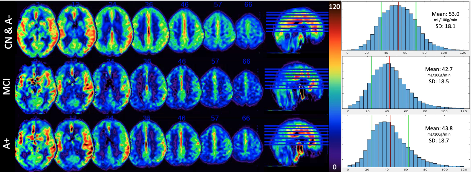
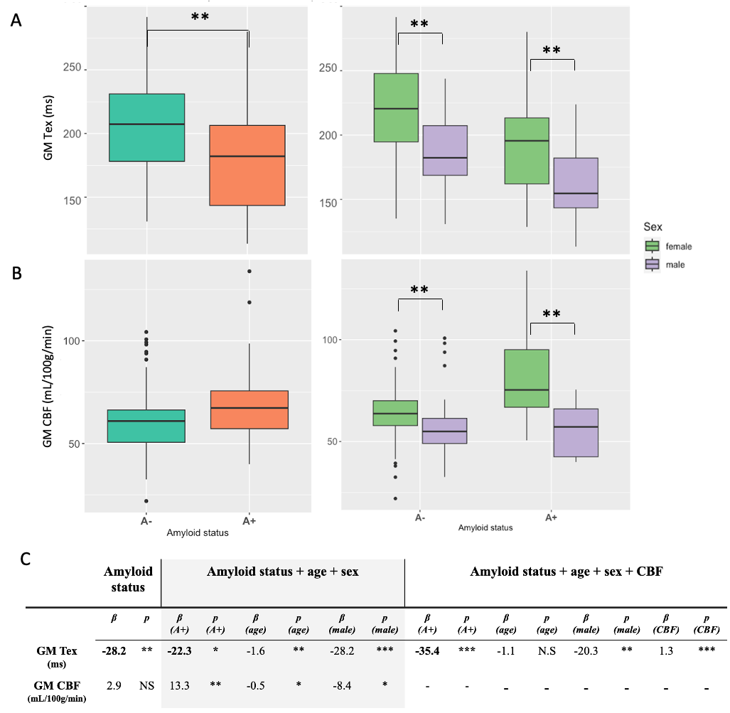
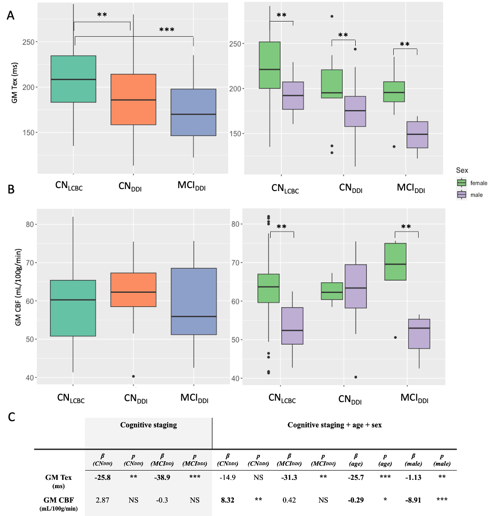
Results: Of the 116 participants, in total, 77 were from the LCBC (64.6±8.4 years, 64% female) and 39 from the DDI (67.7±7.9 years, 51% female) cohorts. DDI included 15 MCIs and 28 A+, of which 12 were both MCI and A+. Across the whole population, GM Tex was negatively correlated with age (r = -0.38, p < 0.001, Figure 1A), whereas for GM CBF, this correlation was not statistically significant (r = -0.26, p = 0.069, Figure 1B). Figures 2 and 3 show, respectively, whole-brain group average Tex and CBF maps from CN A- controls, MCI patients, and A+ subjects. Histograms next to these maps show the GM mean and standard deviation values of the three groups. It can be observed that Tex and CBF values are both higher in the NC A- group than in the MCI or A+ maps.Tex was 15% lower in A+ compared to A- (t=2.75, p=0.01, Figure 4A). CBF was 5% higher in the A+ group than the A- group but did borderline not reach statistical significance (t=-1.94, p=0.06, Figure 4B)9,10. The linear regression analysis showed that amyloid status was associated with BBB water permeability (given by Tex), with higher permeability in A+ compared with A- groups when correcting for age, sex, and CBF (β = -35 s, p < 0.001) (Figure 4C). Moreover, cognitive staging was related to Tex (Figure 5A), even when correcting for age and sex (βMCI = -31.3 s, p < 0.01) (Figure 5C). A similar relationship was not found for CBF (Figure 5B, C).
Discussion: Interestingly, both amyloid positivity and cognitive status were associated with increased BBB water permeability, even when correcting for age and sex. In agreement with previous studies, BBB water permeability was shown to increase with age. These permeability increases might be explained by a normal aging process of increased brain clearance or by BBB dysfunction promoting leakage of toxins to cross from the capillary site to the brain parenchyma. Whether BBB dysfunction is the cause or effect of amyloid deposition cannot be differentiated from these data. These findings encourage the use of BBB-ASL to non-invasively investigate BBB integrity in the early stages of dementia.
Acknowledgements
The DEBBIE project (Developing a non-invasive biomarker for early BBB breakdown in Alzheimer’s disease) is an EU Joint Programme -Neurodegenerative Disease Research (JPND) project. For this DEBBIE substudy, we received funding through the following funding organisations under the aegis of JPND -www.jpnd.eu (BMBF in Germany, NFR in Norway, and ZonMw and Alzheimer Nederland in The Netherlands). The project has received funding from the European Union’s Horizon 2020 research and innovation programme under grant agreement No. 825664.References
1. Montagne, A. et al. APOE4 leads to blood-brain barrier dysfunction predicting cognitive decline. Nature 581, 71–76 (2020).
2. Kress, B. T. et al. Impairment of paravascular clearance pathways in the aging brain. Ann. Neurol. 76, 845–861 (2014).
3. Zeppenfeld, D. M. et al. Association of Perivascular Localization of Aquaporin-4 With Cognition and Alzheimer Disease in Aging Brains. JAMA Neurol. 74, 91–99 (2017).
4. Mahroo, A. et al. Robust Multi-TE ASL-Based Blood-Brain Barrier Integrity Measurements. Front. Neurosci. 15, 719676 (2021).
5. Gregori, J., Schuff, N., Kern, R. & Günther, M. T2-based arterial spin labeling measurements of blood to tissue water transfer in human brain. J. Magn. Reson. Imaging 37, 332–342 (2013).
6. Fladby, T. et al. Detecting At-Risk Alzheimer’s Disease Cases. J. Alzheimers. Dis. 60, 97–105 (2017).
7. Mutsaerts, H. J. M. M. et al. ExploreASL: an image processing pipeline for multi-center ASL perfusion MRI studies. Neuroimage 117031 (2020).
8. Chappell, M. A., Groves, A. R., Whitcher, B. & Woolrich, M. W. Variational Bayesian Inference for a Nonlinear Forward Model. IEEE Trans. Signal Process. 57, 223–236 (2009).
9. Fazlollahi, A. et al. Increased cerebral blood flow with increased amyloid burden in the preclinical phase of alzheimer’s disease. J. Magn. Reson. Imaging 51, 505–513 (2020).
10. Padrela, B. E. et al. Genetic, vascular, and amyloid components of cerebral blood flow in a preclinical population. J. Cereb. Blood Flow Metab. 271678X231178993 (2023).
Figures