0728
Utilizing Synthetic MRI and Deep Learning Reconstruction of DWI to Distinguish Benign from Malignant Breast Lesions1Department of Magnetic Resonance, The First Affiliated Hospital of Zhengzhou University, Zhengzhou, China, 2MR Research China, GE Healthcare, Beijing, China
Synopsis
Keywords: Breast, Cancer, Deep Learning Reconstruction, Diffusion-Weighted Imaging, Breast Diagnosis, Synthetic MRI
Motivation: Breast cancer has emerged as the foremost global malignancy, prompting a growing inclination toward exploring novel non-invasive imaging techniques that obviate the need for contrast agent administration.
Goal(s): Enhancing breast diagnostics without reliance on contrast agents.
Approach: Expanding on the foundation of deep learning-based DWI reconstruction, coupled with Synthetic MRI, as a viable alternative to traditional contrast-enhanced diagnostic methodologies, the focus lies in pinpointing valuable parameters for differential diagnosis.
Results: The fusion of deep learning-reconstructed DWI and Synthetic MRI yields an impressive AUC (Area Under the Curve) of 0.995 in distinguishing between benign and malignant breast pathologies.
Impact: The integration of deep learning-reconstructed DWI with Synthetic MRI not only carries substantial diagnostic significance in discerning between benign and malignant breast conditions but also exhibits the promise of supplanting conventional contrast-enhanced methodologies.
Introduction:
Breast lesion screening has long been a cornerstone of women's health, with a persistent focus on improving detection methods. While MRI has proven to be a valuable tool for breast imaging, it does come with a relatively high false positive rate, necessitating the often-invasive procedure of biopsy for confirmation. This underscores the pressing need for more effective biomarkers that can mitigate the requirement for invasive biopsies. Additionally, traditional breast tumor MRI scans typically rely on contrast agents, some of which have been associated with the formation of deposits and are not suitable for patients with compromised kidney function. Consequently, the shift toward non-contrast MRI scans is becoming an evident trend. A noteworthy advancement in this direction is the adoption of Synthetic MRI technology, which is already in commercial use in clinical settings. This innovative quantitative technique provides comprehensive information on T1, T2, and PD mapping in a scan, greatly assisting in tissue differentiation and lesion diagnosis [1]. It has found widespread application in clinical research. Furthermore, diffusion-weighted imaging (DWI) is a standard scanning technique for breast evaluation, and, when coupled with deep learning-based reconstruction methods [2,3], it has demonstrated remarkable capabilities in enhancing image signal-to-noise ratios and reducing artifacts. Considering the potential of Synthetic MRI to replace traditional contrast-enhanced scans and be used in conjunction with DWI, it holds the promise of meeting the evolving demands of breast screening while simultaneously decreasing the necessity for biopsies, which would significantly benefit patients. Hence, the objective of our current study is to explore the efficacy of combining Synthetic MRI and deep learning-based reconstructed DWI to distinguish between benign and malignant breast lesions, with the ultimate aim of reducing the burden of biopsies in breast lesion diagnosis.Materials and methods
MRI data was uniformly acquired using a 3.0 Tesla MRI scanner (SIGNA Premier, GE Healthcare, WI, USA) in this study. A cohort of 111 female patients, aged between 27 and 65, all with various breast lesions, underwent comprehensive breast MRI examinations inclusive of Synthetic MRI and Diffusion-Weighted Imaging (DWI). The DWI employed a b value of 800 s/mm². The quantitative parameters obtained from Synthetic MRI encompassed T1map, T2map, and proton density (PD), while the Apparent Diffusion Coefficient (ADC) map was derived from both conventional DWI and DWI reconstructed using deep learning techniques. The analysis focused on assessing the diagnostic significance of Synthetic MRI and DWI. Notably, the valuable parameters identified were incorporated into a model, which was subsequently subjected to a comprehensive evaluation to determine its diagnostic efficacy. Furthermore, the study deliberated on whether this model could enhance the accuracy of breast lesion diagnoses.Results and discussion
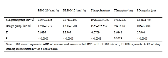
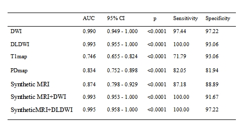
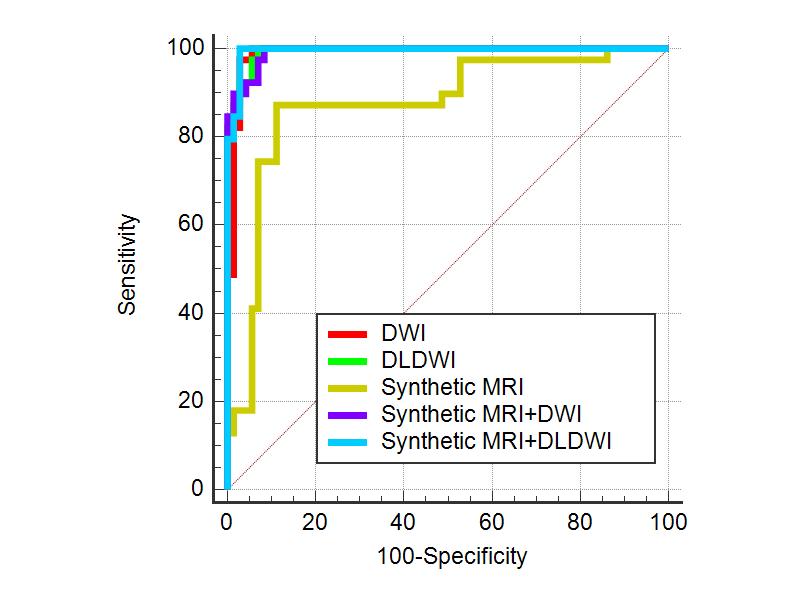
In the malignant group, the ADC values were consistently lower than those in the benign group, as indicated in Table 1, and these differences were found to be statistically significant. However, there was no significant distinction in T2 mapping between benign and malignant lesions (P>0.05). The T1 mapping of malignant lesions measured 1928.3 ms, which was significantly higher than the 1564 ms observed in benign lesions (P < 0.05). Furthermore, the PD mapping of malignant lesions was 82.41 pu, significantly smaller than the 106 pu value observed in benign lesions (P < 0.05).The area under the curve (AUC) for the differential diagnosis, ranked from highest to lowest, is as follows: DLDWI (0.993), DWI (0.99), T1mapping (0.746), and PDmapping (0.834) (see Table 2).T1 mapping and PD mapping parameters in Synthetic MRI were combined to form the Synthetic MRI group for diagnostic purposes. Notably, the fused Synthetic MRI group exhibited greater diagnostic significance with an AUC of 0.874 (refer to Fig. 1 and Table 2).Additionally, the ADC map derived from conventional DWI and significant parameters from Synthetic MRI constituted the Synthetic MRI+DWI group. Furthermore, the ADC map derived from deep learning-reconstructed DWI, along with the significant parameters from Synthetic MRI, comprised the Synthetic MRI+DLDWI group. The results demonstrated that the AUC for the Synthetic MRI+DLDWI group was exceptional at 0.995, surpassing that of each individual indicator and other combinations. Furthermore, the AUC of the Synthetic MRI+DLDWI group exceeded that of the Synthetic MRI+DWI group, suggesting the utility of deep learning reconstruction (DLR) in enhancing diagnostic accuracy (see Figure 1 and Table 2).
Conclusions
Utilizing a synthetic MRI in conjunction with deep learning-reconstructed DWI to construct a multifactor model not only enhances diagnostic accuracy but also holds the potential to reduce the incidence of unnecessary biopsies. This approach can serve as a viable alternative to traditional contrast agent-dependent MRI, thereby alleviating the burden on patients.Acknowledgements
No acknowledgment.References
[1] Kazama T et al., Life (Basel) . 2022 Aug 25;12(9):1307. doi: 10.3390/life12091307.
[2]Minjae Kim et al., Thin-Slice Pituitary MRI with Deep Learning–based Reconstruction: Diagnostic Performance in a Postoperative Setting,Radiology: Volume 298: Number 1—January 2021
[3] Kevin M. Koch et al., Analysis and Evaluation of a Deep Learning Reconstruction Approach with Denoising for Orthopedic MRI,Radiology: Artificial Intelligence Volume 3: Number 6—2021
Figures