0721
Motion-robust distortion-free breast diffusion-weighted MRI using DW-PROPELLER with deep learning reconstruction1Research and Scientific Affairs, GE Healthcare, Menlo Park, CA, United States, 2Radiology, University of Washington, Seattle, WA, United States, 3Radiology and Biomedical Imaging, University of California, San Francisco, San Francisco, CA, United States, 4Fred Hutchinson Cancer Center, Seattle, WA, United States, 5GE Healthcare, Boston, MD, United States, 6GE Healthcare, Menlo Park, CA, United States, 7GE Healthcare, Houston, TX, United States
Synopsis
Keywords: Breast, Breast
Motivation: EPI-based DWI suffers from ghosting, chemical shift, and distortion artifacts. FSE-based DW-PROPELLER has been shown to overcome the above artifacts but at the cost of longer scanner time.
Goal(s): To evaluate the combination of DW-PROPELLER with a deep learning (DL)-based reconstruction to provide motion-robust distortion-free high spatial resolution breast DWI.
Approach: Phantom and in-vivo breast images were acquired using DW-PROPELLER followed by both conventional and DL reconstruction.
Results: DW-PROPELLER with DL showed less distortion, less chemical shift artifacts, and increased SNR and sharpness compared with multi-shot DW EPI in both phantom and in-vivo breast imaging.
Impact: This work demonstrated the feasibility of using a deep learning-based approach to improve image sharpness, reduce noise, and chemical shift artifacts for motion-robust and distortion-free high spatial resolution diffusion-weighted breast imaging.
Introduction
Non-contrast diffusion-weighted imaging (DWI) has shown the potential to improve the detection of breast cancer and further increase the diagnostic specificity over contrast-enhanced MRI1. Single shot echo planar imaging (EPI) is the most common readout for breast DWI due to the short acquisition time but at the expense of ghosting, chemical shift artifacts, and distortions2. Multiplexed sensitivity-encoding (MUSE) is a multishot DW EPI reconstruction that demonstrates better distortion performance and enables higher resolution, but requires additional calculation to remove the data inconsistency from shot-to-shot phase variations3. Alternatively, the fast spin echo (FSE)-based DW-PROPELLER is less sensitive to field inhomogeneities and exhibits fewer artifacts common to the EPI-based techniques4. However, the long scan time restricts the clinical application of DW-PROPELLER. Recent developments in deep convolutional neural networks have provided substantial improvements in SNR and perceived spatial resolution. In this work, we demonstrate the feasibility of combining DW-PROPELLER with a deep learning (DL)-based reconstruction to provide motion-robust distortion-free high spatial resolution breast DWI imaging.Methods
Human in vivo imaging was performed using a 16-channel breast coil for this IRB-approved and HIPAA-compliant study. Three normal subjects were imaged using a non-contrast breast MR protocol including T2 flex, bilateral multi-shot DW-EPI (MUSE) and followed by unilateral DW-PROPELLER on a 3T scanner (Signa Premier, GE HealthCare, Waukesha, WI). The acquisition parameters for DW-PROPELLER included: repetition time (TR)= 2200 ms; echo time (TE)= 48.74 ms; field of view (FOV)= 20 cm; flip angle = 110; receiver bandwidth = +/- 50 kHz; NEX= 5; acquisition matrix = 96 x 96 x 11. MUSE included: TR= 9500 ms; TE= 52.4 ms; FOV= 38 cm; flip angle = 90; receiver bandwidth = +/- 250 kHz; NEX= 5; acquisition matrix = 192 x 192 x 50. Both DW-PROPELLER and MUSE acquired at 2 x 2 mm in-plane and 3mm through-plane resolution. The same imaging protocol was also used to scan a resolution/breast DWI phantom containing a resolution plate with arrays of circles of varying diameters (Premium Breast, CaliberMRI, Boulder, CO), with MUSE and DW-PROPELLER acquired at in-plane resolution of 2 x 2 mm (Low-Res) and 1.6 x 1.6 mm (High-Res) with NEX=2 and 4. Two sets of DW PROPELLER images were generated from the same raw data with the DL recon and conventional reconstruction methods. The DL Recon uses a deep convolutional residual encoder network trained to reconstruct images with reduction in noise and artifacts5.Results
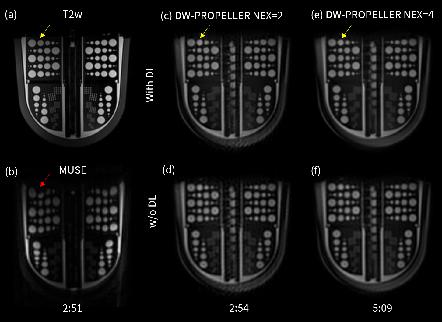
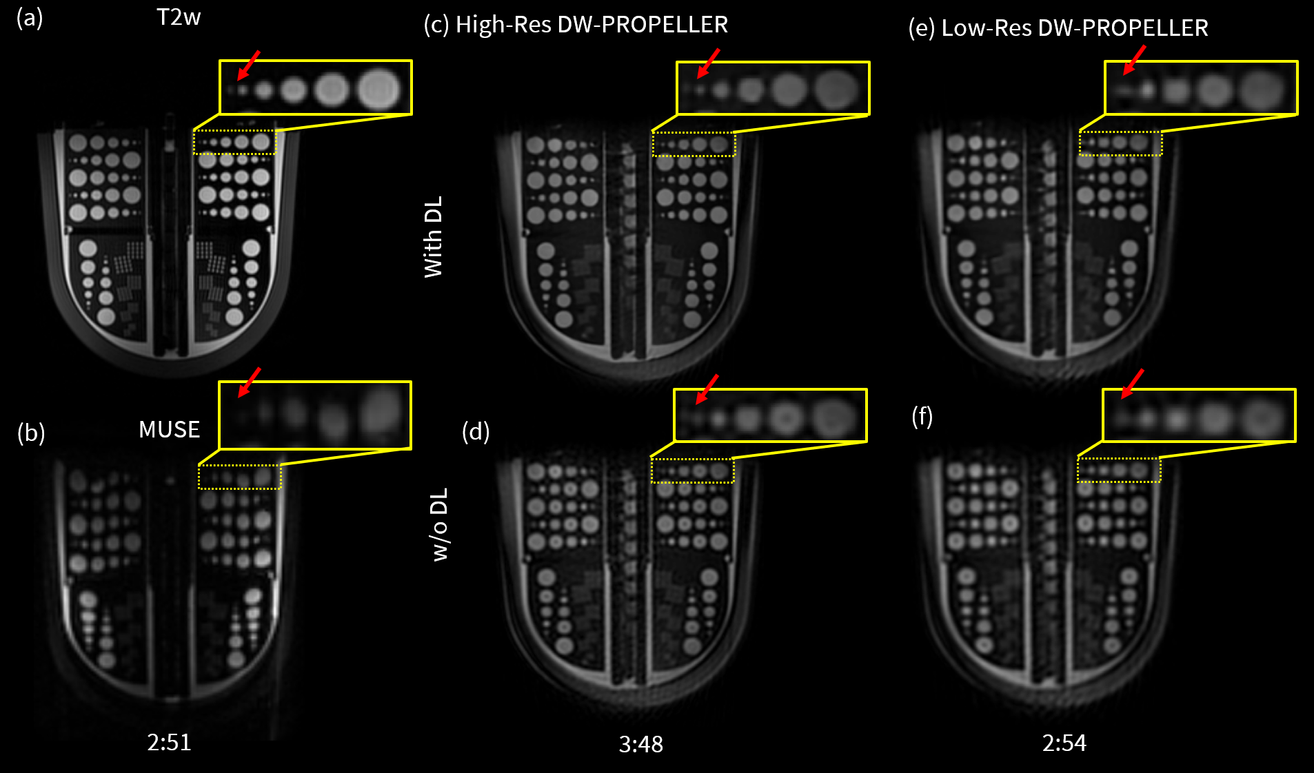
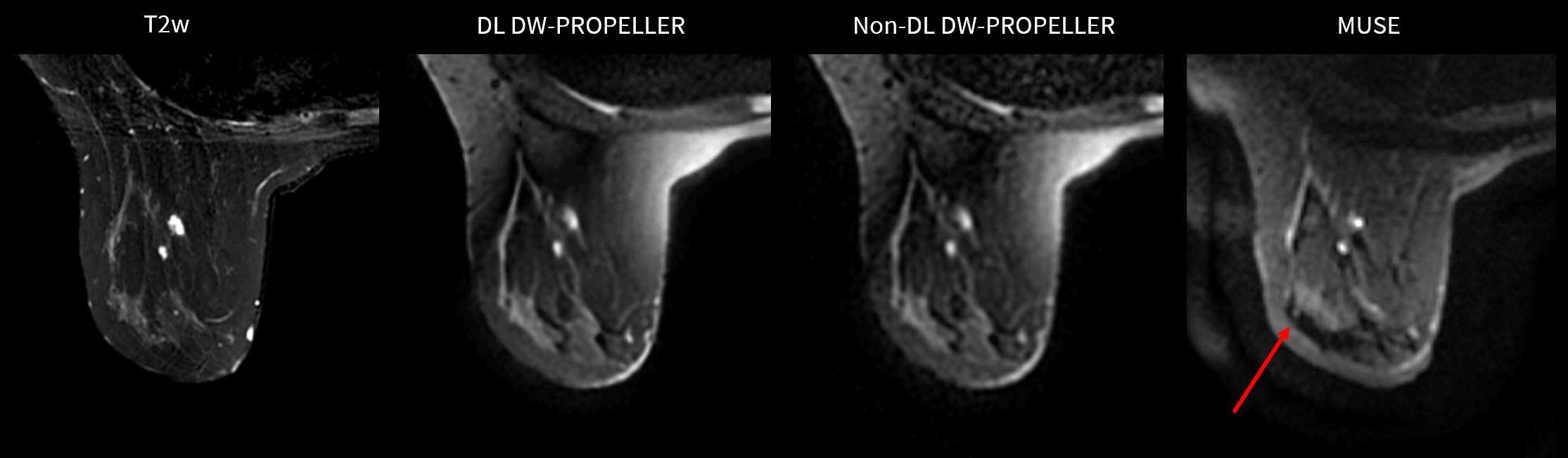
Images from phantom scanning are shown in Figure 1 comparing T2w, multi-shot DW-EPI (MUSE), and DW-PROPELLER with NEX=2 and 4 with and without DL. Compared to MUSE (Fig. 1b), DW-PROPELLER exhibits less geometric distortion. However, conventional DW-PROPELLER shows slightly lower SNR (Fig. 2d). Apparent noise is lower in the high NEX image (Fig. 2f) but at the cost of longer scan time. With DL reconstruction, DW-PROPELLER with NEX=2 (Fig. 1c) displays increased SNR and edge sharpness even compared with DW-PROPELLER with NEX=4 without DL (Fig. 1f). Figure 2 demonstrates the utilization of DL to improve clinical in-plane resolution without sacrificing too much extra scan time, where High-Res DW-PROPELLER with DL (Fig. 2c) can better distinguish 1mm and 2mm holes compared with Low-Res DW-PROPELLER (Fig. 2e-f) and shows better visibility in the 2mm holes. From each of the DWI-PROPELLER images shown in Figure 2, a corresponding line profile across the top resolution pattern is plotted in Figure 3. Note increased edge sharpness with the DL High-Res. Figure 4 shows representative DW images in a normal volunteer, with visibly increased SNR, reduced geometric distortion, reduced chemical shift artifact, and improved overall sharpness using DL in DW-PROPELLER. Note that fat saturation inhomogeneity is an issue that can be seen in all DW-PROPELLER imagesDiscussion and Conclusion
DW-PROPELLER mitigates geometric distortion and chemical shift artifacts typically seen on multi-shot DW-EPI images that may obscure important visualization of fibroglandular tissue and axillary structures. However, DW-PROPELLER with chemical fat saturation can suffer from sensitivity to B0 and B1 variations, causing fat suppression to fail. Still, this work demonstrates that a deep learning network can be trained to improve SNR, and in-plane resolution to provide motion-robust distortion-free breast DW images. This technique may pave the way for scanning more challenging clinical cases such as patients with lesions located at the chest wall and axillary region where EPI-based DW images often have signal drop-out and geometric distortion. Future work will focus on improving fat suppression and increasing slice coverage.Acknowledgements
No acknowledgement found.References
- Diffusion MRI of the breast: Current status and future directions - Iima - 2020 - Journal of Magnetic Resonance Imaging - Wiley Online Library. Accessed November 8, 2023. https://onlinelibrary.wiley.com/doi/abs/10.1002/jmri.26908
- Le Bihan D, Poupon C, Amadon A, Lethimonnier F. Artifacts and pitfalls in diffusion MRI. J Magn Reson Imaging. 2006;24(3):478-488. doi:10.1002/jmri.20683
- Chen N kuei, Guidon A, Chang HC, Song AW. A robust multi-shot scan strategy for high-resolution diffusion weighted MRI enabled by multiplexed sensitivity-encoding (MUSE). NeuroImage. 2013;72:41-47. doi:10.1016/j.neuroimage.2013.01.038
- Multishot diffusion‐weighted FSE using PROPELLER MRI - Pipe - 2002 - Magnetic Resonance in Medicine - Wiley Online Library. Accessed November 8, 2023. https://onlinelibrary.wiley.com/doi/full/10.1002/mrm.10014
- Wang X, Ersoz A, Litwiller D, Ma J, Stafford J, Bayram E. Robust Diffusion-Weighted Imaging with Deep Learning-Based DW PROPELLER Reconstruction. In: ; :3919. doi:10.58530/2022/3919
Figures