0711
Diffusion MRI of the Abdomen with Motion-robust Diffusion Encoding, Multi-shot Readout, and Optimized Slice-specific Shimming1Medical Physics, University of Wisconsin-Madison, Madison, WI, United States, 2Radiology, University of Wisconsin-Madison, Madison, WI, United States, 3GE Healthcare, Menlo Park, CA, United States, 4GE Healthcare, Boston, MA, United States, 5GE Healthcare, Waukesha, WI, United States
Synopsis
Keywords: Liver, Diffusion/other diffusion imaging techniques
Motivation: Single-shot and multi-shot M1-optimized diffusion imaging (MODI) are recent DWI methods used to mitigate motion and distortion artifacts, yet they often experience chemical shift-based fat suppression failures in the abdomen.
Goal(s): To optimize fat suppression in multi-shot MODI-DWI of the abdomen.
Approach: Slice-specific chemical shift-encoded (CSE) data-informed optimization of shims is combined with single-shot and multi-shot MODI-DWI in 7 subjects imaged at 3T.
Results: Improved fat suppression and water signal excitation were observed alongside the motion and distortion artifact reduction provided by multi-shot MODI-DWI. Unwanted fat signal was reduced through this technique in areas of interest such as the liver, spleen, and ribcage.
Impact: Motion-robust, low-distortion DWI of the abdomen, with reliable fat suppression is demonstrated by combining multi-shot EPI, M1-optimized DW waveforms, and an optimized slice-by-slice shimming approach. This combined method may enable improved detection and staging of cancer in the abdomen.
Introduction
Diffusion weighted imaging (DWI) of the abdomen has broad current and potential applications related to the detection and staging of cancer, as well as the characterization of diffuse disease. Several technical challenges hinder the clinical utilization of DWI, including its sensitivity to physiological motion, image distortions, and inadequate fat suppression. M1-optimized diffusion imaging (MODI) gradient waveforms, in combination with multi-shot EPI (MUSE) techniques1 enable motion-robust, reduced-distortion DWI2. However, this technique is still limited by bright signals from unsuppressed fat that may consequently obscure organs of interest.Chemical shift-based fat suppression often fails in the presence of B0 inhomogeneities in the abdomen3,4. Alternative methods such as T1-based fat signal nulling can be robust to B0 inhomogeneity but lead to reduced signal-to-noise efficiency5. Slice-specific shimming for DWI using advanced chemical shift-encoding (CSE)-based information has been recently proposed6 and could provide improved fat suppression and water signal strength in multi-shot MODI-DWI of the abdomen.
The purpose of this work was to combine slice-by-slice shimming with multi-shot MODI-DWI acquisitions and evaluate the ability of this approach to provide motion-robust, low-distortion DWI of the abdomen with reliable fat suppression.
Methods
Subjects: 7 healthy volunteers (4 male/3 female) under IRB approval and informed written consent.MRI Acquisition: Images were acquired at 3T (Signa Premier, GE Healthcare) using an anterior array coil (Air Coil, GE Healthcare) and a posterior embedded table coil. All DWI images were acquired with respiratory triggering. Subject data were acquired with multiple DWI gradient waveforms, including: 1) single shot Monopolar DWI, 2) single shot MODI-DWI, 3) multi-shot Monopolar DWI, 4) multi-shot MODI-DWI. MODI-DWI was acquired using an M1 ~0.63 s/mm fixed across all b-values. Two b-values (50 & 500 s/mm2) were acquired for each sequence, with number of repetitions=[1,2] and 3 diffusion directions. Monopolar-DWI was acquired with all the same parameters, but M1 was higher and varied across b-values. All DWI sequences included spatial-spectral excitation for water excitation/fat suppression. Other DWI acquisition parameters included: 6mm slice thickness; 2mm spacing; 32 slices. In addition, CSE-MRI was acquired prior to DWI to enable slice-specific shimming. CSE-MRI was acquired in a single end-expiration breath-hold, with the following parameters: 128×128 matrix size, TR=6.2ms, TE1=0.93ms, ΔTE=0.77ms, 6 echoes, and otherwise identical prescriptions as the DWI sequences. Co-localized fat-only and water-only images, B0 field maps, and R2* maps were generated from the CSE acquisition to calculate optimized shims.
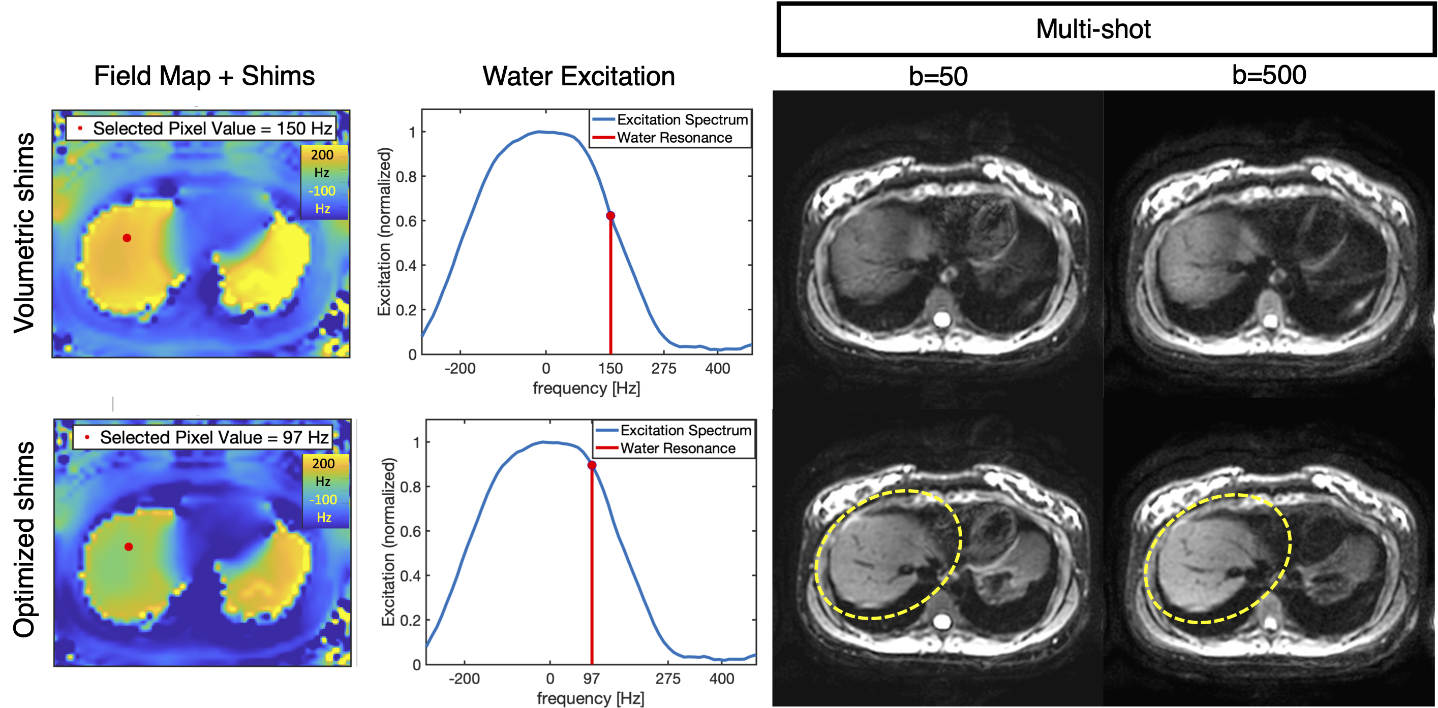
Optimization of slice-specific shim values: With slice-specific knowledge of the fat-only image, water-only image, and B0 field maps, the method predicts the water and fat excitation from the DWI spectral selectivity profile, measured empirically through phantom excitation data, over the field of view and optimizes three “shim” parameters (the X/Y shim values and excitation center frequency) at each slice to maximize the water signal excitation and minimize the fat signal excitation.
To evaluate the shimming performance, each DWI acquisition described above was obtained twice: once using conventional volumetric shimming, and once using the proposed slice-specific optimized shimming.
Results
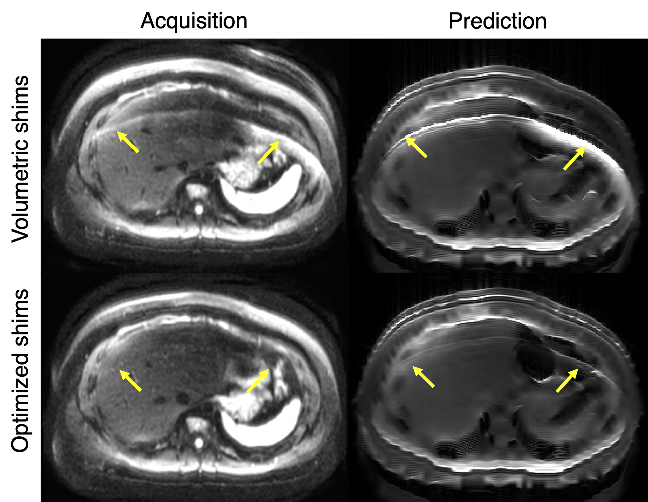
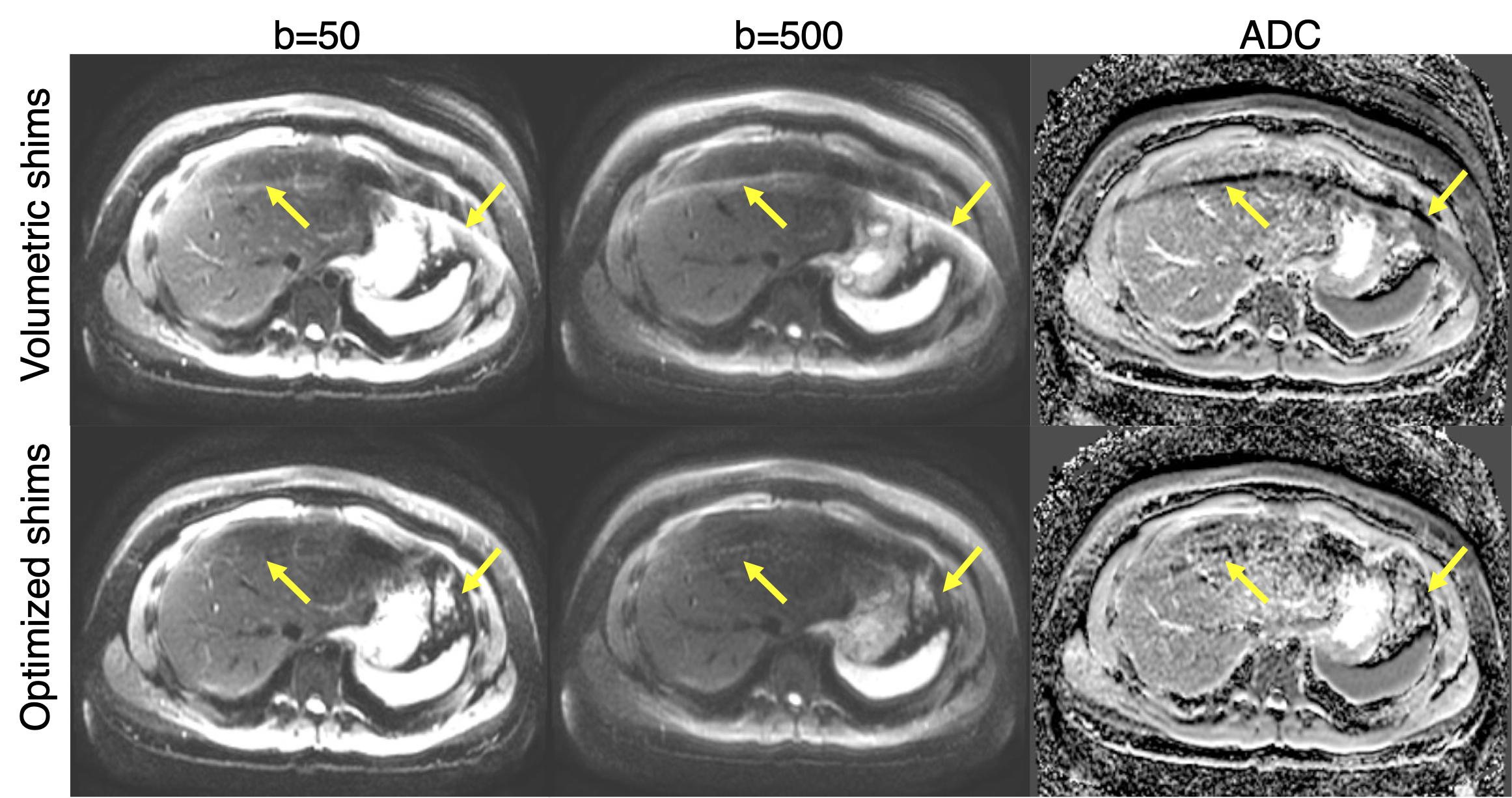
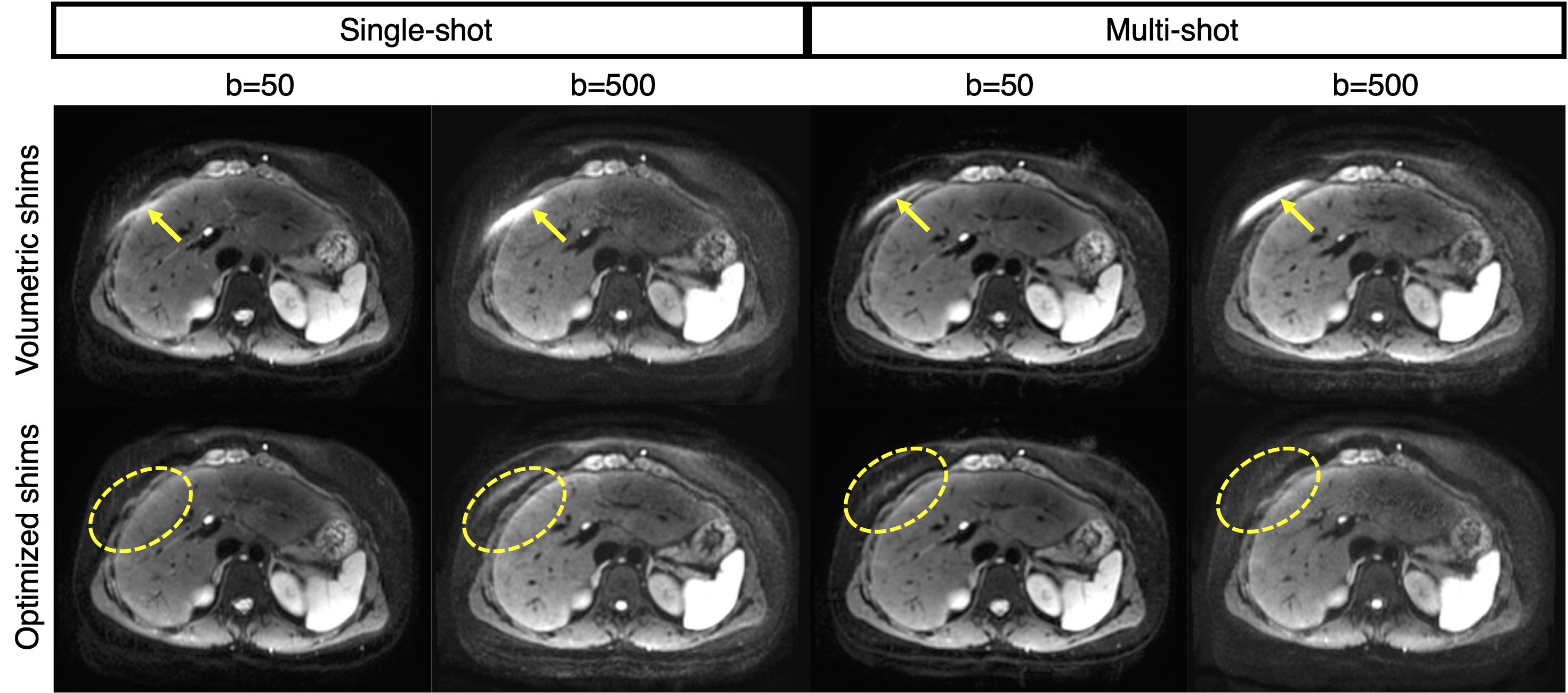
Acquired data from the 7 volunteers was successfully analyzed and contained no major artifacts. Liver images demonstrated fat suppression failures in multiple slices of volumetrically-shimmed DWI sequences per volunteer. CSE-based predictions used in shim optimization were representative of the water and fat relative signal intensities and distributions in acquired DWI data (Figure 1). CSE-based shimming exhibited decreased fat suppression failure artifact intensity when compared with conventional volumetric shimming, and ADC map quality was consequently enhanced (Figure 2). In combination with multi-shot MODI-DWI acquisitions, optimized CSE-based shimming enabled motion-robust, reduced-distortion DWI with improved fat suppression (Figure 3). CSE-based optimized shimming removed fat suppression failures that obscured spleen and ribcage signal in addition to liver (Figure 4). Near the liver dome, water signal gain was observed for both b-value images in CSE-based optimized shim acquisitions (Figure 5).Discussion
This work has demonstrated the potential to leverage CSE-based slice-specific optimized shimming, in combination with motion-robust (MODI) diffusion waveforms and multi-shot readouts to produce abdominal DWI with high image quality and reduced distortions.This technique may enable robust fat suppression in DWI, without the SNR efficiency penalty of T1-based fat nulling methods. This is particularly important in motion-robust, multi-shot DWI due to the SNR losses introduced by MODI (longer TE) and multi-shot readouts (reduced sampling efficiency with multiple shots).
A limitation of this work is that it relied on a low number of healthy volunteers. Additional studies in patients are needed as future work.
In conclusion, multi-shot MODI-DWI benefits from slice-specific dynamic shimming optimized using CSE data to maximize water excitation and fat signal suppression.
Acknowledgements
The authors acknowledge support from the NIH (R01-EB030497), the University of Wisconsin-Madison Office of the Vice Chancellor for Research and Graduate Education with funding from the Wisconsin Alumni Research Foundation, as well as from the UW Departments of Radiology and Medical Physics. GE Healthcare also provides research support to the University of Wisconsin.References
1. Chen NK, Guidon A, Chang HC, Song AW. A robust multi-shot scan strategy for high-resolution diffusion weighted MRI enabled by multiplexed sensitivity-encoding (MUSE). Neuroimage. 2013;72:41-47. doi:10.1016/j.neuroimage.2013.01.038
2. Geng R, Zhang Y, Rice J, et al. Motion-robust, blood-suppressed, reduced-distortion diffusion MRI of the liver. Magn Reson Med. 2023;89(3):908-921. doi:10.1002/mrm.29531
3. Roberts, N. T., Hinshaw, L. A., Colgan, T. J., Ii, T., Hernando, D., & Reeder, S. B. (2021). B0 and B1 inhomogeneities in the liver at 1.5 T and 3.0 T. Magnetic Resonance in Medicine, 85(4), 2212–2220. https://doi.org/10.1002/mrm.28549
4. Grande, F. Del, Santini, F., Herzka, D. A., Aro, M. R., Dean, C. W., Gold, G. E., & Carrino, J. A. (2014). Fat-suppression techniques for 3-T MR imaging of the musculoskeletal system. Radiographics, 34(1), 217–233. https://doi.org/10.1148/rg.341135130
5. Haase, A., Frahm, J., Hanicke, W., & Matthaei, D. (1985). 1H NMR chemical shift selective (CHESS) imaging. Physics in Medicine and Biology, 30(4), 341–344. https://doi.org/10.1088/0031-9155/30/4/008
6. Tollefson A, Wu G, Lan P, Guidon A, Van Der Heijden RA, Tamada D, Pirasteh A, Hernando D. Slice-by-slice Dynamic Shimming Based on a Chemical Shift-Encoded Acquisition to Improve Fat Suppression in DWI. Proc. Intl. Soc Mag. Reson. Med. 33 (2023) Abstract 3409.
Figures