0705
Assessment of rhabdomyolysis-induced acute kidney injury with chemical exchange saturation transfer MRI1School of Biomedical Engineering, Southern Medical University, Guangzhou, China, 2Guangdong Provincial Key Laboratory of Medical Image Processing & Guangdong Province Engineering Laboratory for Medical Imaging and Diagnostic Technology, Southern Medical University, Guangzhou, China, 3Department of Rehabilitation Medicine, Zhujiang Hospital, Southern Medical University, Guangzhou, China, 4Medical Imaging Center, Nanfang Hospital, Southern Medical University, Guangzhou, China, 5Institute of Translational Physiology, Charite-Universitatsmedizin Berlin, Berlin, Germany, 6Philips Healthcare, Guangzhou, China, 7Philips Healthcare, Shenzhen, China, 8Berlin Ultrahigh Field Facility (B.U.F.F.), Max Delbruck Center for Molecular Medicine in the Helmholtz Association, Berlin, Germany
Synopsis
Keywords: Small Animals, CEST & MT
Motivation: There is a gap in exploring the progression of acute kidney injury (AKI) caused by rhabdomyolysis (RM).
Goal(s): Investigate the feasibility of CEST MRI for assessing the progression of RM-induced AKI in a mouse model.
Approach: The RM-AKI model was established by intramuscular injection of glycerol solution and examined longitudinally by CEST (days 1, 3, 7, 15, 30). CEST quantification parameters and their diagnostic performance were compared.
Results: MTR presented a significant difference, and showed the best diagnostic performance for AKI and moderate negative correlations with pathological changes.
Impact: MTR has potential clinical utility for assessing renal diseases.
Introduction
Rhabdomyolysis(RM)-induced acute kidney injury (AKI), is a clinical syndrome of rapid decline in renal function caused by RM, which leads to a mortality rate of up to 37%(1). The assessment of RM-induced AKI remains challenging because clinical diagnostic indicators, such as BUN and SCr, have low sensitivity as they only detect reductions in glomerular filtration rate(2). CEST is sensitive to the concentration and the exchange rate of endogenous metabolites, and their environment(3). Previous studies have shown that CEST is sensitive to pathophysiological changes during ischemia-reperfusion and in an unilateral ureteral obstruction model(4,5). In this study, we investigated the feasibility of CEST-MRI for the assessment of renal injury and the progression of RM-induced AKI.Methods
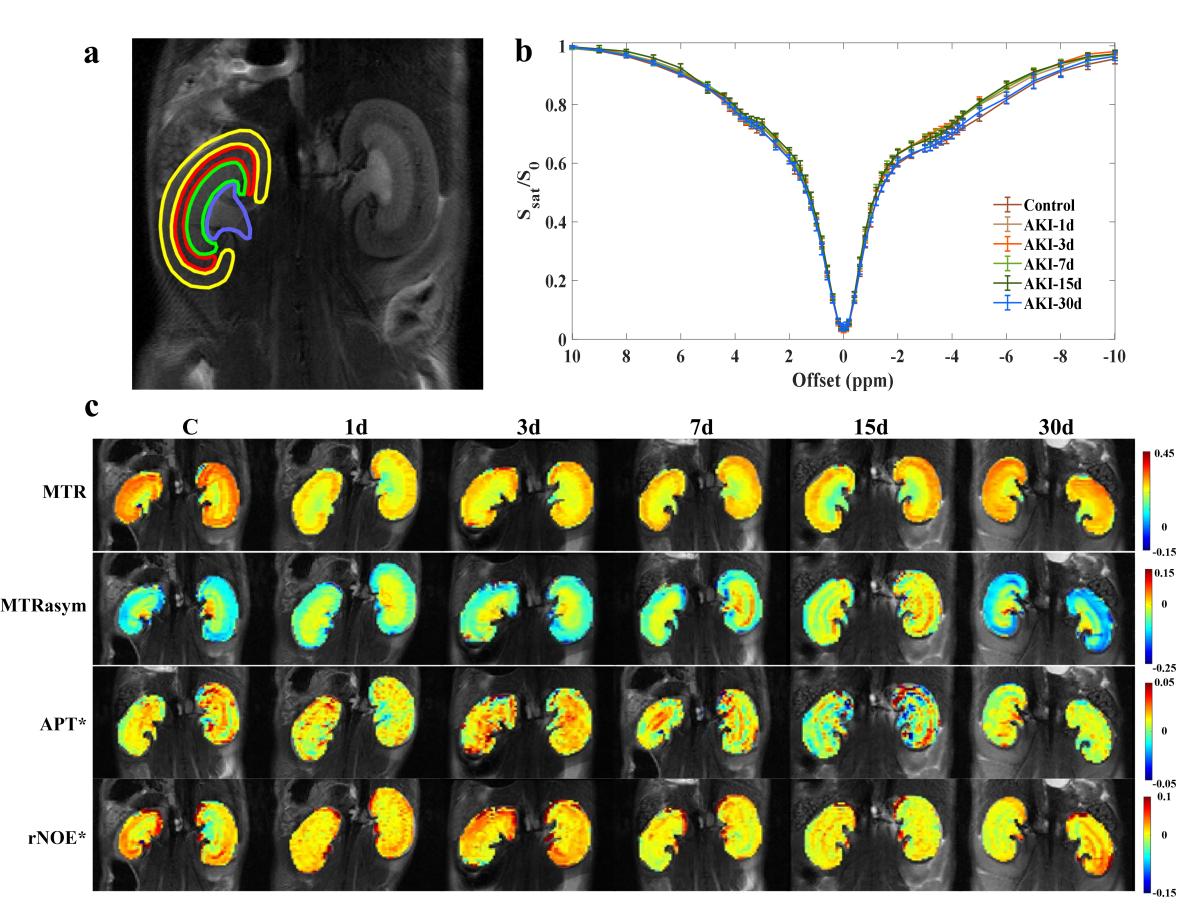
All C57BL/6J mice (male, 8-10 weeks) were randomly assigned to either an AKI group or a healthy control group. The AKI group received a bilateral intramuscular injection of 50% glycerol solution (7.5 mL/kg body weight, n=30). The control group was administered the same volume of 0.9% normal saline (n=10). Mice were longitudinally imaged on days 1, 3, 7, 15, and 30 after injection. CEST-MRI was performed on a 7.0-T animal MRI scanner (Bruker BioSpec, Ettlingen, Germany) with a 40 mm volume coil for RF transmission and a 4-channel phased surface coil for reception. For each scan, data was collected using RARE imaging with the RF power of 1.5 μT and the duration of 3 second for CEST preparation. Saturation images were obtained at 53 frequency offsets, which were unevenly distributed between -10 ppm to 10 ppm and mainly concentrated around ± 3.6 ppm. CEST data was quantified using the MTR(3.6ppm), MTRasym(3.6ppm), APT*, and rNOE*. Student’s t-test was used to compare the difference between control and AKI groups. Pearson correlation coefficients were obtained for comparisons between the CEST parameters, physiologic characteristics, blood parameters, and fibrosis score. ROC curve analysis was used to compare the diagnostic performance.Results
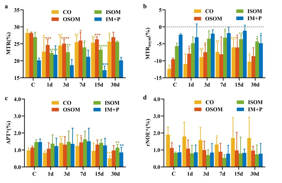
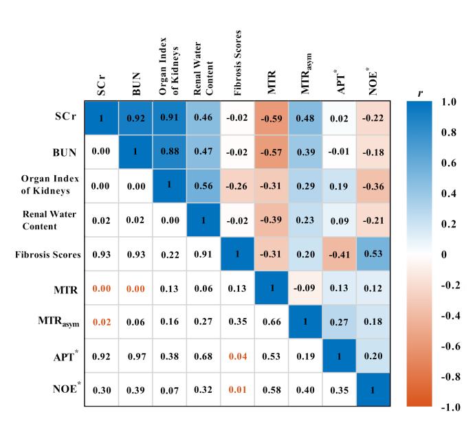
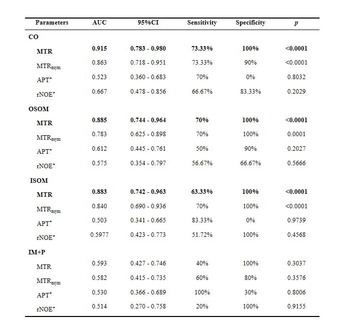
Substantial signal reductions were observed at frequency offsets of 3.6 and -3.6 ppm, as shown in Fig. 1b. The CEST parameter maps obtained for the control group nicely display the renal structure. For the RM-induced AKI group this structure got diminished with the progression of the disease.(Fig. 1c). Following induction, the MTR in cortex, OSOM, and ISOM decreased significantly, peaked on day 1, and then steadily recovered. With AKI progression, the MTRasym increased significantly and reached a maximum on day 7 in ISOM, and day 15 in cortex and OSOM. The APT* and rNOE* images did not exhibit overt changes. According to the Pearson correlation, the MTR had the strongest correlation with pathological changes (especially in BUN and SCr) for RM-induced AKI. The ROC curve analysis showed the MTR exhibited the best diagnostic performance in cortex (AUC = 0.915), OSOM (AUC = 0.885), and ISOM (AUC = 0.883).Discussion
The signal at 3.6 ppm was generally attributed to APT effect, while the -3.6 ppm signal was linked to the rNOE effect. Myoglobin is released in significant quantities as a result of RM, which indirectly induces blood flow and reduces the rNOE signal. The increase in the water content generated by AKI inflammation leads to APT signal reduction. When RM induces renal damage, tubular epithelial cells undergo apoptosis, which lowers the both APT and rNOE signal. The APT signal is also affected by the pH alterations in glycerol-induced AKI mice. The appearance, distribution, and reversibility of this AKI model are similar to the clinical RM phenomenon, particularly the lesions seen in patients experiencing acute renal failure owing to crush injury. Therefore, we inferred that endogenous CEST would be helpful for the assessment of comparable renal pathologies as caused by allogeneic transfusion and crush syndrome in humans.Conclusion
Longitudinally CEST-MRI in a mouse model of RM-induced AKI revealed that the MTR metric is sensitive to pathophysiological changes associated with RM-induced renal injury in the acute stage and its gradual recovery. The APT and rNOE effects at ±3.6 ppm are related to pathological changes in RM-induced AKI. The MTR value obtained from CEST-MRI can potentially be used to assess renal injury in RM.Acknowledgements
This work was supported by the National Natural Science Foundation of China (No. U21A6005), KeyArea Research and Development Program of Guangdong Province (Nos. 2018B030340001 and 2018B030333001), and Guangdong Basic and Applied Basic Research Foundation (Nos. 2020A1515110577 and 2019A1515111182).References
1. Simpson JP, Taylor A, Sudhan N, et al. Rhabdomyolysis and acute kidney injury: creatine kinase as a prognostic marker and validation of the McMahon Score in a 10-year cohort: A retrospective observational evaluation. Eur J Anaesthesiol 2016;33:906-12.
2. Singh R, Dodkins J, Doyle JF, et al. Acute Kidney Injury Biomarkers: What Do They Tell Us? Contrib Nephrol 2018;193:21-34.
3. van Zijl PC, Yadav NN. Chemical exchange saturation transfer (CEST): what is in a name and what isn't? Magn Reson Med 2011;65:927-48.
4. Irrera P, Consolino L, Cutrin JC, et al. Dual assessment of kidney perfusion and pH by exploiting a dynamic CEST-MRI approach in an acute kidney ischemia-reperfusion injury murine model. NMR Biomed 2020;33:e4287.
5. Stabinska J, Singh A, Haney NM, et al. Noninvasive assessment of renal dynamics and pH in a unilateral ureter obstruction model using DCE MR-CEST urography. Magn Reson Med 2023;89:343-55.
Figures