0703
Imaging of thorax and diaphragm movement in mechanically ventilated mice and rats1Radiology and Nuclear Medicine, Amsterdam UMC, Amsterdam, Netherlands, 2Intensive Care, Erasmus MC, Rotterdam, Netherlands, 3Physiology, Amsterdam UMC, Amsterdam, Netherlands, 4Biomedical Engineering & Physics, Amsterdam UMC, Amsterdam, Netherlands, 5Intensive Care, Radboudumc, Nijmegen, Netherlands, 6MR Solutions, Guildford, United Kingdom, 7BioMedical Engineering and Imaging Institute, Icahn School of Medicine at Mount Sinai, New York, NY, United States
Synopsis
Keywords: Biology, Models, Methods, Preclinical, Diaphragm, Small Animals, Thorax, 3D CINE imaging
Motivation: The pathophysiology of diaphragm dysfunction in mechanically ventilated patients is not fully understood and adequate animal models are required to accommodate further research.
Goal(s): Our goal was to develop a method to image 3D thoracic movement during mechanical ventilation of mice and rats at different pressure levels of mechanical ventilation.
Approach: With our setup we visualized the movement of the thorax in mice and rats using self-gated 3D pseudo-radial k-space sampling.
Results: Imaging was feasible in both animal types and increase of pressure resulted in a decrease of mean diaphragm excursion of 0.9 and 1.3 mm in mouse and rat respectively.
Impact: Our proposed setup allows controlled mechanical ventilation and MR imaging of 3D thorax movement in mice and rats. This can be used to study the pathophysiology behind mechanical ventilation-induced respiratory muscle dysfunction, and ultimately guiding clinical practice in respiratory care.
Introduction
Mechanical ventilation is the cornerstone of treatment in the intensive care unit. Unfortunately, it also results in diaphragm dysfunction in 80% of the patients, which is associated with increased morbidity and mortality1,2. The pathophysiology behind diaphragm dysfunction is not fully understood, but ventilator settings play a crucial role3. One important setting is positive end-expiratory pressure (PEEP): the pressure that remains in the lungs after expiration, to keep the lungs open for proper oxygenation. In a rat model of mechanical ventilation, high PEEP decreased the number of sarcomeres in series in the diaphragm, by moving the diaphragm caudally and thereby shortening the muscle4. This places the diaphragm at an unfavorable force-length relationship3. We have replicated these experiments using a mouse mechanical ventilation model. Interestingly, the outcomes differed from those seen in the rat model, with PEEP leading to an increased number of sarcomeres in series.We hypothesize that the difference in results between rats and mice is caused by differences in thoracic movement. To study this, we aimed to develop MRI protocols to measure both mechanically ventilated rats and mice using a 3D self-gated pseudoradial gradient echo sequence. We show the feasibility of our new MR protocol by demonstrating 3D CINE imaging of the thorax at different PEEP levels.
Methods
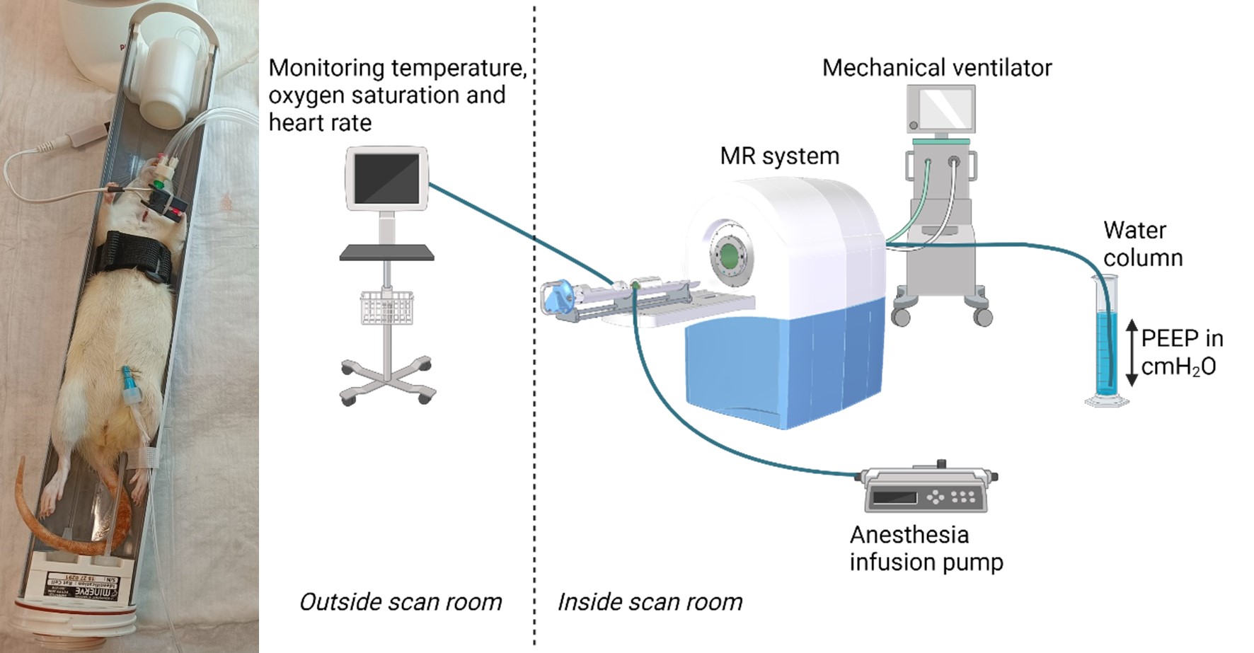
Experimental setupAn overview of the setup is provided in Figure 1. Rats (Wistar) and mice (C57BL/6) were anesthetized with a mix of ketamine, atropine and dexmedetomidine. Next, a tracheostomy was performed to enable mechanical ventilation of the animal. Anesthesia was provided by peritoneal infusion of a mix of ketamine and dexmedetomidine (rats) or ketamine, dexmedetomidine and atropine (mice). Animals were placed in a 7T MR system (MR Solutions, Guildford, UK) while ventilated with an MR-compatible ventilator (CWE MRI-1). PEEP was applied by submerging the expiratory tube in a water column outside the magnet. Imaging was repeated at three PEEP levels: 0, 4 and 8 cmH2O, while other ventilation parameters remained constant.
Sequence and image reconstruction
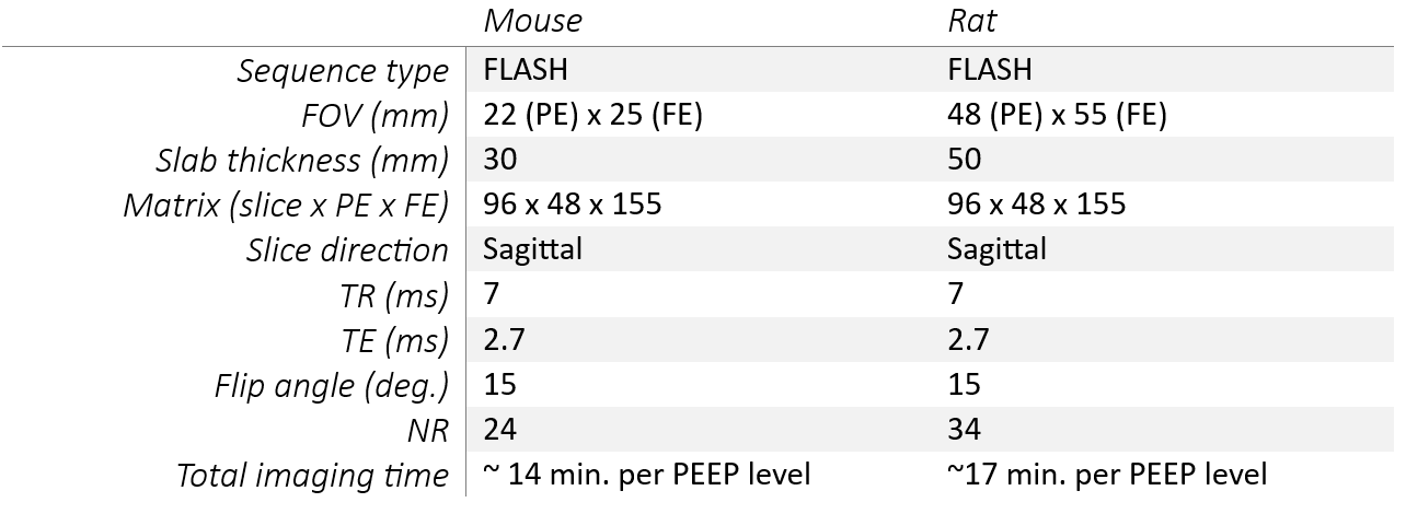
3D CINE imaging of the thorax was performed using a 3D gradient echo sequence with pseudoradial k-space filling and additional recording of a navigator signal resulting from the slice selection rewinder gradient every TR (Table 1). Retrospective binning and reconstruction of the data in 10 respiratory time frames was performed using in-house developed software5, including compressed sensing using BART6. Additionally, esophageal pressure measurements were performed to measure the compliance of the thorax in both animal models.
Analysis
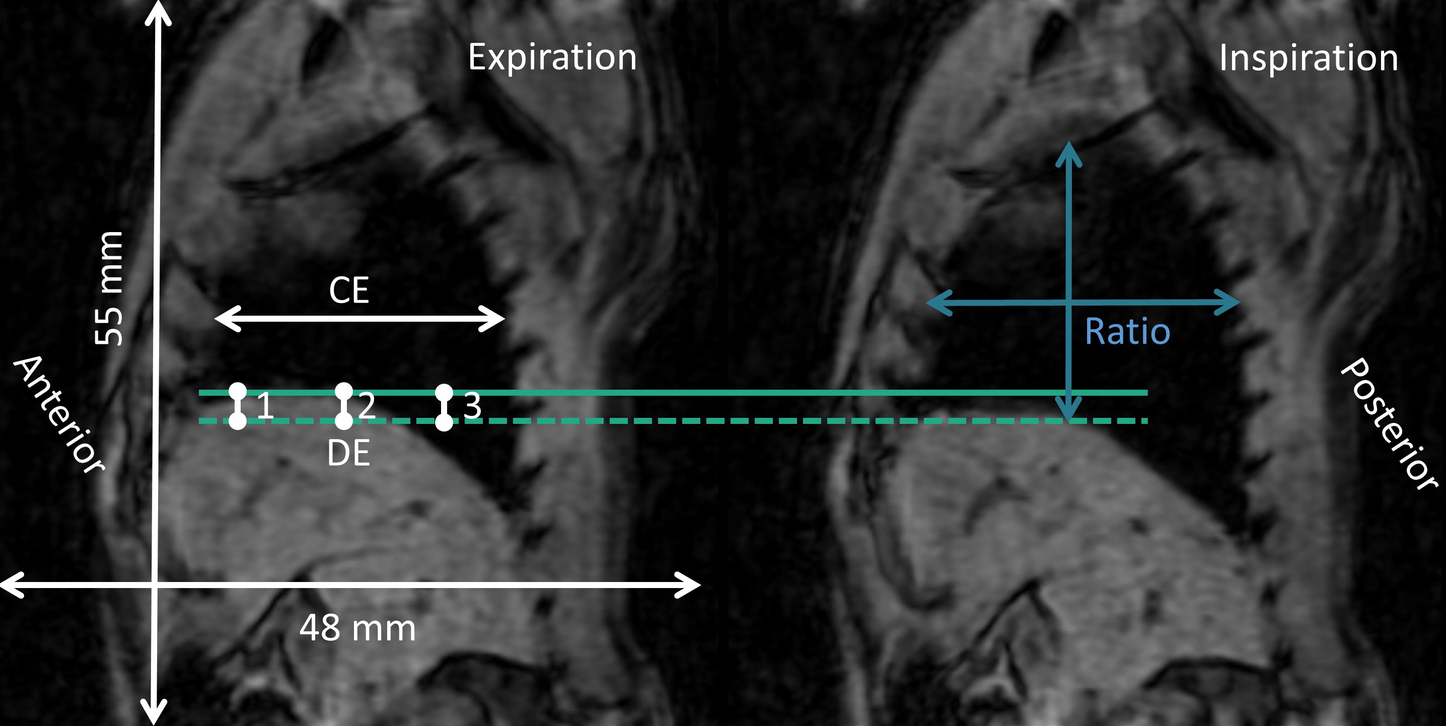
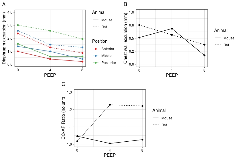
Diaphragm excursion (DE) was quantified in Matlab (R2022b) at all PEEP levels at three different locations and chest wall excursion (CE) was measured halfway the lung (Figure 2). Secondly, the ratio between craniocaudal and anterior-posterior distance (CC-AP ratio) at expiration was determined for every PEEP level (Figure 2).
Results
Examples of 3D CINE imaging are provided in Figure 3. Image quality is sufficient to visualize thoracic movement in both animal types. DE was larger in rats compared to mice and decreased with increasing PEEP for both animal types (Figure 4). CE was similar for both animal types and generally decreased with increasing PEEP levels (Figure 4). In mice, CE first increased and then decreased at 8 cmH2O PEEP. The CC-AP ratio increased with increasing PEEP levels for rats, but remained constant in mice (Figure 4).Discussion
Our study shows that 3D imaging with retrospective gating allows to measure thorax movement in two animal models during mechanical ventilation. An advantage of our setup is that retrospective reconstruction of the thorax movement is facilitated by the set respiratory frequency. These techniques enable us to answer our research question, but also have potential to be used in upcoming research involving cardiac or thoracic imaging in pre-clinical animal research.Preliminary results indicated that change in DE and CE with increasing PEEP level was similar in both animal models, while the CC-AP ratio was larger in the rat at higher PEEP levels, indicating elongation of the lungs and thorax in the rat. This may indicate that mainly the shape of the thorax is different in rats compared to mice. These results may partly explain histological differences between rat and mouse diaphragm.
Future research will focus on 3D image analysis to study spatial differences in thorax movement. This also allows for investigating the change in diaphragm curvature, which is crucial for diaphragm function and remodeling7,8. Lastly, we will artificially decrease chest wall compliance by placing an elastic restrictive band around the thorax to assess the effects of chest wall compliance on diaphragm excursion, because diseases and age of patients can affect chest wall compliance.
Acknowledgements
This work was supported by a ZonMW grant (nr. 09120011910004).References
- Dres M, Goligher EC, Heunks LMA, Brochard LJ. Critical illness-associated diaphragm weakness. Intensive Care Medicine. 2017;43(10):1441-52
- Heunks L, Ottenheijm C. Diaphragm-Protective Mechanical Ventilation to Improve Outcomes in ICU Patients? Am J Respir Crit Care Med. 2018;197(2):150-2.
- Jansen D, Jonkman AH, Vries HJ, Wennen M, Elshof J, Hoofs MA, et al. Positive end-expiratory pressure affects geometry and function of the human diaphragm. J Appl Physiol (1985). 2021;131(4):1328-39.
- Lindqvist J, van den Berg M, van der Pijl R, Hooijman PE, Beishuizen A, Elshof J, et al. Positive End-Expiratory Pressure Ventilation Induces Longitudinal Atrophy in Diaphragm Fibers. American Journal of Respiratory and Critical Care Medicine. 2018;198(4):472-85.
- Daal MRR, Strijkers GJ, Calcagno C, Garipov RR, Wust RCI, Hautemann D, Coolen BF. Quantification of Mouse Heart Left Ventricular Function, Myocardial Strain, and Hemodynamic Forces by Cardiovascular Magnetic Resonance Imaging. J Vis Exp. 2021(171).
- Uecker MT, Jon; Ong, Frank; Holme, Christian; Lustig, Michael. BART: version 0.4.01. Zenodo; 2017.
- Boriek AM, Black B, Hubmayr R, Wilson TA. Length and curvature of the dog diaphragm. J Appl Physiol (1985). 2006;101(3):794-8.
- Greybeck BJ, Wettergreen M, Hubmayr RD, Boriek AM. Diaphragm curvature modulates the relationship between muscle shortening and volume displacement. Am J Physiol Regul Integr Comp Physiol. 2011;301(1):R76-82.
Figures