0701
MRI with cancer cell membrane coated Mn (II) nanoprobes in precise diagnosis of early pancreatic cancer1Affiliated hospital of Jiangnan University, Wuxi, China, 2GE Healthcare, MR Research, Beijing, China
Synopsis
Keywords: Probes & Targets, Tumor
Motivation: gadolinium widely used in MRI clincally show low detecting efficiency for early pancreatic cancer(<2 cm). It is thus urgent to develop safe and effective contrast agents to address this issue.
Goal(s): This study aims to investigate the potential of cancer cell membrane coated Mn(II) nanoprobes (PMNP@CMs) in MRI for precise diagnosis of early pancreatic cancer.
Approach: PMNP@CMs were prepared for MRI of pancreatic cancer to explore the imaging effects in vivo and in vitro compared with gadolinium.
Results: PMNP@CMs showed more predominant MRI effect than gadolinium and may be considered as safe and efficient contrast agents to realize diagnosis for early pancreatic cancer.
Impact: This work has developed a nano magnetic resonance imaging contrast agent (PMNP@CMs) that is more advantageous than gadolinium and can achieve precise diagnosis of early pancreatic cancer.
Introduction
Pancreatic cancer is a challenging issue in the field of cancer medicine due to its high malignancy, late detection, and treatment complexity1, 2. Timely detection in the early stage of pancreatic cancer can significantly improve the survival rate of patients. However, gadolinium widely used in MRI clinically have low specificity and detection rates for early pancreatic cancer (<2 cm) identification, and also exert nephrotoxicity3. In the present study, manganese ions with high paramagnetic moment were chelated with polydopamine nanoparticles to form a nanocontrast agent (Mn2+@polydopamine nanoparticles, PMNPs) for MRI. To improve its targeting and selectivity for pancreatic cancer, the PMNPs were wrapped with engineered pancreatic cancer cell membranes, resulting in cancer cell membrane-coated PMNPs (PMNP@CMs). We thus aimed to investigate the potential of PMNP@CMs in MRI for precise diagnosis of early pancreatic cancer.Materials and methods
Preparation of PMNP@CMs: PMNPs were prepared by the mixture of H2O, ethanol, NH3·H2O and MnCl2. To coat CM vesicles onto the surface of PMNPs, PMNPs solution was mixed with CM vesicles and the mixture was sonicated for 5 min. Cell and animal studies: For MR imaging, pancreatic cancer cells (PANC-01, and PATU8988), normal pancreatic ductal epithelial cells (hTERT-HPNE), and liver cancer cells (HepG2) were used in this study. All animal experiments were in accord with Institutional Animal Care and Use Committe. Mice involved in the experiment were Male BALB/c nude mice. MRI imaging: MRI experiments in vitro and in vivo were performed using a 3.0 T MR scanner (Architect, GE Healthcare Systems) with 12- or 32-channel phased array coil and 4-channel high-resolution animal coil. The following imaging sequences with scan parameters were applied: TR (repetition time)/TE (echo time) of 520/14 ms, section thickness of 1 mm, field of view (FOV) of 60×60 mm2, and matrix of 256×256 for fast-spin-echo (FSE) based T1-weighted imaging; TR/TE of 500/10 ms, section thickness of 3 mm, FOV of 200×200 mm2, and matrix of 256×256 for synthetic MRI of magnetic resonance image compilation (MAGiC) sequence; TR/TE of 2000/82 ms, section thickness of 1 mm, FOV of 90×90 mm2, and matrix of 256×256 for the T1 fs Cube sequence. Data processing: Quantitative T1 maps were obtained from vendor-provided MAGiC post-processing software. Through 3D MIP post-processing software, 3D MR images were obtained including coronal and sagittal reconstructions. All MR images required delineation of region of interest (ROI). ROI was drawn as the largest rectangle within the target region. Data analysis was performed by GraphPad Prism 8.0 and SPSS statistics 26.0 software.Results
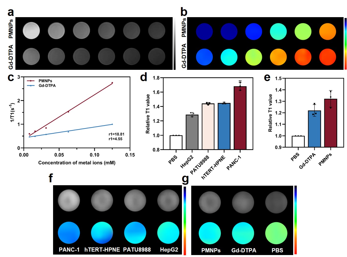
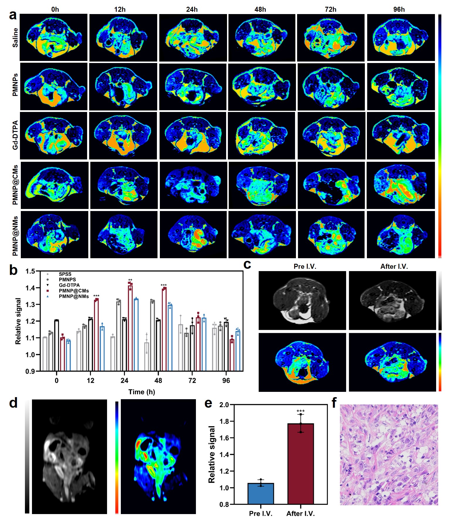
The experimental results showed that the T1-weighted effect of PMNPs was remarkable (Fig. 1a,b). Compared with gadolinium (Gd-DTPA), the R1 value of PMNPs was 4.1 times higher than that of Gd-DTPA (Fig. 1c). This indicates that the PMNPs can serve as a highly sensitive T1 contrast agent. After wrapped with cancer cell membranes, PMNP@CMs showed excellent T1-weighted effect with a relative T1 value of 1.7 in cell experiments, significantly higher than PMNPs and Gd-DTPA groups (p<0.01)(Fig. 1d, f). PMNP@CMs exhibited more prominent imaging in pancreatic cancer cells compared to other tumor cells and normal pancreatic ductal epithelial cells (p<0.01)(Fig. 1e, g). Also, PMNP@CMs possessed good hemocompatibility and could be stably stored in several types of aqueous solutions for at least 7 days.Furthermore, a xenograft mouse model of early pancreatic cancer was established to evaluate the potential of PMNP@CMs in vivo. The results showed that the nanoparticles could highly accumulated in the tumor and reached the peak after 24 h of intravenous administration (Fig.2a). Meanwhile, the tumor signal ratio of PMNP@CMs was significantly higher than other contrast agent groups (p<0.01) and improved by 17% as compared with that of Gd-DTPA (Fig.2b). After the injection of PMNP@CMs, the MR images demonstrated clear visualization of the ultra-small xenograft carcinoma (<1 mm) in axial and coronal images (Fig.2c, d) and the quantitative data showed significant difference (p<0.001)(Fig.2e). H&E staining showed the presence of heterogeneous tumor cells, which confirmed the occurrence of pancreatic cancer (Fig.2f). Moreover, PMNP@CMs did not affect the body weight, major organs, and biochemical indexes of mice during the 7-day test period.
Discussion and conclusions
PMNP@CMs showed better T1-weighted effect in MRI of pancreatic cancer in vitro and in vivo compared with gadolinium. The finding suggested that high sensitivity of PMNP@CMs allowed for identifying microscopic pancreatic cancer, thus realizing early and precise diagnosis for pancreatic cancer. It also demonstrated good stability and biosafety. To sum up, this study provides a new possibility for detecting pancreatic cancer at an early stage, suggesting that PMNP@CMs may be used as a safe and efficient contrast agent to address these issues.Acknowledgements
Funding: This project was supported by Jiangsu provincial commission of health and family planning (Grants No. H2019087).References
1. Ryan DP, Hong TS, Bardeesy N. Pancreatic adenocarcinoma. N Engl J Med. 2014; 371(11): 1039-1049.
2. Siegel RL, Miller KD, Fuchs HE, et al. Cancer statistics, 2021. CA Cancer J Clin. 2021; 71(1): 7-33.
3. Yi Z, Luo Z, Barth ND, et al. In vivo tumor visualization through MRI off-on switching of NaGdF4 -CaCO3 nanoconjugates. Adv Mater. 2019; 31(37): e1901851.
Figures