0699
Biophysical analysis of hepatocellular carcinoma and tumor niche in an orthotopic mouse model with diffusion MRI and MR elastography1Radiology, Charité – Universitätsmedizin Berlin, Berlin, Germany, 2Department of Veterinary Medicine, Institute of Animal Welfare, Animal Behavior and Laboratory Animal Science, Freie Universität Berlin, Berlin, Germany, 3iPATH.Berlin, Charité – Universitätsmedizin Berlin, Berlin, Germany, 4Institute of Medical Informatics, Charité – Universitätsmedizin Berlin, Berlin, Germany
Synopsis
Keywords: Preclinical Image Analysis, Elastography, Cancer, HCC
Motivation: The biomechanical interplay between hepatocellular carcinoma (HCC) and the hosting liver is poorly understood.
Goal(s): To characterize the development of HCC and its interactions with the surrounding liver using imaging-based biophysical properties.
Approach: We investigated longitudinally HCC and the host liver in an orthotopic mouse model using MR elastography (MRE) and diffusion-weighted imaging (DWI).
Results: During tumor development, the host liver became softer with reduced viscosity and restricted water diffusivity while HCC became stiffer, less viscous and restricted water diffusivity.
Impact: Preclinical MRE is a useful tool to study biomechanical properties of tumors and the tumor environment. In a mouse model of hepatocellular carcinoma, we showed for the first time how liver tumors shape their biomechanical niche in the hosting liver.
Introduction
Hepatocellular carcinoma (HCC) is the sixth most common cancer worldwide and the third leading cause of cancer-related deaths1,2. Our understanding of the complexity of HCC has progressed over the years with a current focus on the intricate interplay between the tumor and the tumor hosting liver. These interactions yield important changes in tissue composition and structure, which could be non-invasively examined with multiparametric imaging (mp-MRI) and multifrequency MR elastography (MRE). MRE quantifies the mechanical properties of soft tissues in vivo, such as shear wave speed (SWS in m/s) surrogating stiffness, and penetration rate (PR in m/s) representing inverse viscosity3. Although MRE has demonstrated its potential in the characterization of tumors in different organs including the liver4-8, only a few studies have taken the host liver into account by investigating hepatic biomechanical changes during cancer progression9. Therefore, we used a syngeneic orthotopic HCC mouse model10 to investigate the changes in the biomechanical properties and apparent diffusion coefficient (ADC) in tumor and host liver by MRE and mp-MRI.Methods
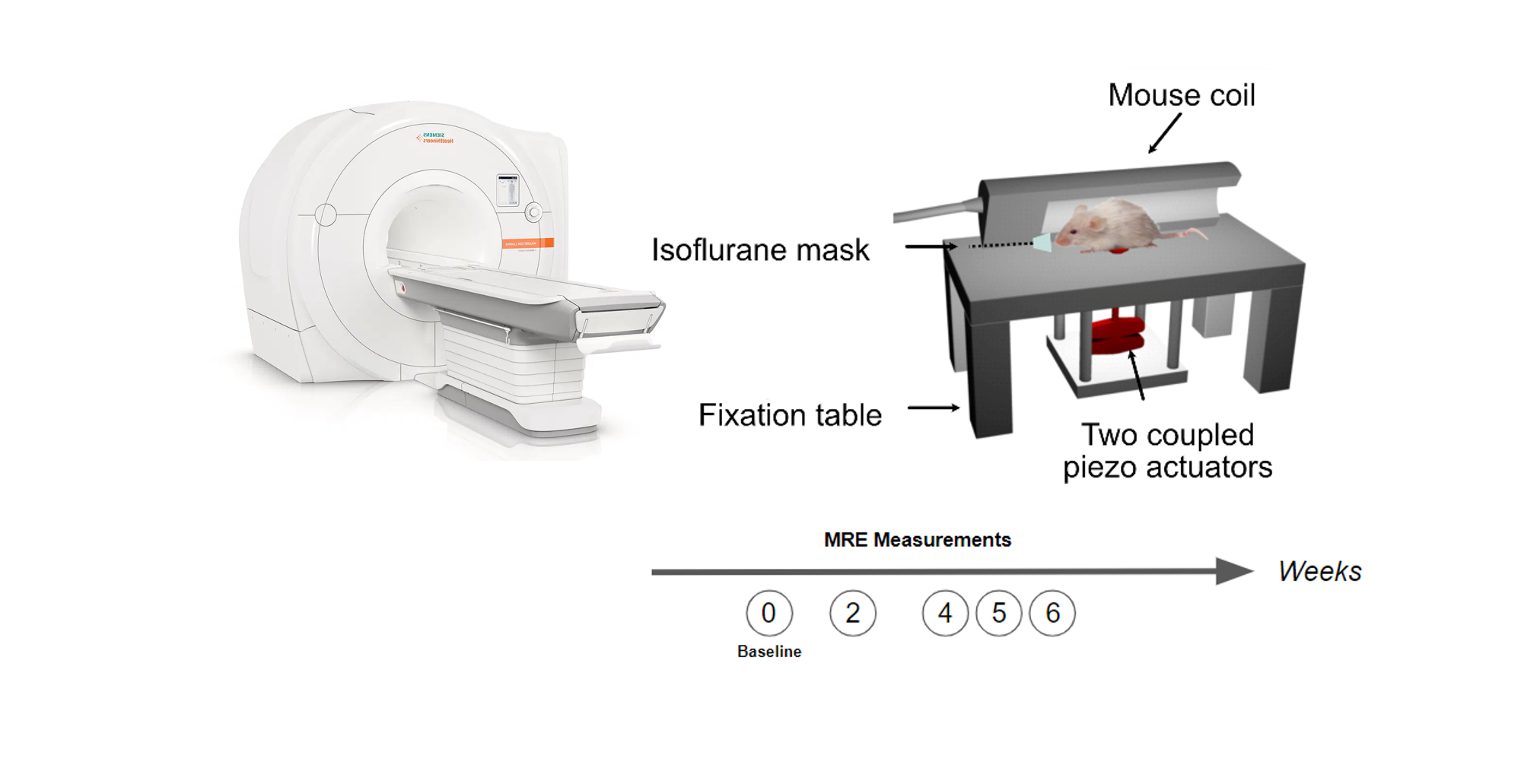
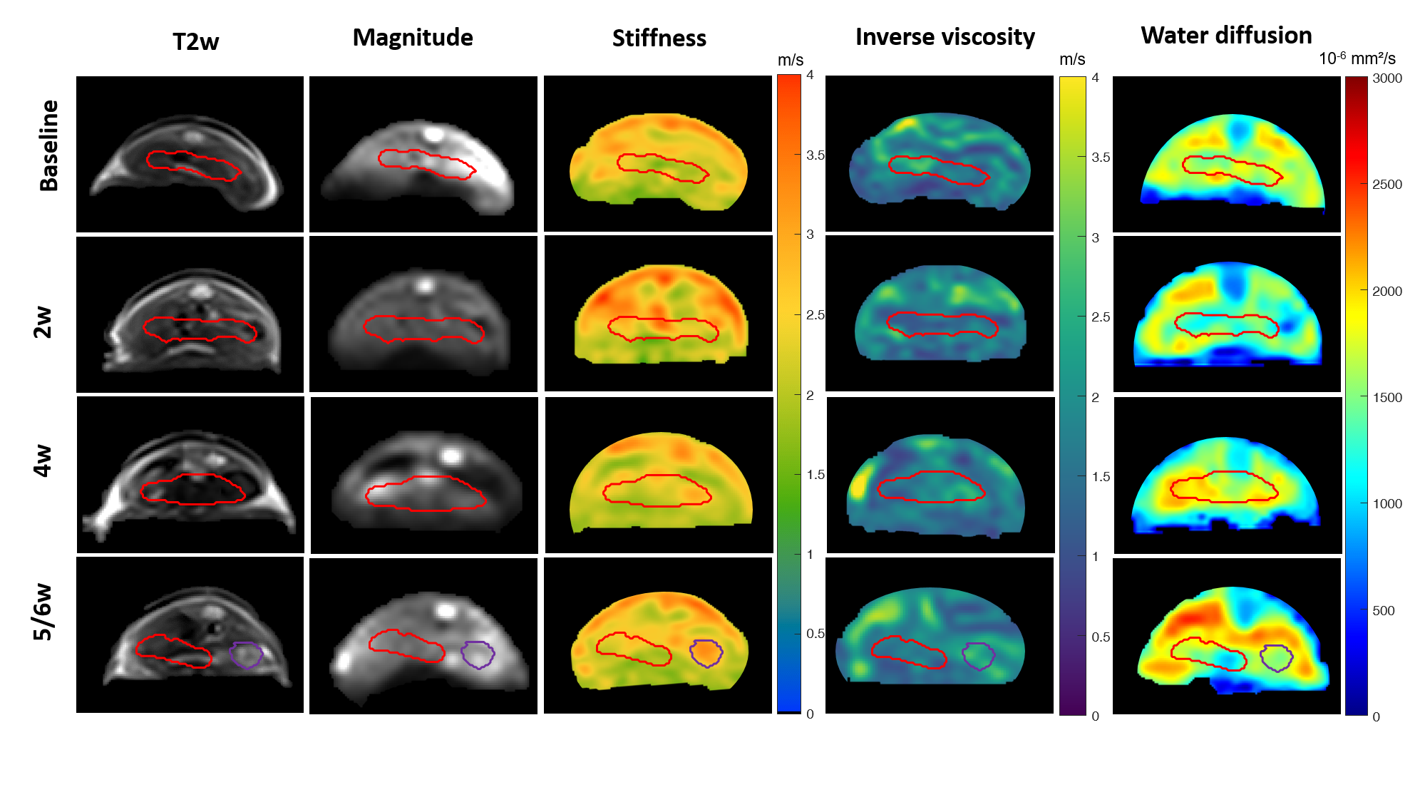
Fourteen adult female BALB/c mice underwent surgical implantation of mouse HCC cells (BNL 1ME A.7R.1 - TIB-75)10,11 directly into the liver. Imaging scans were conducted one week before the implantation (baseline), then at two (2w), four (4w), five- or six-weeks (5/6w) post-surgery. Measurements were conducted in a 3T clinical MRI scanner (Magnetom Lumina, Siemens, Germany). Twenty-one axial T2w images with a resolution of 0.25x0.25x1.2 mm³ (TR=2500 ms, TE=77 ms) were acquired. Multifrequency MRE was performed using a single-shot spin-echo echo-planar sequence with eight wave dynamics. Twenty-one 1.2-mm-thick slices with a resolution of 1.0 x 1.0 mm were acquired. Shear waves of 300, 400 and 500 Hz were induced in the liver using a custom-made setup with two-coupled piezo-actuators (Figure 1). Eleven axial DWI slices (resolution: 0.8 x 0.8 x 2.0 mm³) were acquired using four b-values (0/50/400/800 s/mm²). MRE data were processed using wave-number based inversion algorithm (k-MDEV) which provided maps of SWS and PR3 (Figure 2). MRE magnitude and DWI images were interpolated using rigid registration to T2w images as reference. For both MRE and DWI, regions of interest (ROIs) were manually delineated using ITK-SNAP12. Histopathology was conducted for both liver and tumor.Results
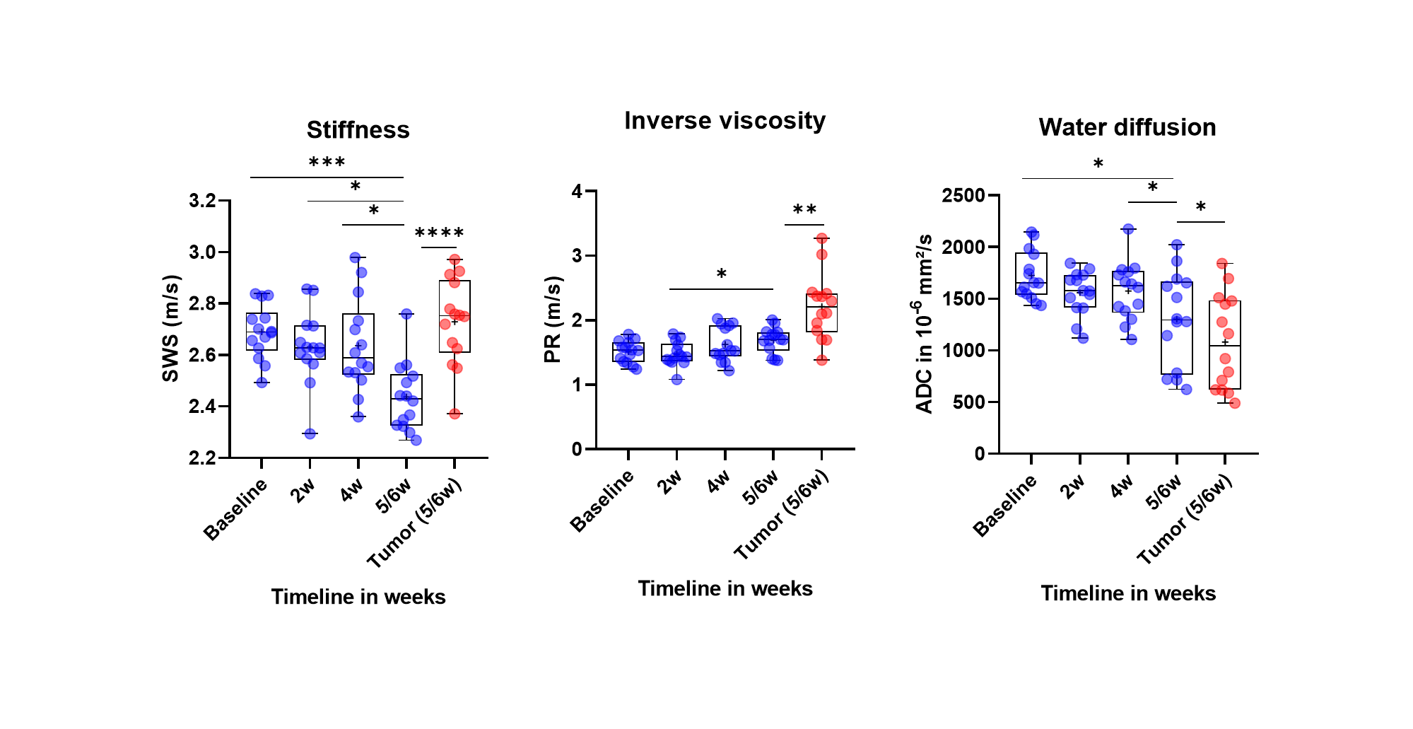
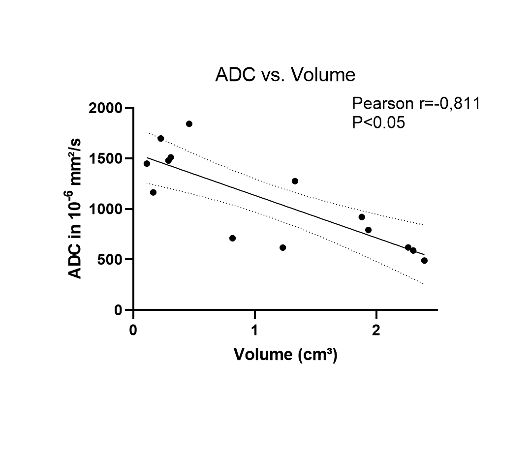
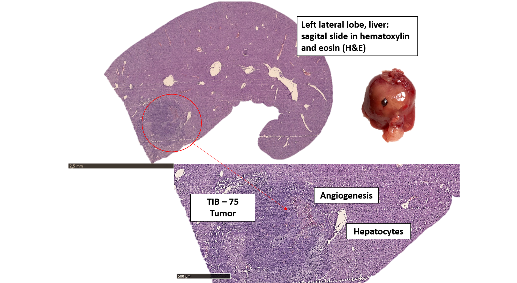
At 5/6w, tumors were clearly visible in all 14 mice. The average tumor volume was 1.2±0.9 cm3. A significant decrease in liver SWS was observed over time (baseline: 2.7±0.1 m/s, 2w: 2.6±0.14 m/s, 4w:2.6±0.2 m/s, 5/6w 2.5±0.13 m/s, p<0.001.), as seen in Figure 3. In addition, a significant increase in PR was observed (baseline:1.5±0.17 m/s; 2w:1.5±0.19 m/s, 4w:1.6±0.3 m/s, 5/6w:1.7±0.2 m/s, p<0.05). ADC decreased during HCC progression (baseline:1725±237 µm²/s, 2w:1559±212 µm²/s, 4w:1576±278 µm²/s 5/6w:1302±456 µm²/s, p<0.05). Compared to the host liver at 5/6w, all tumors appeared stiffer (SWS_tumor: 2.8±0.2 m/s vs. SWS_liver: 2.5±0.2 m/s, p<0.01) and less viscous (PR_tumor: 2.2±0.5 m/s vs. PR_liver 1.7±0.2 m/s, p<0.01). ADC was also lower in all tumors compared to their surrounding livers (ADC_tumor:1083±460 µm²/s vs. ADC_liver:1302±456 µm²/s, p<0.05). There was a negative correlation between tumor volume and tumor ADC (Pearson r =-0.81, p<0.01) (Figure 4). Histology analysis showed tumor growth with angiogenesis as shown in Figure 5 for one animal at 5/6w.Discussion
This study represents the first in-vivo investigation of the biophysical properties of HCC and the HCC-bearing liver in an orthotopic mouse model. During tumor development, the host liver became softer with reduced viscosity and restricted water diffusivity. We suspected that the implantation of the tumor cells triggered the trauma responses of the liver, resulting in structural adaptation, likely involving inflammatory cell infiltration, extracellular matrix (ECM) modulation, hepatocyte reorganization, and necrosis formation13. It should be noted that in this study, HCC cells were implanted into healthy livers, which contain significantly less collagen than fibrotic livers where HCCs typically develop14. As a result, there may be more structural changes in the liver, leading to alterations in hepatic biophysical properties. In 5/6 w, the tumors exhibited higher stiffness, reduced viscous properties, and hindered water diffusivity than the host livers. This could be attributed to increased cellularity within the tumor, transforming HCC into a more elastic and solid mass as compared to the surrounding liver, consistent with prior studies15. The negative correlation detected between tumor volume and water diffusivity also reflected growth-related increase of cellularity, as previously reported in literature16.Conclusion
Our study presents compelling evidence that in vivo biophysical properties are sensitive to structural alterations in the liver during HCC progression. The biophysical characteristics of both the tumors and their host livers provide insight into their interactions, which require validation through further histopathological and biochemical analyses.Acknowledgements
The authors acknowledge support from the German Research Foundation (DFG)-SFB1340 Matrix in Vision and GRK2260, as well as the Central Biobank Charité (ZeBanC) for their role in digitizing histological slides.References
1. Ferlay J, et al. Global Cancer Observatory: Cancer Today. Lyon, France: International Agency for Research on Cancer; 2020. Available from: https://gco.iarc.fr/today. [Accessed 2023-11-07].
2. McGlynn, K.A., et al. Epidemiology of Hepatocellular Carcinoma. Hepatology. 2021; 73(1): p 4-13.
3. Tzschatzsch H, et al. Tomoelastography by multifrequency wave number recovery from time-harmonic propagating shear waves. Med Image Anal. 2016;30: p. 1-10.
4. Dittmann, F., et al. Tomoelastography of the abdomen: Tissue mechanical properties of the liver, spleen, kidney, and pancreas from single MR elastography scans at different hydration states. Magn Reson Med, 2017; 78(3): p. 976-983.
5. Guo, Jing; et al. MR Elastography in Cancer. Investigative Radiology.2023; 58(8): p 578-586.
6. Venkatesh SK, et al. MR elastography of liver tumors: preliminary results. AJR Am J Roentgenol. 2008; 190(6):1534-40.
7. Guo, J., et al. Mechanical characterization of a mouse GL261 glioma model using MR elastography. Proceedings of the 23rd Annual Meeting of ISMRM (ed. I.P.o.t.s.A.M.of ISMRM), Toronto, 2015: p. 1101.
8. Hennedige TP, et al. Comparison of magnetic resonance elastography and diffusion-weighted imaging for differentiating benign and malignant liver lesions. Eur Radiol. 2016;26(2): p. 398-406.
9. Riegler J, et al. Tumor elastography and its association with collagen and the tumor microenvironment. Clin Cancer Res. 2018;24(18): p. 4455-4467.
10. Das DK, et al. A "patient-like" orthotopic syngeneic mouse model of hepatocellular carcinoma metastasis. J Vis Exp. 2015;(105): p. e52858.
11. Chang CJ, et al. Combined GM-CSF and IL-12 gene therapy synergistically suppresses the growth of orthotopic liver tumors. Hepatology. 2007;45(3): p. 746-754
12. Yushkevich, P.A., et al. User-guided 3D active contour segmentation of anatomical structures: significantly improved efficiency and reliability. Neuroimage, 2006; 31(3): p. 1116-28.
13. Sas Z, et al. Tumor Microenvironment of Hepatocellular Carcinoma: Challenges and Opportunities for New Treatment Options. Int J Mol Sci. 2022;23(7): p. 3778
14. Llovet JM, et al. Hepatocellular carcinoma. Nat Rev Dis Primers. 2021; 7(1): p. 6
15. Shahryari, M., et al. Tomoelastography Distinguishes Noninvasively between Benign and Malignant Liver Lesions. Cancer Res, 2019; 79(22): p. 5704-5710.
16. Shankar S, et al. Role of Diffusion Weighted Imaging (DWI) for Hepatocellular Carcinoma (HCC) Detection and its Grading on 3T MRI: A Prospective Study. J Clin Exp Hepatol. 2016;6(4): p. 303-310
Figures