0698
NaGdF4-Based Magnetic Resonance Nanoprobes for Qualitative Inflammation Imaging in Glioma: Hot or Cold?1Department of Radiology, Shanghai Fourth People's Hospital, School of Medicine, Tongji University, Shanghai, China, 2Department of Orthopaedics, Shanghai Key Laboratory for Prevention and Treatment of Bone and Joint Diseases, Ruijin Hospital, Shanghai Jiao Tong University School of Medicine, Shanghai, China
Synopsis
Keywords: Probes & Targets, Molecular Imaging
Motivation: It is in urgent need to develop an imaging method to reveal the intrinsic ‘cold’ or ‘hot’ status of tumor microenvironment for glioma patients, which would offer guidance for planning therapeutic regimen, and thus maximize the therapeutic efficacy and reduce unnecessary treatment.
Goal(s): To visualize the inflammatory status of glioma tumor microenvironment non-invasively using myeloperoxidase responsive NaGdF4-based nanoprobes under MRI.
Approach: Different glioma models with different inflammatory status were created and imaged with our nanoprobes under 11.7 T at T1WI (n = 6 each group).
Results: MPO-enriched ‘hot’ gliomas showed patchy hypointense T1 signal while MPO-rare ‘cold’ gliomas presented moderate hyperintensity in T1WI.
Impact: Depending on the level of MPO in tumor microenvironment, nanoprobes will get self-assembled in various degree, thus altering T1 relaxation time. By using this, it is promising to monitor the tumor inflammatory status for glioma patients thus guide clinical treatment.
Introduction
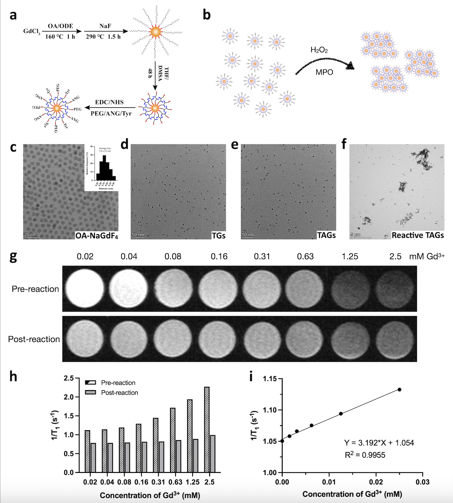
Immunotherapy for glioblastoma (GBM) has been a research focus area since the concept that central nervous system is immune privileged has been challenged. However, the outcomes of GBM immunotherapy in clinical trials have been disappointing1. Tumors that exert pro-inflammatory response to immunotherapy are referred ‘hot’, whereas tumors that remain immunologically quiescent are referred ‘cold’2. Multiple studies suggest that GBMs are mostly cold tumors3, 4. Hence, immunotherapy for GBM aims to convert ‘cold’ tumors into ‘hot’ and thus reverse local immunosuppressive tumor microenvironment (TME)5. Not all GBM patients benefit from ongoing immunotherapies and MRI is commonly used to evaluate the therapeutic efficacy of GBM patients.The clinical routine MRI cannot truly reflect the responsiveness of immunotherapies for GBM patients in short time follow-ups. Thus, it is in urgent need to develop a novel imaging method to reveal the intrinsic ‘cold’ or ‘hot’ status of TME for GBM patients, which would offer guidance for planning the therapeutic regimen, and thus maximize the therapeutic efficacy and reduce unnecessary treatment.The ‘cold’ or ‘hot’ status of GBM is related to the anti- or pro-inflammatory process and tumor infiltrating lymphocytes in the TME. In GBM, tumor-associated macrophages (TAMs) account for up to 30-50% of all infiltrating immune cells6, 7. It is reported that M1-like TAM is presented in large amount in ‘hot’ TME, which indicates better tumor outcomes8. Myeloperoxidase (MPO), an oxidant-producing enzyme that catalyzes H2O2 to form hypochlorous acid and other reactive oxygen species, is mainly secreted in host defense by M1-like TAMs9-11. In this study, by utilizing the different secreting level of MPO between ‘cold’ and ‘hot’ tumor, we aim to non-invasively visualize the inflammatory status in GBM TME using MRI based MPO responsive nanoprobes. NaGdF4 nanoparticles (NPs) is a well-developed T1 contrast agent with high biocompatibility12. By modifying the surface of NaGdF4 NPs with tyramine (Tyr) containing phenolic hydroxyl, the obtained NPs may be oxidized in the presence of MPO and H2O2, which will be self-assembled responsively, thus altering the T1 relaxation time and present altered image contrast in T1WI13. Angiopep-2 (ANG) has been used to target glioma14. Therefore, we hypothesize that the tyramine and ANG modified NaGdF4 NPs, which is named as TAGs in this study, can non-invasively and real-timely reflect the ‘cold’ or ‘hot’ status of GBM by MRI.
Methods
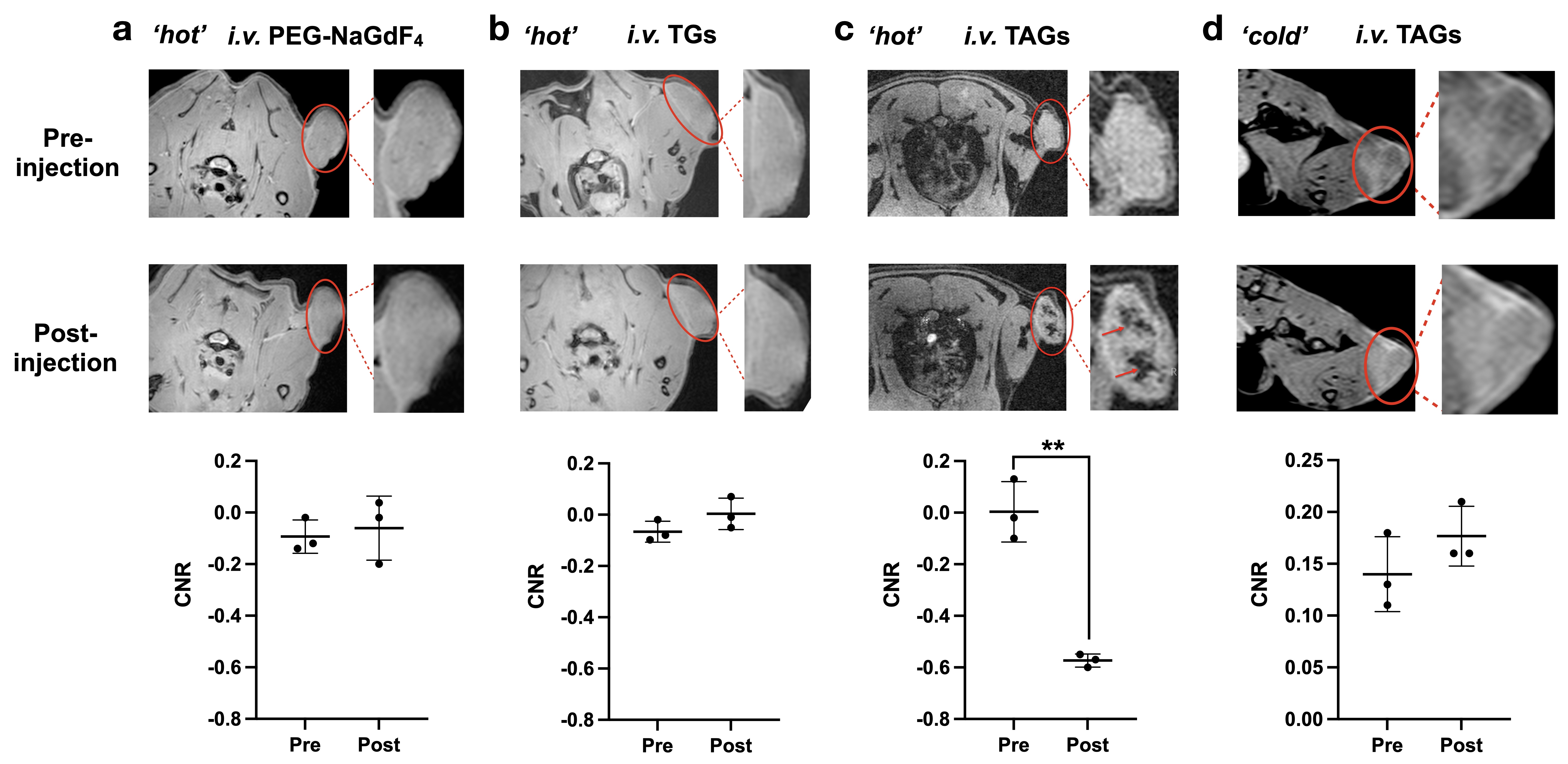
MRI based Tyr-ANG-NaGdF4 nanoprobes, named as TAGs, with surface modification of ANG to target glioma and phenolic hydroxyl to sense MPO were developed, aiming to noninvasively visualize the ‘hot’ or ‘cold’ TME inflammatory status of GBM based on MRI. In vitro MPO responding ability was conducted by incubating the desired solution of different Gd3+ concentrations with an excess of H2O2 and MPO. Transmission electron microscopy (TEM) and MRI were performed before and after the MPO catalytic reaction. C57BL/6 mice and SD rats with subcutaneously transplanted glioma model using GL261 cells and C6 cells, respectively, were created. In vivo MR imaging was performed on the two models after tail vein injection of TAGs at a concentration of 10 mg Gd/kg under 11.7 T at T1WI (n = 3 each group). After imaging, tumors were harvested for flow cytometric analysis, immunohistochemistry staining, MPO activity assay and bio-TEM.Results
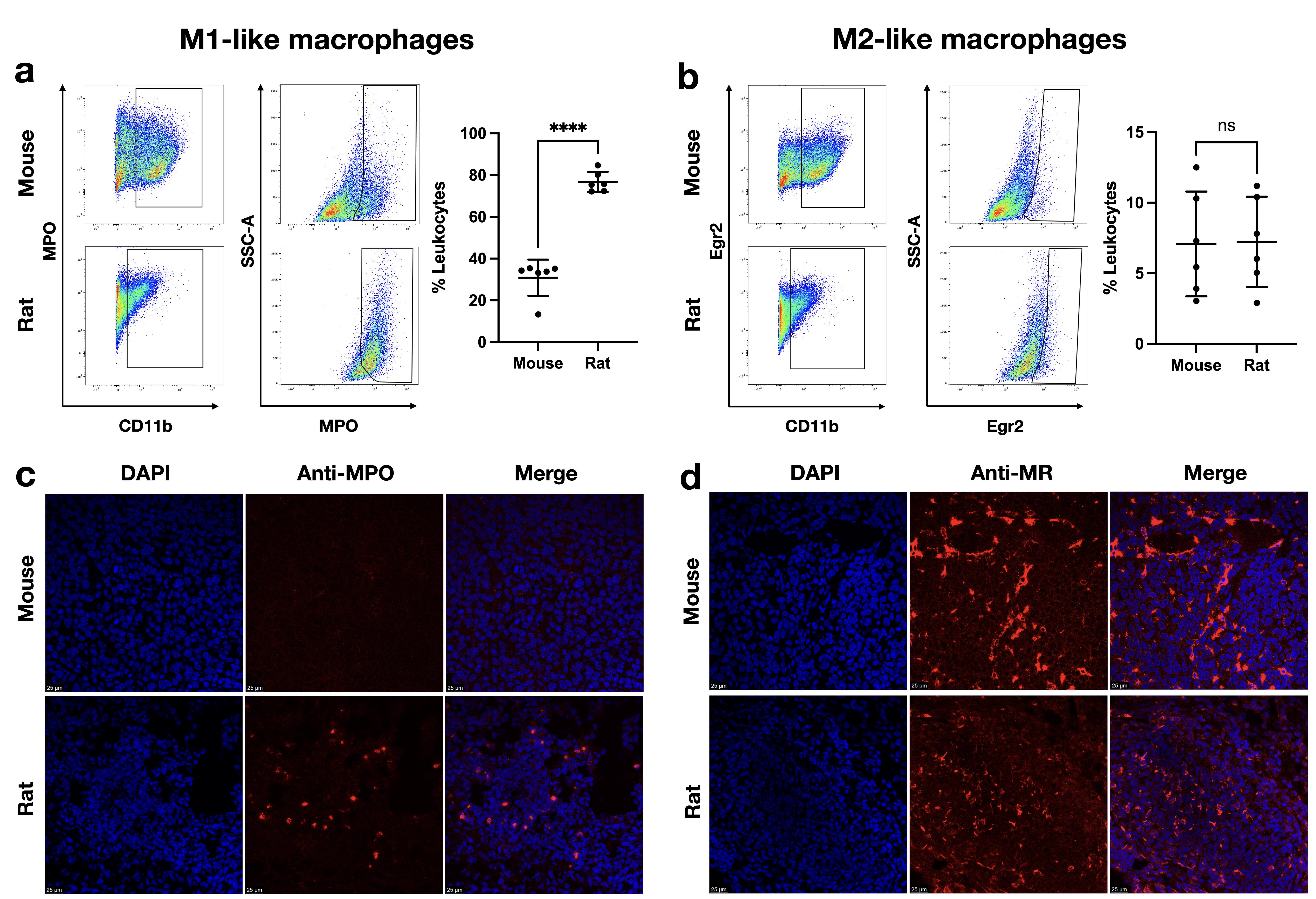
Cellular and in vivo toxicity confirm that TAG nanoprobes are of desirable high biocompatibility. After incubating with H2O2 and MPO, TAG nanoprobes show oligomerization under TEM with T1 relaxation time lengthened at 11.7 T. C6 SD rats with subcutaneous glioma show patchy darkened T1 signal within the tumor after injection of TAGs, confirmed by following biological analysis with high MPO level and abundant M1-like macrophages, suggesting MPO-enriched ‘hot’ TME inflammatory status. GL261 mouse models show moderate hyperintensity in T1-weighted MRI, which confirmed with low MPO level and few M1-like macrophages, signifying the MPO-rare ‘cold’ TME within the tumor. PEG-NaGdF4 nanoparticles without MPO targeting show faint T1 enhancement for both the C6 SD rat and GL261 mouse models.Conclusion
The synthesized TAGs can qualitatively reflect and differentiate the inflammatory status of GBM non-invasively by sensing MPO in TME using MRI. Depending on the level of MPO in the TME, the TAG nanoprobes will get self-assembled in various degree, thus altering the T1 relaxation time and present altered image contrast in T1WI. It was found that ‘hot’ GBMs showed patchy hypointense T1 signal while ‘cold’ GBMs only showed moderate enhanced T1 signal. By using the MPO responsive nanoprobes, it is promising to monitor the TME inflammatory status for glioma patients thus guide clinical treatment especially during immunotherapy.Acknowledgements
This work was supported by the National Natural Science Foundation of China (82102114, 82272061), and the Foundation of National Facility for Translational Medicine (Shanghai) (TMSF-2021-4-003).References
1. R. Medikonda, G. Dunn, M. Rahman, P. Fecci, M. Lim, A review of glioblastoma immunotherapy, J. Neurooncol. 151 (2021) 41-53.
2. M. Lim, Y. Xia, C. Bettegowda, M. Weller, Current state of immunotherapy for glioblastoma, Nat. Rev. Clin. Oncol. 15 (2018) 422-442.
3. I. Parney, K. Petruk, C. Hao, W. Roa, J. Turner, D. Ramsay, Cytokine and cytokine receptor mRNA expression in human glioblastomas: evidence of Th1, Th2 and Th3 cytokine dysregulation, Acta. Neuropathologica. 103 (2002) 171-178.
4. M. Weller, M. van den Bent, J.C. Tonn, R. Stupp, M. Preusser, E. Cohen-Jonathan-Moyal, R. Henriksson, E. Le Rhun, C. Balana, O. Chinot, M. Bendszus, J.C. Reijneveld, F. Dhermain, P. French, C. Marosi, C. Watts, I. Oberg, G. Pilkington, B.G. Baumert, M.J.B. Taphoorn, M. Hegi, M. Westphal, G. Reifenberger, R. Soffietti, W. Wick, G. European Association for Neuro-Oncology Task Force on Gliomas, European Association for Neuro-Oncology (EANO) guideline on the diagnosis and treatment of adult astrocytic and oligodendroglial gliomas, Lancet Oncol. 18 (2017) e315-e329.
5. B.D. Liebelt, G. Finocchiaro, A.B. Heimberger, Principles of immunotherapy, Handb. Clin. Neurol. 134 (2016) 163-181.
6. D. Hambardzumyan, D.H. Gutmann, H. Kettenmann, The role of microglia and macrophages in glioma maintenance and progression, Nat. Neurosci. 19 (2016) 20-27.
7. A.S. Basheer, F. Abas, I. Othman, R. Naidu, Role of inflammatory mediators, macrophages, and neutrophils in glioma maintenance and progression: mechanistic understanding and potential therapeutic applications, Cancers (Basel) 13 (2021) 4226.
8. E.M. Garrido-Martin, T.W.P. Mellows, J. Clarke, A.P. Ganesan, O. Wood, A. Cazaly, G. Seumois, S.J. Chee, A. Alzetani, E.V. King, C.C. Hedrick, G. Thomas, P.S. Friedmann, C.H. Ottensmeier, P. Vijayanand, T. Sanchez-Elsner, M1hot tumor-associated macrophages boost tissue-resident memory T cells infiltration and survival in human lung cancer, J. Immunother. Cancer 8 (2020) e000778.
9. J. Wang, N. Jalali Motlagh, C. Wang, G.R. Wojtkiewicz, S. Schmidt, C. Chau, R. Narsimhan, E.G. Kullenberg, C. Zhu, J. Linnoila, Z. Yao, J.W. Chen, D-mannose suppresses oxidative response and blocks phagocytosis in experimental neuroinflammation, Proc. Natl. Acad. Sci. U S A 118 (2021) e2107663118.
10. A. Strzepa, K.A. Pritchard, B.N. Dittel, Myeloperoxidase: a new player in autoimmunity, Cell. Immunol. 317 (2017) 1-8.
11. N. Jalali Motlagh, C. Wang, E.G. Kuellenberg, G.R. Wojtkiewicz, S. Schmidt, J.W. Chen, D-mannose slows glioma growth by modulating myeloperoxidase activity, Cancers (Basel) 13 (2021) 6360.
12. H. Xing, S. Zhang, W. Bu, X. Zheng, L. Wang, Q. Xiao, D. Ni, J. Zhang, L. Zhou, W. Peng, K. Zhao, Y. Hua, J. Shi, Ultrasmall NaGdF4 nanodots for efficient MR angiography and atherosclerotic plaque imaging, Adv. Mater. 26 (2014) 3867-3872.
13. M. Querol, J.W. Chen, R. Weissleder, A. Bogdanov Jr, DTPA-bisamide-based MR sensor agents for peroxidase imaging, Org. Lett. 7 (2005) 1719-1722.
14. M. Demeule, J.C. Currie, Y. Bertrand, C. Che, T. Nguyen, A. Regina, R. Gabathuler, J.P. Castaigne, R. Beliveau, Involvement of the low-density lipoprotein receptor-related protein in the transcytosis of the brain delivery vector angiopep-2, J. Neurochem. 106 (2008) 1534-1544.
Figures