0696
In vivo 1H MRS identifies intramyocellular lipids as early prediabetic marker in diet-induced obesity1Heinrich Heine University, Düsseldorf, Germany
Synopsis
Keywords: Small Animals, Metabolism, Diabetes, Lipids, MRS
Motivation: Obesity is one of the main risk factors for type 2 diabetes and is also associated with an increased cardiovascular risk. However, the transition from the early states of impaired glucose intolerance to a more progressive disease stage still remains elusive.
Goal(s): To identify biomarkers and time points for early therapeutic interventions to stop further transition.
Approach: We used an established mouse model exposed for 9 weeks to a diabetogenic diet and longitudinally monitored important organs/tissues by MRI+MRS.
Results: Using MRS we could detect massive accumulation of lipids in all organs which preceded even a sigificant weight gain in DD-fed mice.
Impact: MRS of intramyocellular lipids was most sensitive to reveal in vivo very early alterations in the prediabetic state and, thus, may also be used in humans to identify patients at the transition point from prediabetes to diabetes.
Introduction
Obesity is one of the main risk factors for type 2 diabetes and is also associated with an increased cardiovascular risk1,2. However, the transition from the early states of impaired glucose intolerance to a more progressive disease stage still remains elusive3. Thus, in the present study, we used mice exposed to a diabetogenic diet (DD) to characterize the temporal development of alterations in different organs by comprehensive MRI/MRS phenotyping in order to identify time points, where therapeutic interventions might stop further transition.Methods
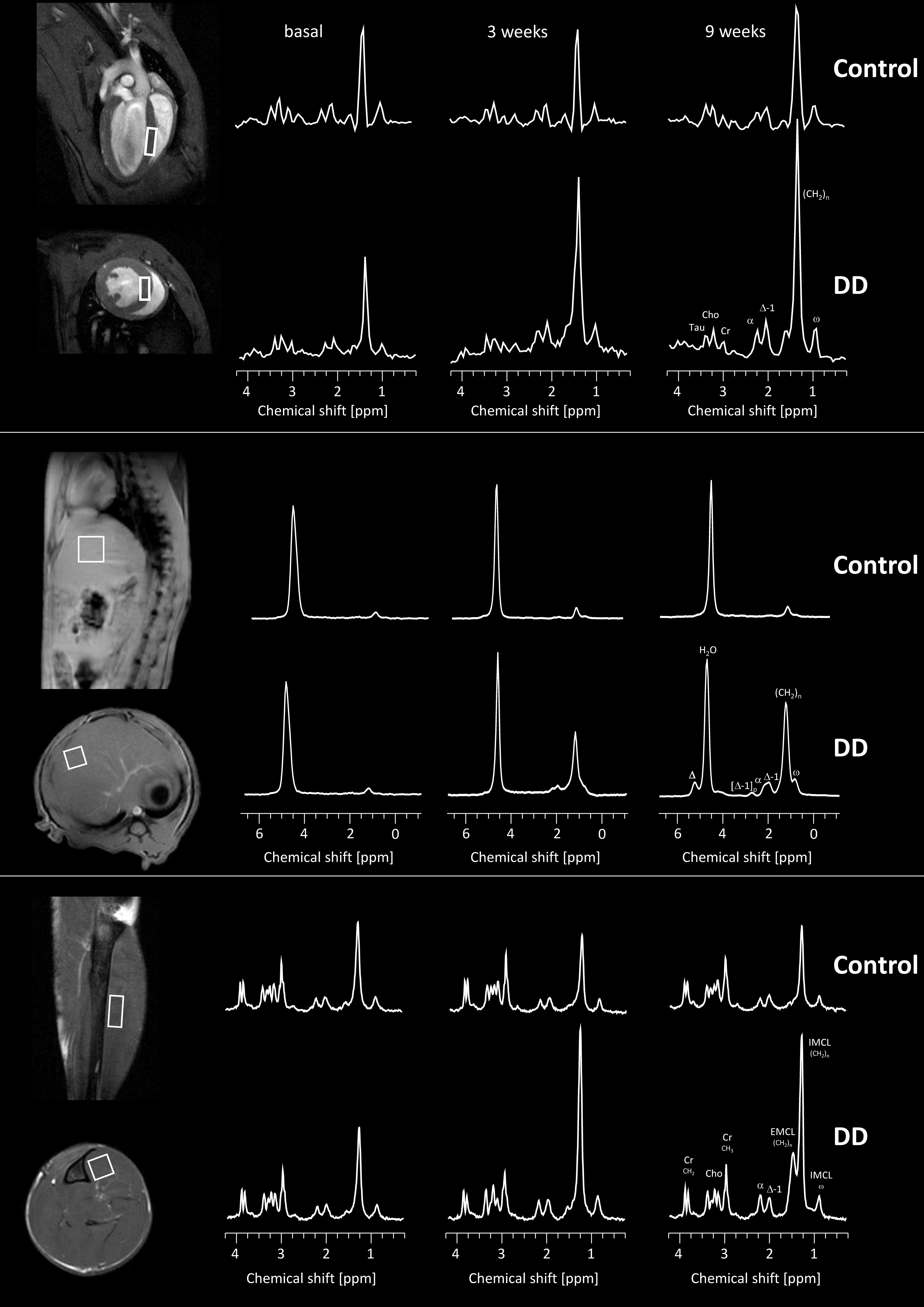
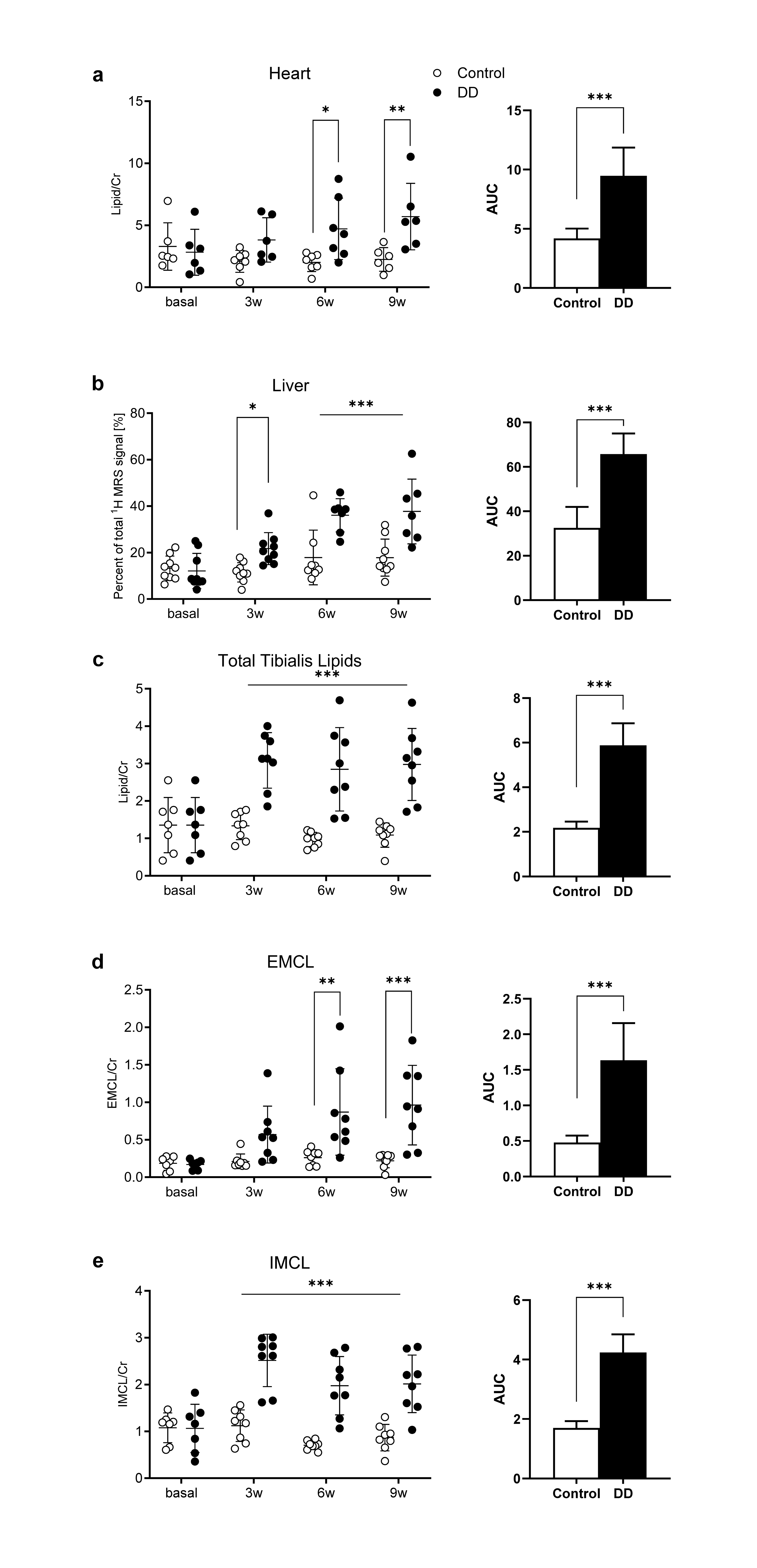
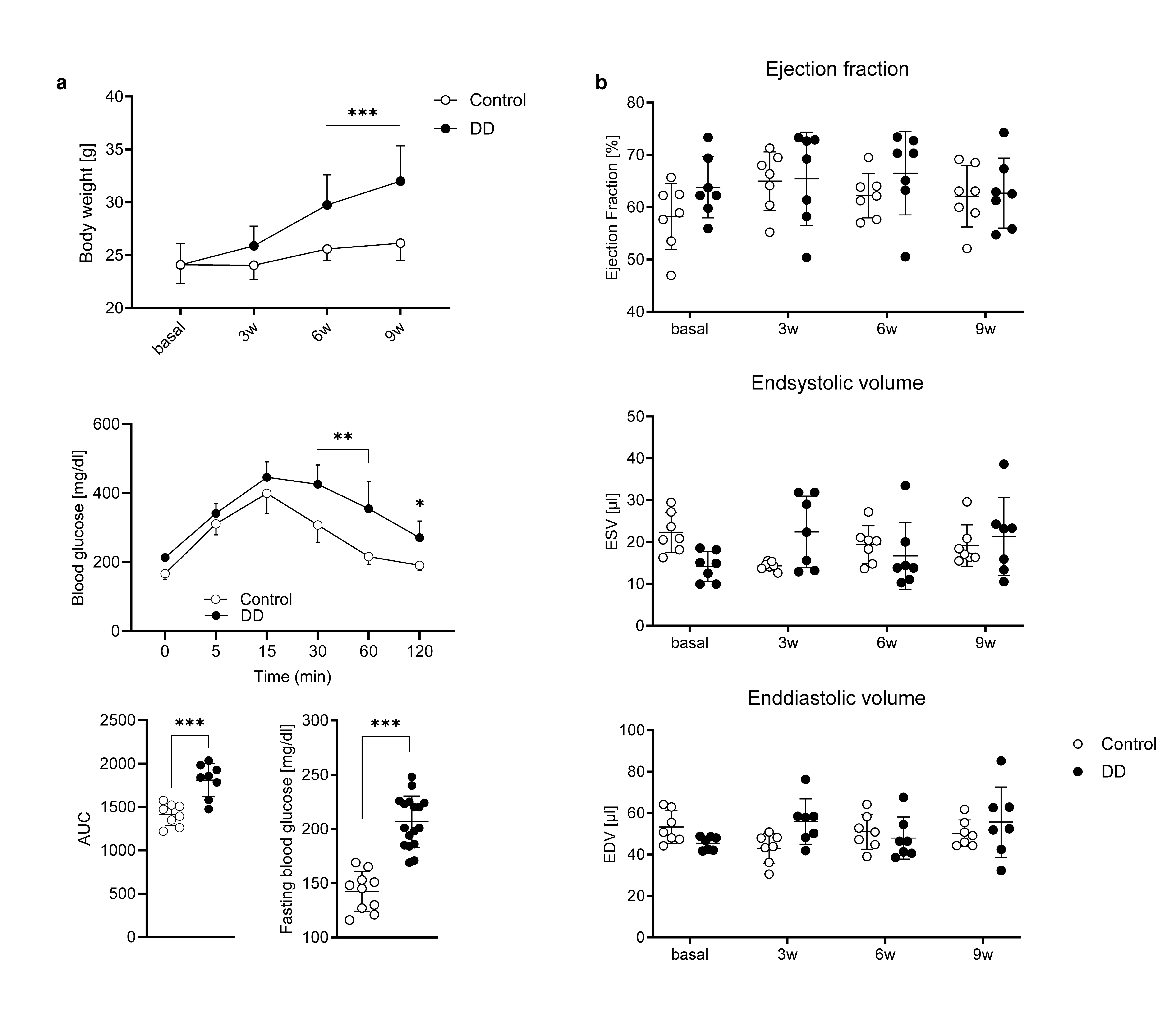
Male, 8-10 week old C57BL/6J mice were fed a DD for 9 weeks while control animals received a matching chow diet. Body weight, fasting blood glucose and intraperitoneal glucose tolerance were determined every 3 weeks. Furthermore, in vivo 1H MRS was used to determine every 3 weeks the lipid content in the heart (PRESS, TAcq 17 min, 1024 averages, 256 data points, ECG/respiration-triggered, water suppression, voxel size 1×2×3 mm3), the extra- and intramyocellular lipids (IMCL+EMCL) in M. tibialis anterior (PRESS, TAcq 18 min., 1024 averages, 2048 data points, water-suppression, voxel size 1.3×1.3×3 mm3) and the lipid content of the liver (PRESS, TAcq 4 min., 256 averages, data 2048 points, ECG/respiration triggered, voxel size 2×2×2 mm3).Results
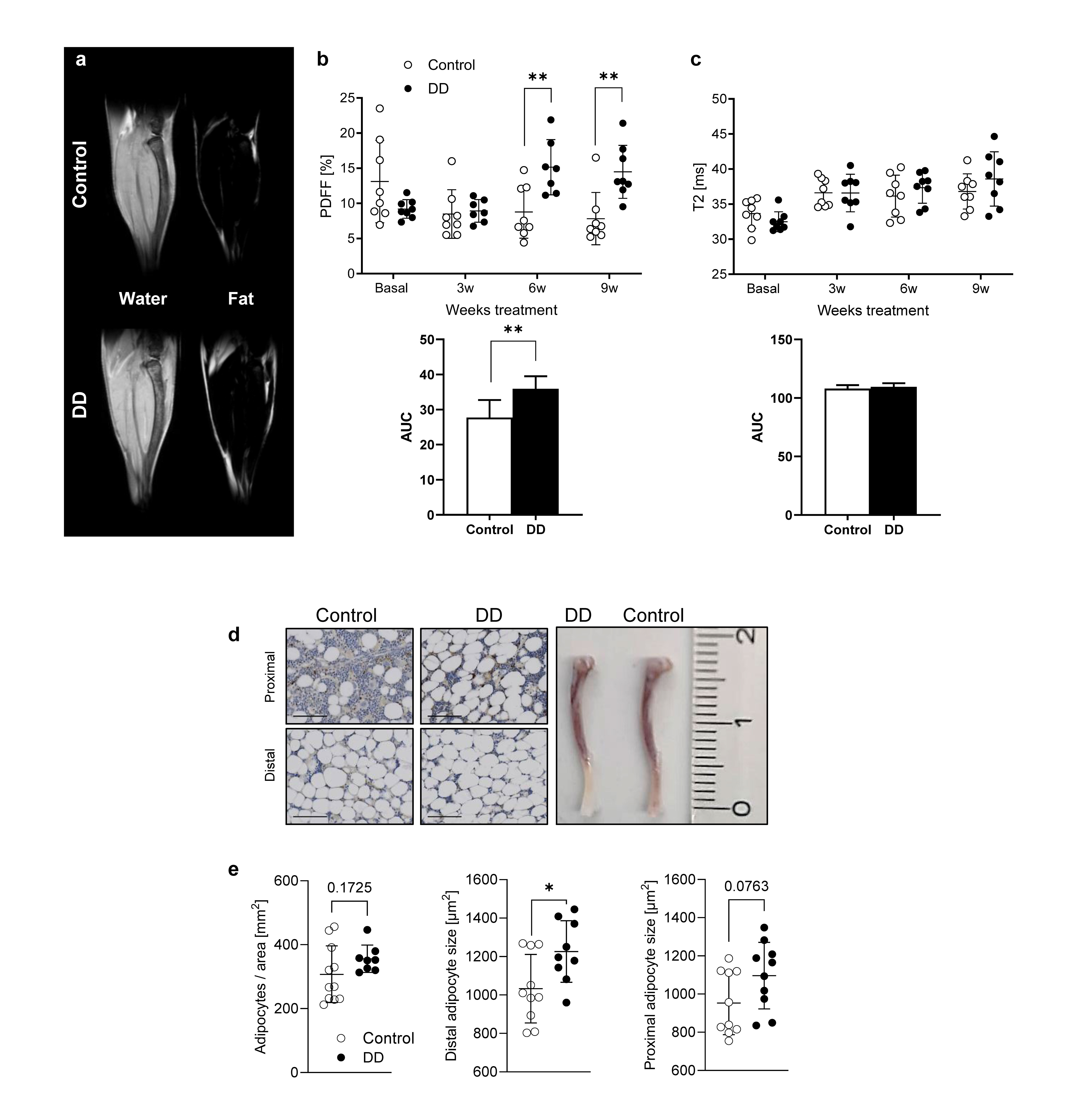
Volume-selective 1H MRS was used to assess the lipid content in organs known to be critically affected by diabetes, i.e. heart, liver and skeletal muscle. Representative baseline 1H MR spectra are illustrated in Figure 1, 2nd column, for each organ. Of note, in spectra from heart and tibialis muscle the dominating water signal was suppressed to allow accurate identification of the low lipid signals. With this, resonances for creatine, taurine, choline and several signals originating from lipids could be unequivocally resolved (Figure 1, top+bottom). While signals for the first metabolites were unaltered during the course of the diet, all lipid peaks were clearly increased in DD mice as compared to controls (Figure 1, 3rd and 4th column). Interestingly, the susceptibility of the investigated organs to the diabetogenic diet was largely differential: the strongest response upon onset of DD was observed for lipids in the tibialis muscle (Figure 1, bottom). Quantification of the in vivo data (Figure 2) revealed that IMCL exhibited already a massive increase after 3 weeks of DD (Figure 2e), staying almost constant thereafter, while EMCL (Figure 2d) increased steadily over time. Of note, the approximately three-fold increase in tibialis IMCL preceded even a sigificant weight gain in DD-fed mice (Figure 3a) and proved to be the most sensitive parameter for the induced metabolic changes. These very early alterations in skeletal muscle were followed by a substantial hepatosteatosis after 6 weeks of feeding (Figure 2b) and also a significant elevation of cardiac lipids in DIO animals (Figure 2a). Despite the almost three-fold fat increase in the heart, functional cardiac parameters were unaffected over the 9 weeks of diet (Figure 3b).Next, we further focused on the bone marrow as hematopoietic niche crucially involved in the normal immune response. Here, we used water/fat separation by multi chemical shift selective imaging (mCSSI4) to determine the bone marrow proton density fat fraction (PDFF) and employed T2 mapping for additional tissue characterization. As can be recognized from representative water/fat images from the calf (Figure 4a), diabetogenic feeding resulted in a substantial fat accumulation particularly in the distal part of the tibia, which reached the level of significance after 6w or when calculating the area-under-the-curve (AUC, Figure 4b). In contrast to the pronounced lipid increase in the bone marrow of DD mice detected by PDFF measurements, parametric maps (T2) of the tibia provided no evidence for any alterations in bone marrow tissue texture (Figure 4c). Of note, the same was true for the adjacent tibialis muscle (not shown), so the direct chemical-shift-based approaches (either mCSSI for the bone marrow and 1H MRS for the muscle) proved most sensitive to these rapid metabolic effects on DD exposure here. Consistent with the DIO-related fat accumulation observed in vivo, histology revealed an increased number as well as size of adipocytes in the tibia, especially in the distal part, due to DD feeding (Figure 4d+e).
Conclusions
Using the diet-induced diabetes model we demonstrate massive accumulation of lipids in all organs studied, in part already at a time, where body weight of mice was not yet altered. Importantly, 1H MRS was most sensitive to reveal in vivo very early alterations in tissue properties in the prediabetic state. Here, in particular intramyocellular lipids may serve in the clincal setting as marker to identify patients at the transition point from prediabetes to diabetes.Acknowledgements
No acknowledgement found.References
- Maggio CA, Pi-Sunyer FX. Obesity and type 2 diabetes. Endocrinology and Metabolism Clinics of North America. 2003;32:805–822.
- Melmer A, Kempf P, Laimer M. The Role of Physical Exercise in Obesity and Diabetes. Praxis (Bern 1994). 2018;107:971–976.
- La Sala L, Pontiroli AE. Prevention of Diabetes and Cardiovascular Disease in Obesity. Int J Mol Sci. 2020;21:E8178.
- Flögel U, Temme S, Jacoby C, Oerther T, Keul P, Flocke V, Wang X, Bönner F, Nienhaus F, Peter K, Schrader J, Grandoch M, Kelm M, Levkau B. Multi-targeted 1H/19F MRI unmasks specific danger patterns for emerging cardiovascular disorders. Nat Commun. 2021;12:5847.
Figures