0695
Sex- and brain region-specific 1H MRS neurometabolic profiles in young rats with hepatic encephalopathy1CIBM Center for Biomedical Imaging, Lausanne, Switzerland, 2Animal Imaging and Technology, EPFL, Lausanne, Switzerland, 3Service of Clinical Chemistry, University of Lausanne and University Hospital of Lausanne, Lausanne, Switzerland, 4Swiss Pediatric Liver Center, Department of Pediatrics, Gynecology and Obstetrics, University Hospitals Geneva and University of Geneva Medical School, Geneva, Switzerland
Synopsis
Keywords: Spectroscopy, Metabolism, MRS, hepatic encephalopathy, sex-difference, SPECIAL
Motivation: The lack of preclinical studies on potential sex-differences in the pathophysiology of hepatic encephalopathy (HE) prevents a comprehensive understanding of the disease.
Goal(s): To study the effect of sex on the neurometabolic profiles measured with 1H MRS.
Approach: We compared 1H MRS metabolite concentrations in the hippocampus and striatum of young male versus female rats with HE.
Results: We observed overall stronger neurometabolic effects of the disease on male versus female HE rats, including weight loss and decrease in brain antioxydants levels (Asc, GSH), likely hampering the capacity of male animals to counteract oxidative stress, important player in the pathophysiology of HE.
Impact: Sex-differences, often overlooked in preclinical studies, investigated here showed distinct 1H MRS neurometabolic profiles in female versus male rats with hepatic encephalopathy (HE), opening a new window of investigation for patients with HE.
Introduction
Type C hepatic encephalopathy (HE) is a decompensating event occurring in 30 to 40% of the patients with chronic liver disease (CLD), and characterized by motor and cognitive impairment that can evolve into coma and death1. Neurometabolic 1H-MRS fingerprints of the disease have been described both in patients2 and the bile-duct-ligated (BDL) rat model of type C HE3.So far, preclinical studies on HE almost exclusively focused on male animals and on the adult brain. Clinical studies suggested that children suffer from poorer outcome after liver transplantation than adults4,5, also supported by stronger neurometabolic changes in young versus adult male BDL rats6,7.
In clinical populations, sex-difference in the prevalence and response to HE have been reported, cirrhotic women being more likely to develop HE and with a higher mortality rate than men8. Young girls with biliary atresia are also more vulnerable to cognitive and skills delays before liver transplantation compared to young boys9.
The aim of this study is thus to explore the potential sex-differences in 1H-MRS neurometabolic profiles of young male and female BDL rats, in the striatum and hippocampus.
Methods
The BDL rat model of type C HE was used, surgery performed 21 days after the rats’ birth to study the developing brain. BDL rats were compared to SHAM-operated rats at the same age.Liver enzymes and systemic blood biomarkers were measured longitudinally to follow disease progression.
MR experiments were performed at week 6 post-surgery in hippocampus (11.2µL) and striatum (15.6µL). 4 groups were compared: female SHAM (N=4), female BDL (N=3), male SHAM (N=3), male BDL (N=5).
1H-MRS acquisitions were conducted on a Bruker 14.1T scanner with the semi-adiabatic SPECIAL sequence (TE/TR=9.3/4000ms, 240 shots). The basis set was simulated (NMRScope-B/jMRUI10), the macromolecules acquired in the striatum with double-inversion-recovery (TI1/TI2=2200/850ms) and residual metabolites removed11. Water-referenced metabolite concentrations were corrected for water (water T2 - striatum: 29 ms, hippocampus: 30.4 ms) versus metabolites T2. Metabolites with relative CRLB below 15% for all rats and in both brain regions were reported. Group-differences were tested with a two-way ANOVA (disease and sex effects), corrected with Bonferroni multiple-comparison post-hoc test, for each metabolite and blood marker individually. ANOVA significant interaction, of interest here, highlights where the disease affects male and female rats differently.
Results and Discussion
Characteristic changes in blood biomarkers with disease progression (increased bilirubin, ammonia, GOT, GPT and decreased Glc) did not differ between males and females (Fig 1), suggesting a similar systemic condition. HE was associated with significant weight loss in male (-29%) but not in female rats (Fig 1), in line with recent findings in adult BDL rats12. In patients with CLD, sarcopenia and thus the lower capacity of muscles to clear ammonia, leads to more frequent overt HE episodes and higher mortality rates13.Fig 2 confirms good spectral quality in the 4 groups. The quantification results in the striatum (Fig 3) and hippocampus (Fig 4) confirmed previously reported changes in young BDL rats6: increased Gln (striatum:+56%, hippocampus:+115%, males/females averaged) and decreased Ins (striatum:-30% hippocampus:-28%). tCho decrease was significantly stronger in males than females in both brain regions (striatum, male:-42%, female:-12%, hippocampus, male:-51%, female:-18%). Although not significant, a trend of stronger Gln increase in male versus female rats was observed in both brain regions (striatum, male:+68%, female:+44%, hippocampus, male:+143%, female:+88%). In both brain regions, Tau and tCr were higher in healthy males versus females and did not change with disease.
Hippocampus showed additional significant differences between groups compared to striatum: decreased Asp, GABA, Glu and increased Lac were found in BDL rats at the group level (combining sexes), but did not survive multiple-comparison tests, requiring a large sample size.
Very importantly, in the hippocampus, Asc and GSH, the two major brain antioxydants, remained constant in females but decreased significantly in males with HE (Asc:-13%, GSH:-24%). Decreased brain antioxydant levels in male BDL rats could hint towards their stronger vulnerability to brain oxidative stress (OS), playing an important role in pathophysiology of HE14,15, compared to females. This observation is supported by a recent study showing that female adult BDL rats have higher blood albumin levels compared to male BDLs, protecting females against systemic OS12.
Conclusion
We report the first comparison of 1H-MRS neurometabolic profiles between male and female HE rats. Our results suggest stronger biochemical effects of the disease in male rats, developing weight loss and decreased brain antioxydant levels hampering their capacity to counteract OS. Understanding why, on the other hand, women in clinical HE population suffer from poorer outcome than men could pave the way to a better understanding of the disease.Acknowledgements
This project was supported by the European Union's Horizon 2020 research and innovation program under the Marie Sklodowska-Curie grant agreement No 813120 (INSPiRE-MED), the SNSF projects no 310030_173222, 310030_201218 and the Leenaards and Jeantet Foundations. We acknowledge access to the facilities and expertise of the CIBM Center for Biomedical Imaging founded and supported by Lausanne University Hospital (CHUV), University of Lausanne (UNIL), Ecole polytechnique fédérale de Lausanne (EPFL), University of Geneva (UNIGE) and Geneva University Hospitals (HUG).References
1. Vilstrup H, Amodio P, Bajaj J, et al. Hepatic encephalopathy in chronic liver disease: 2014 Practice Guideline by the American Association for the Study of Liver Diseases and the European Association for the Study of the Liver. Hepatology. 2014;60(2):715-735. doi:10.1002/hep.27210
2. Kreis R, Farrow N, Ross BrianD. Diagnosis of hepatic encephalopathy by proton magnetic resonance spectroscopy. The Lancet. 1990;336(8715):635-636. doi:10.1016/0140-6736(90)93439-V
3. Braissant O, Rackayová V, Pierzchala K, Grosse J, McLin VA, Cudalbu C. Longitudinal neurometabolic changes in the hippocampus of a rat model of chronic hepatic encephalopathy. J Hepatol. 2019;71(3):505-515. doi:10.1016/j.jhep.2019.05.022
4. Sorensen LG, Neighbors K, Martz K, Zelko F, Bucuvalas JC, Alonso EM. Longitudinal Study of Cognitive and Academic Outcomes after Pediatric Liver Transplantation. The Journal of Pediatrics. 2014;165(1):65-72.e2. doi:10.1016/j.jpeds.2014.03.032
5. Devictor D, Desplanques L, Debray D, et al. Emergency liver transplantation for fulminant liver failure in infants and children. Hepatology. 1992;16(5):1156-1162. doi:10.1002/hep.1840160509
6. Rackayova V, Braissant O, Rougemont AL, Cudalbu C, McLin VA. Longitudinal osmotic and neurometabolic changes in young rats with chronic cholestatic liver disease. Sci Rep. 2020;10:7536. doi:10.1038/s41598-020-64416-3
7. Simicic D, Rackayova V, Braissant O, et al. Neurometabolic changes in a rat pup model of type C hepatic encephalopathy depend on age at liver disease onset. Metab Brain Dis. 2023;38(6):1999-2012. doi:10.1007/s11011-023-01210-w
8. Pemmasani G, Tremaine WJ, Suresh Kumar VC, et al. Sex differences in clinical characteristics and outcomes associated with alcoholic hepatitis. Eur J Gastroenterol Hepatol. 2023;35(10):1192-1196. doi:10.1097/MEG.0000000000002612
9. Caudle SE, Katzenstein JM, Karpen S, McLin V. Developmental assessment of infants with biliary atresia: differences between boys and girls. J Pediatr Gastroenterol Nutr. 2012;55(4):384-389. doi:10.1097/MPG.0b013e318259ed20
10. Starčuk Z, Starčuková J. Quantum-mechanical simulations for in vivo MR spectroscopy: Principles and possibilities demonstrated with the program NMRScopeB. Anal Biochem. 2017;529:79-97. doi:10.1016/j.ab.2016.10.007
11. Simicic D, Rackayova V, Xin L, et al. In vivo macromolecule signals in rat brain 1H-MR spectra at 9.4T: Parametrization, spline baseline estimation, and T2 relaxation times. Magnetic Resonance in Medicine. 2021;86(5):2384-2401. doi:10.1002/mrm.28910
12. Macedo de Oliveira M, Monnet-Aimard A, Bosoi CR, Tremblay M, Rose CF. Sex is associated with differences in oxidative stress and susceptibility to severe hepatic encephalopathy in bile-duct ligated rats. Journal of Neurochemistry. 2022;162(4):337-351. doi:10.1111/jnc.15661
13. Bhanji RA, Moctezuma-Velazquez C, Duarte-Rojo A, et al. Myosteatosis and sarcopenia are associated with hepatic encephalopathy in patients with cirrhosis. Hepatol Int. 2018;12(4):377-386. doi:10.1007/s12072-018-9875-9
14. Pierzchala K, Simicic D, Sienkiewicz A, et al. Central nervous system and systemic oxidative stress interplay with inflammation in a bile duct ligation rat model of type C hepatic encephalopathy. Free Radical Biology and Medicine. 2022;178:295-307. doi:10.1016/j.freeradbiomed.2021.12.011
15. Simicic D, Cudalbu C, Pierzchala K. Overview of oxidative stress findings in hepatic encephalopathy: From cellular and ammonium-based animal models to human data. Analytical Biochemistry. 2022;654:114795. doi:10.1016/j.ab.2022.114795
Figures
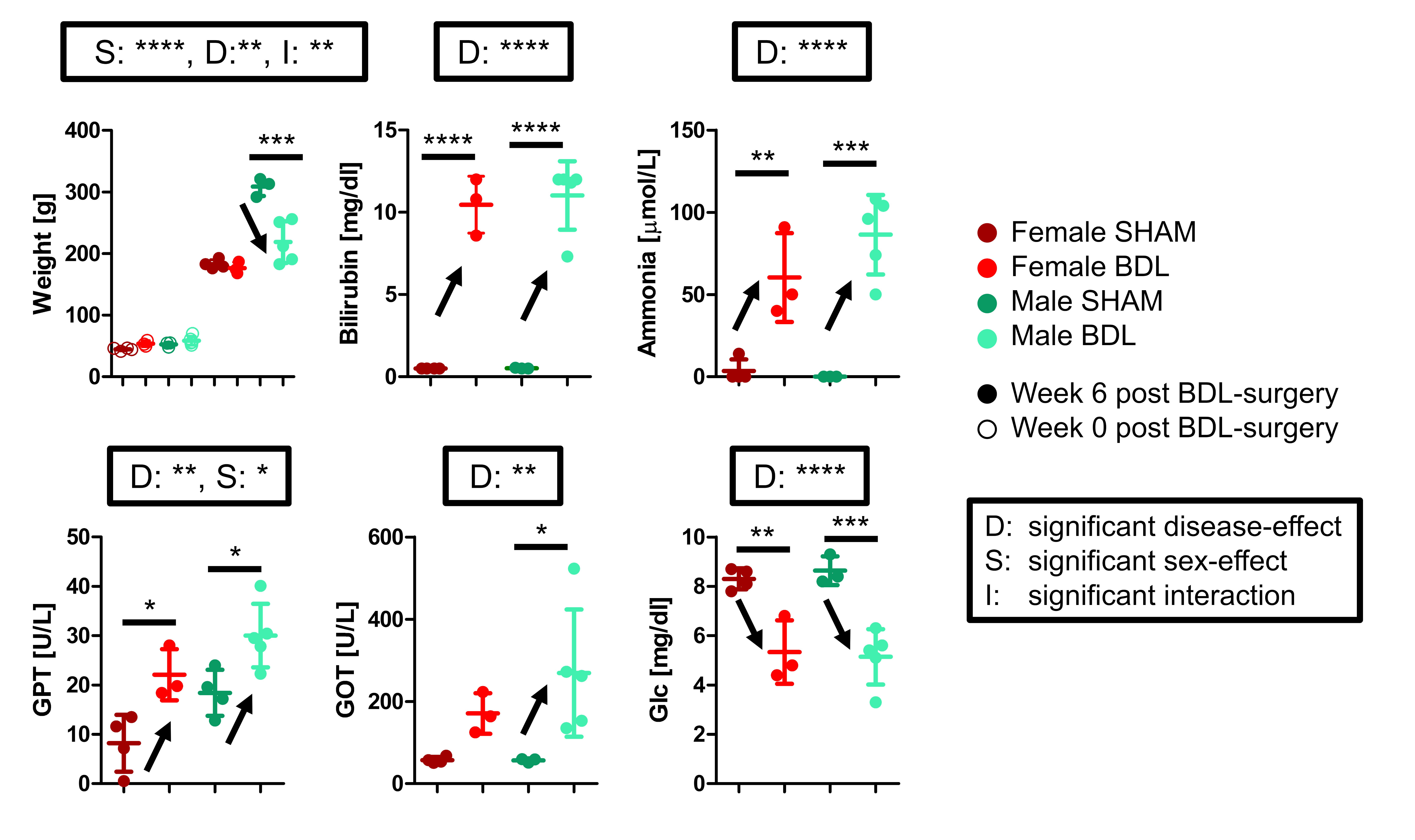
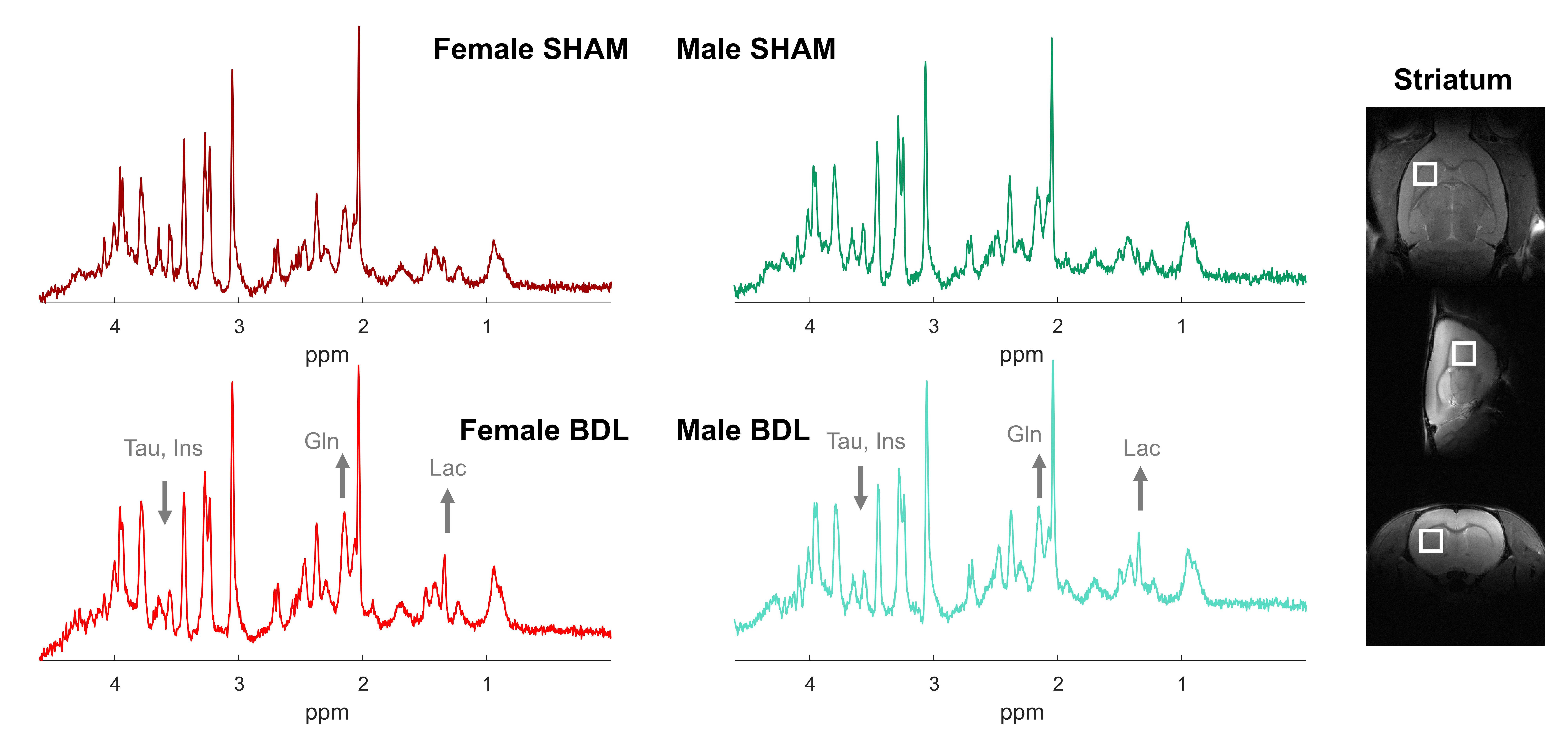
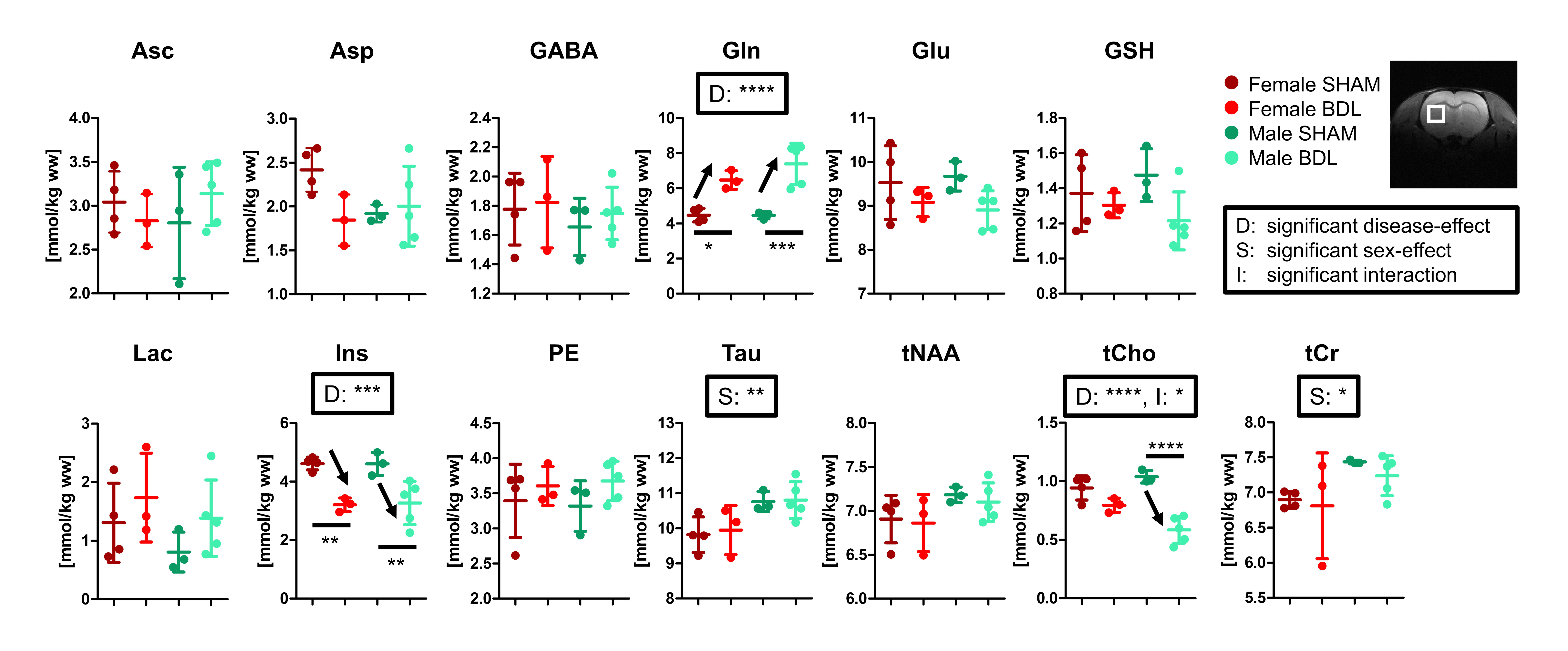
Fig 3 – Water-referenced metabolite concentrations in the striatum in each group. Individual animal values, mean and SD are displayed. The black square on top of each graph indicates the results of the group statistics (legend on the right), and the black lines/stars/arrows the differences that survived the multiple-comparison test. *: p<0.05, **: p<0.01, ***: p<0.001, ****: p<0.0001.
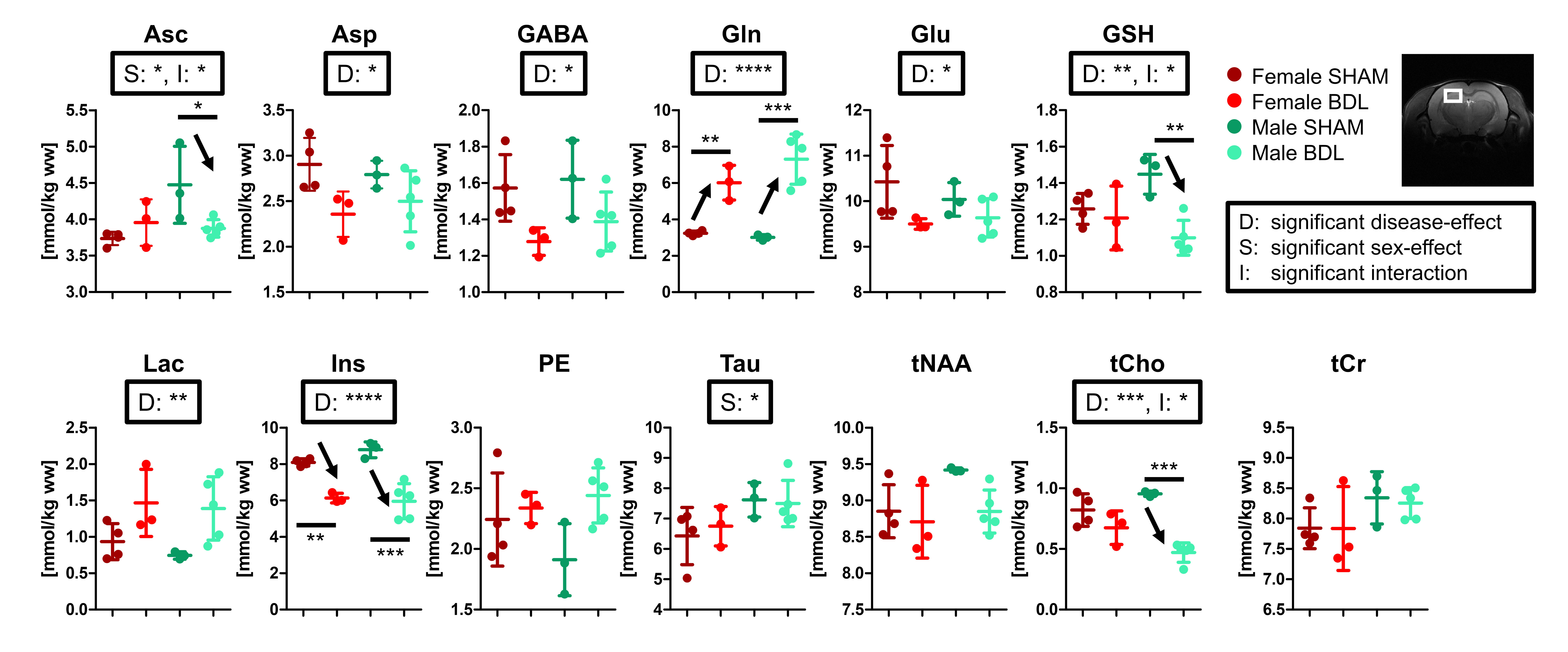
Fig 4 – Water-referenced metabolite concentrations in the hippocampus in each group. Individual animal values, mean and SD are displayed. The black square above each graph indicates the results of the group statistics (legend on the right), and the black lines/stars/arrows the differences that survived the multiple-comparison test. *: p<0.05, **: p<0.01, ***: p<0.001, ****: p<0.0001.