0693
Common coordinate framework of infant marmoset brain based on ultra-high-resolution diffusion MRI1Department of Radiology, Children's Hospital of Philadelphia, Philadelphia, PA, United States, 2Department of Bioengineering, University of Pennsylvania, Philadelphia, PA, United States, 3Department of Anatomy and Neurobiology, Shandong University, Jinan, China, 4Department of Radiology, University of Pennsylvania, Philadelphia, PA, United States, 5Department of Anatomy and Neurobiology, University of Wisconsin - Madison, Madison, WI, United States, 6Department of Neurology, University of California San Francisco, San Francisco, CA, United States
Synopsis
Keywords: Large Animals, Nonhuman Primates, Normal development, normal development, large animals-nonhuman primates, ultra-high resolution diffusion MRI, common coordinate framework
Motivation: Integrating a spatially resolved and molecularly defined cell atlas with studies of developing brain function, neurophysiology, and behavior will require an anatomical common coordinate framework (CCF). Ultra-high-resolution diffusion-MRI (dMRI) improves anatomical determinations and provides rich contrasts and microstructural information.
Goal(s): To build the first dMRI-based anatomical CCF for infant marmoset brains.
Approach: Ultra-high resolution dMRI at 9.4T was performed on a 10-month-old marmoset brain. Anatomical regions were delineated.
Results: An ultra-high-resolution CCF for the infant marmoset brain at isotropic 0.1mm diffusion MR imaging resolution, characterized by comprehensive labels of fine neuroanatomical structures and coordinate framework.
Impact: The first infant marmoset brain CCF will allow integrating spatially resolved, molecularly defined cell atlas with studies of developing brain function, neurophysiology, and behavior. It will provide insights into evolution and human-specific features of brain development relevant to brain disorders.
Purpose
A common coordinate framework (CCF) is an anatomical atlas into which microstructural, cellular, genetic, functional, and neurophysiological information can be integrated. Creating a CCF for infant marmoset brains will provide insights into evolution and uncover human-specific features of brain development relevant to brain disorders. Traditional histology-based brain atlases are 2D. Small developmental brains require higher resolution imaging. Ultra-high resolution diffusion MRI (dMRI) provides a unique opportunity for defining the first 3D infant marmoset brain CCF. We present the first ultra-high resolution (0.10×0.10×0.10mm3) dMRI-based infant marmoset brain CCF. We compare long range fibers for the developmental marmoset brain with long-range fibers in a cross-species age-matched human subject to demonstrate the evolutionary importance of our CCF.Methods
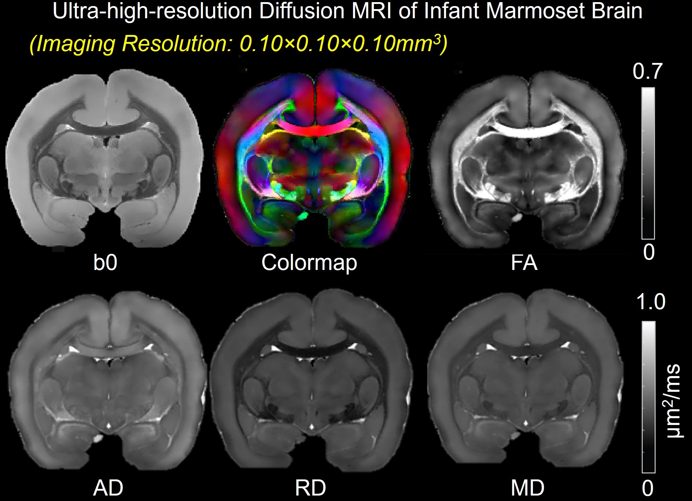
Acquisition of ultra-high-resolution dMRI data of 10-month-old postmortem marmoset brainA Bruker 9.4T scanner was used for ultra-high-resolution dMRI. A 2D spin echo diffusion sequence was used. DMRI parameters were b =1500 s/mm2, 30 unique gradient directions, TE = 36 ms, TR = 8.6s, FOV = 25mm×32mm, imaging matrix = 180×320 ×190 for an imaging resolution of 0.10×0.10×0.10 mm3.Repetition number=4.
Acquisition of dMRI data of cross-species age-matched 3-year-old human brain
A 3-year-old human were scanned on a 3T Philips Achieva system with sedation1. DMRI was acquired using a single-shot echo-planar imaging (EPI) sequence with b =1000 s/mm2, 30 unique gradient directions, TE=100ms, TR=9.3s, FOV= 256 × 256 mm2. imaging matrix =128 × 128, slice thickness=2mm, slice number=70. Repetition number=2.
Diffusion tensor imaging (DTI) processing
Marcenko-Pastur principle component analysis (PCA) denoising2 was performed on the raw dMRI data with patch radius=2. Automated image registration (AIR)3 was conducted on raw DWIs. The tensor fitting was conducted with DtiStudio4.
Creation of CCF
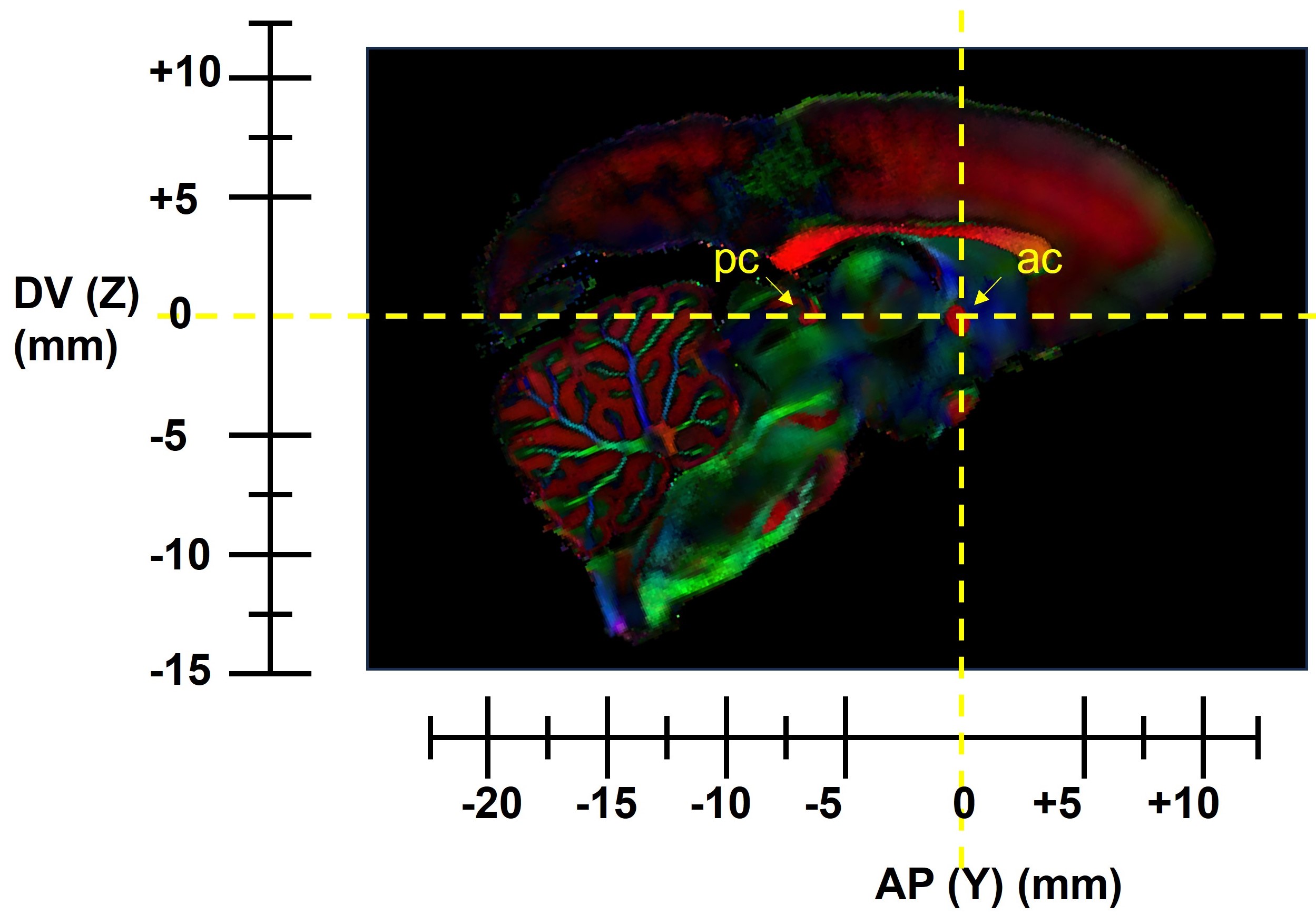
The origin of the CCF is chosen at the mid-sagittal plane of the anterior commissure (ac). A line connecting the posterior commissure and ac in the medial sagittal plane is defined as the anterior posterior (AP) axis, and the dorsal-ventral (DV) axis is perpendicular to the AP axis in the mid-sagittal plane. (Fig. 2). Manual delineation of brain regions was performed by an experienced neuroanatomist based on DTI-derived contrasts and a high resolution dMRI based adult marmoset atlas5.
DTI tractography in six tract groups
Streamline tractography6 was used for DTI fiber tracking of all ex-vivo marmoset and in-vivo human brain DTI data. The tractography protocol for tracing five categories of white matter (WM) tracts in human brain7 was also used to trace tracts in the marmoset brains.
Results
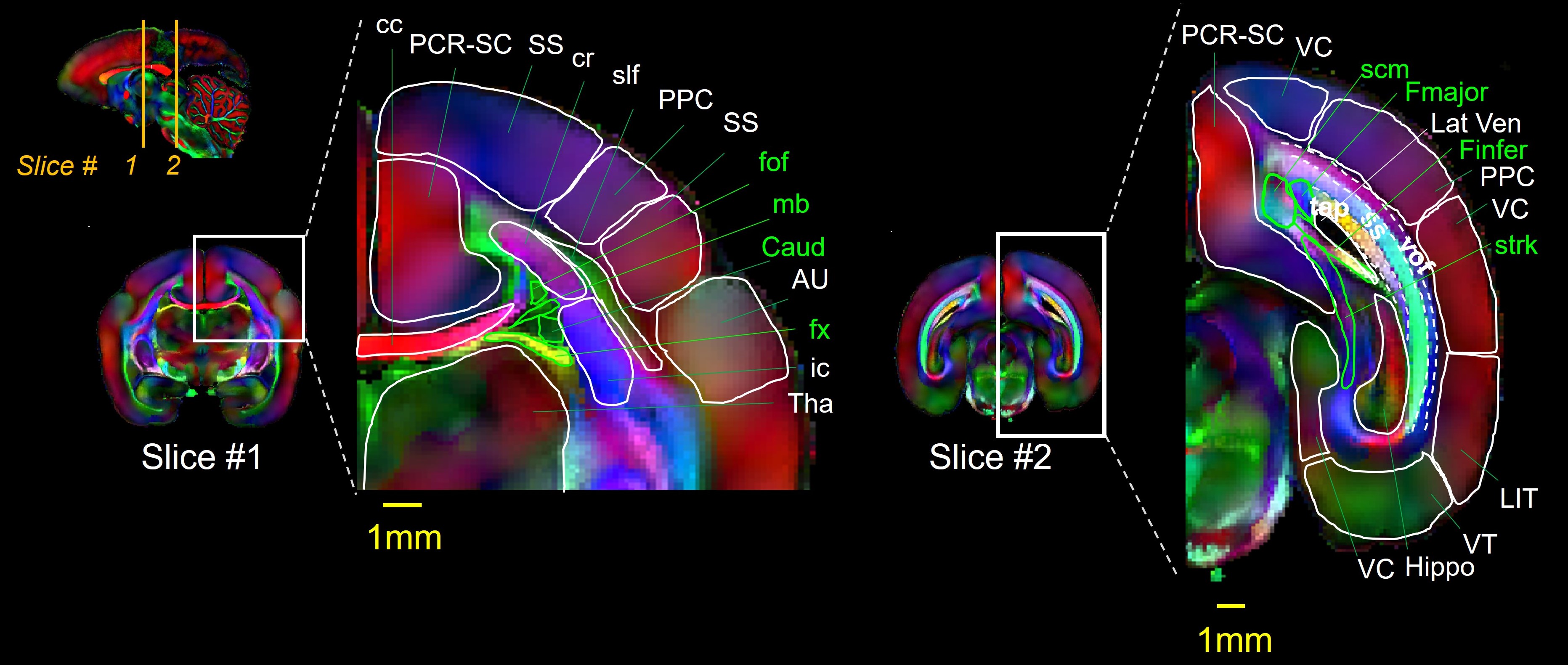
Infant marmoset brain CCFDTI derived maps from the first ultra-high resolution (0.10×0.10×0.10 mm3) dMRI dataset for a 10-month-old marmoset is shown in Fig. 1. In the space of this dataset, we defined the first developmental marmoset CCF (Fig. 2) The CCF is conveniently oriented for the registration of 2D histological, cellular, and genetic data. Representative slices from our developmental atlas are shown in Fig. 3 with the power to delineate fine adjacent WM structures such as the occipitofrontal fasciculus (fof) and Muratoff bundle (mb) (left panel).
Evolution under brain development framework
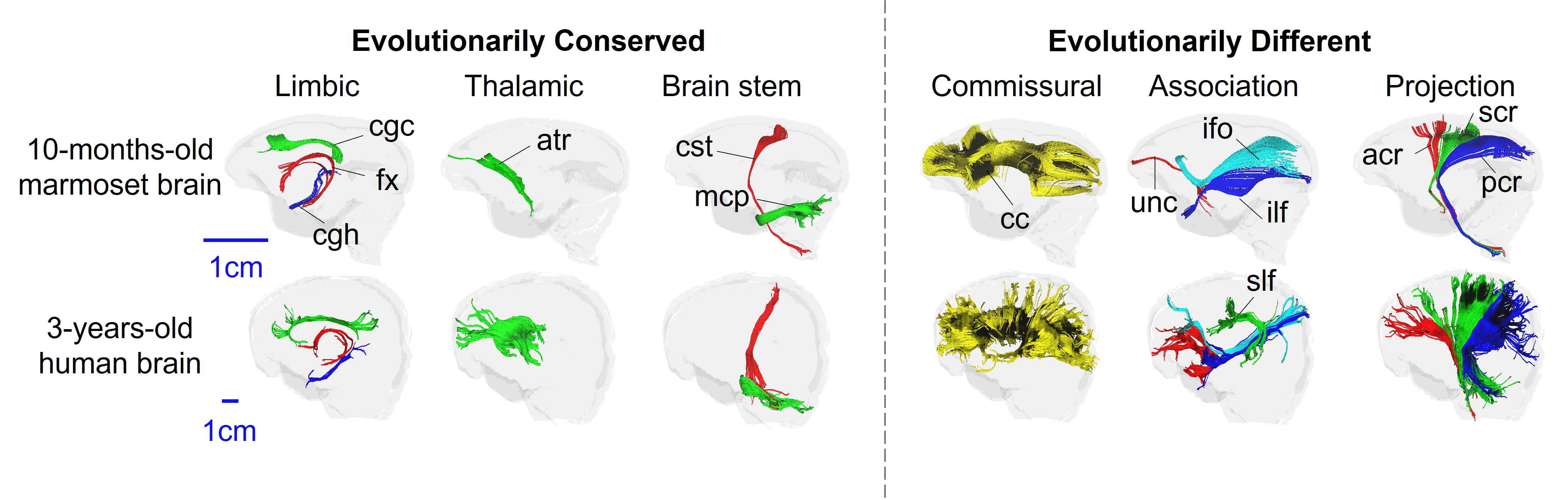
As shown in Fig. 4, DTI tractography results show evolutionarily conserved WM tract structures for the limbic, thalamic, and brain stem tract groups associated with brain functions universal to humans and marmosets such as physiological functions, whereas commissural, association, and projection tract groups with more advanced functions specific to humans such as language are evolutionarily different. Superior longitudinal fasciculus (slf) relevant to advanced language functions cannot be traced in the developmental 10-month-old marmoset brain, whereas it can be clearly delineated in the cross-species age-matched 3-year-old human brain. The anterior corona radiata (acr) does not extend far into the prefrontal lobe in the marmoset brain in contrast to the acr in the 3-year-old human brain.
Discussion and conclusion
We built the first ultra-high resolution dMRI-based infant marmoset brain CCF at the 0.10×0.10×0.10mm3 isotropic diffusion MR imaging resolution. DTI-derived contrasts show clear delineation of adjacent small WM tracts. The detailed anatomical atlas built based on the DTI-derived maps serve as a common space for mapping marmoset brain development from cellular, genetic, and functional perspectives and will provide insights into human evolution and uncover human-specific features of brain development relevant to brain disorders. As a first example of the evolutionary importance of our CCF, comparison of tractography results to a developmental human brain at equivalent age show evolutionarily distinct development of commissural, association, and projection tracts associated with more advanced cognitive functions and evolutionarily conserved tract structures in other tract groups. Ultra-high resolution dMRI acquisition on fetal, neonate, and adolescent marmoset brains and CCFs for these developmental stages are underway.Acknowledgements
This study is funded by NIH NIH R01MH092535, R01MH125333, R01EB031284, R01MH129981, R21EB009545, R21MH123930, UM1MH130991 and P50HD105354.References
1. Yu Q., Peng Y., Kang H., Peng Q., Ouyang M., Slinger M.,… Huang H. Differential white matter maturation from birth to 8 years of age. Cerebral Cortex, 2020, 30(4), 2674-2690.
2. Veraart J, Fieremans E, Novikov DS. Diffusion MRI noise mapping using random matrix theory. Magnetic Resonance in Medicine. 2016 doi: 10.1002/mrm.26059.
3. Woods RP, Grafton ST, Holmes CJ, Cherry SR, Mazziotta JC Automated image registration: i. General methods and intrasubject, intramodality validation. J Comput Assist Tomogr 1998 22:139–152
4. Jiang H, Van Zijl PC, Kim J, Pearlson GD, Mori S DtiStudio: resource program for diffusion tensor computation and fiber bundle tracking. Comput Methods Programs Biomed, 2006,81:106–116
5. Liu C, Ye FQ, Newman JD, Szczupak D, Tian X, Yen C, ... & Silva A resource for the detailed 3D mapping of white matter pathways in the marmoset brain. Nature neuroscience,2020. 23(2), 271-280.
6. Mori S, Crain BJ, Chacko VP, & Van Zijl PC. Three‐dimensional tracking of axonal projections in the brain by magnetic resonance imaging. Annals of Neurology: 1999, 45(2), 265-269
7. Wakana S., Caprihan A., Panzenboeck M, Fallon JH, Perry M, Gollub RL, ... & Mori S. Reproducibility of quantitative tractography methods applied to cerebral white matter. Neuroimage,2007, 36(3), 630-644.
8. Feng L, Jeon T, Yu Q, Ouyang M, Peng Q, Mishra V, ... & Huang H. Population-averaged macaque brain atlas with high-resolution ex vivo DTI integrated into in vivo space. Brain Structure and Function, (2017) 222, 4131-4147.
Figures