0691
Effect of nicotinamide riboside on glutamate in a 5XFAD mouse model of Alzheimer’s disease as measured by glutamate CEST MRI1Department of Bioengineering, University of Pennsylvania, Philadelphia, PA, United States, 2Department of Radiology, University of Pennsylvania, Philadelphia, PA, United States, 3Department of Physiology, University of Pennsylvania, Philadelphia, PA, United States
Synopsis
Keywords: Biomarkers, Alzheimer's Disease
Motivation: Nicotinamide riboside (NR) supplementation has increased in popularity for treating neurodegenerative diseases and is attributed to elevated NAD+ levels. Effective monitoring of NR-mediated changes may highlight metabolic underpinnings of NR in dementia.
Goal(s): This study uses glutamate-CEST MRI to monitor changes in glutamate levels following NR supplementation in wild-type and 5XFAD mouse models of AD.
Approach: Mice (WT and AD) were treated with NR or a vehicle (placebo) for 12 weeks followed by GluCEST MRI.
Results: There was a significant GluCEST increase in AD mice compared to WT. Following NR, GluCEST decreased in AD mice, primarily in the hippocampus.
Impact: NR supplementation may help alleviate excitotoxicity in AD, thereby preventing neuronal cell death/degeneration. GluCEST provides an effective method for assessing changes in glutamate levels, allowing for monitoring excitotoxicity in patients presenting symptoms of AD and the effects of NR treatment.
Introduction
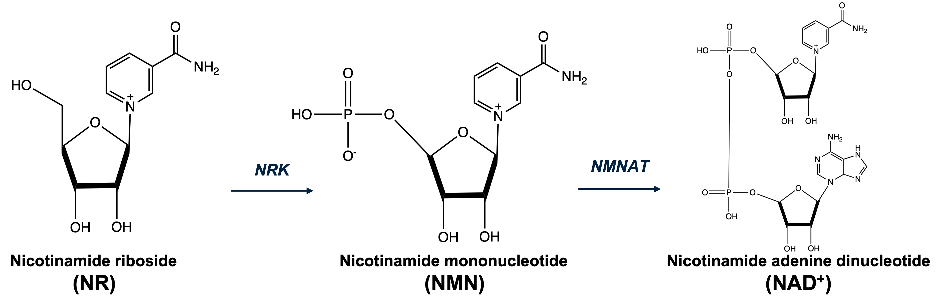
In addition to the major hallmarks of Alzheimer’s disease (AD) - including amyloid-beta plaques, neurofibrillary tangles, and glucose hypometabolism1,2 – perturbation to the glutamatergic system and its effects on neuronal plasticity has been proposed as a potential mechanism for the development of AD3. The main mechanisms underlying excitotoxicity include: (1) overstimulation of N-methyl-d-aspartate receptors (NMDARs), which are highly permeable to Ca2+ and allow an excessive and prolonged influx of Ca2+ in excitotoxic conditions, leading to neuronal damage and death, and (2) the impaired re-uptake of glutamate by glial cells due to decrease glutamate transporter activity and expression as well as competitive binding of toxic amyloid-beta to astrocytic glutamate transporters4. Nicotinamide riboside (NR), a precursor to nicotinamide adenine dinucleotide (NAD+), has been shown to prevent axonal degeneration and neuronal cell death through conversion to NAD+, a molecule important for redox homeostasis5. The increase in NAD+ rescues neuronal cells from oxidate stress under conditions of excitotoxicity and increased Ca2+ influx. This study uses the 5XFAD mouse model of AD, which recapitulates many human AD phenotypes with a relatively early and aggressive presentation, to measure the effect of NR supplementation on glutamate concentrations using glutamate CEST MRI6.Methods
Wild-type (WT) B6SJLF1/J (n = 20) and 5XFAD (n = 18) mice were obtained from Jackson Labs (Maine). NR was supplemented in drinking water at a concentration of 3g/L, delivering a dose of ~ 500 mg/kg/day starting at 12 weeks of age. This dose was selected base on prior studies showing physiologically meaningful effects in mice and is consistent with the dosing that has been successfully employed in AD models7,8. The mice, aged from 3 to 9 months of age, were examined in the following four groups: (1) WT + vehicle, (2) WT + NR, (3) AD + vehicle, (4) AD + NR. The age range was chosen to reflect the period in which 5XFAD mice progress from asymptomatic to symptomatic. MR imaging experiments were performed on a 9.4T magnet interfaced with Bruker Avance III HD using a 20mm 1H transceiver volume coil. Mice were anesthetized with isoflurane and secured using a custom head restrainer for imaging. An anatomical T2-weighted TurboRARE sequence was acquired for slice-selection, and glutamate CEST (GluCEST) was performed with the following parameters: Sat. dur.: 1s, sat. delay: 6s, offsets: 2.4ppm to 3.6ppm in ss: 0.2ppm, TR: 8ms, TE: 4ms, FOV: 20x20mm2, slice-thickness: 1mm, in-plane resolution: 0.156x0.156µm2. Nine brain regions (gray and white matter) were evaluated following atlas-based registration using a template created by Dorr et al. Two-way ANOVA was used for statistical analysis to evaluate the effect of genotype and supplementation on GluCEST.Results
Eight of the nine brain regions showed a statistically significant change (two-way ANOVA, p<0.05) in GluCEST with respect to disease-type (DT), with 5XFAD mice exhibiting higher GluCEST contrast than WT. These brain regions included sub-regions of the hippocampus (dentate gyrus and striatum granulosum), cerebral cortex, thalamus, hypothalamus, and white matter regions including the corpus callosum, fimbria, and cerebral peduncle. The hippocampus did not show a significant change with respect to with respect to WT but showed a significant decrease w.r.t. NR supplementation. Furthermore, the dentate gyrus of hippocampus showed a significant increase in GluCEST in AD mice followed by a significant decrease in GluCEST with supplementation. No significant interaction term (effect of DT on supplementation and vice versa) was observed for any of the regions. Representative GluCEST maps show increased glutamate levels in gray matter regions for AD mice, particularly the hippocampus, cortex, thalamus, and hypothalamus. With NR supplementation, there is a decrease in glutamate levels, with noticeable changes in AD mice compared to WT mice.Discussion
NR supplementation may alleviate excitotoxicity present in AD pathology, with a significant decrease in glutamate levels in the hippocampal region as observed in 5XFAD mice. The hippocampus, a brain region integral to memory formation and retrieval, is a structure well-known to lose synaptic plasticity and undergo atrophy in AD pathology9. The observed changes in GluCEST may reflect the early deposition of amyloid-beta fibrils and the disruption of glutamate transmitter cycling and neuronal cell death due to excess glutamate levels. Along with the NMDAR antagonist memantine10, an FDA approved therapeutic for excitotoxicity, NR may confer additional neuroprotective effects and potentially serve as an alternative therapeutic in reducing excitotoxicity present in AD and other neurodegenerative diseases.Acknowledgements
Research reported in this publication was supported by the National Institute of Biomedical Imaging and Bioengineering of the National Institutes of Health under award Number P41EB029460, the National Institute of Aging of the National Institutes of Health under Award Number R01AG063869, and the national Institute of Diabetes and Digestive and Kidney Diseases of the National Institutes of Health under Award Number R01DK098656.References
1. Breijyeh Z, Karaman R. Comprehensive review on alzheimer’s disease: causes and treatment. Molecules 2020;25 doi: 10.3390/molecules25245789.
2. Mosconi L, Pupi A, De Leon MJ. Brain glucose hypometabolism and oxidative stress in preclinical Alzheimer’s disease. Ann. N. Y. Acad. Sci. 2008;1147:180–195 doi: 10.1196/annals.1427.007.
3. Esposito Z, Belli L, Toniolo S, Sancesario G, Bianconi C, Martorana A. Amyloid β, glutamate, excitotoxicity in Alzheimer’s disease: are we on the right track? CNS Neurosci. Ther. 2013;19:549–555 doi: 10.1111/cns.12095.
4. Vaur P, Brugg B, Mericskay M, et al. Nicotinamide riboside, a form of vitamin B3, protects against excitotoxicity-induced axonal degeneration. FASEB J. 2017;31:5440–5452 doi: 10.1096/fj.201700221RR.
5. Wang R, Reddy PH. Role of glutamate and NMDA receptors in alzheimer’s disease. J Alzheimers Dis 2017;57:1041–1048 doi: 10.3233/JAD-160763.
6. Cai K, Haris M, Singh A, et al. Magnetic resonance imaging of glutamate. Nat. Med. 2012;18:302–306 doi: 10.1038/nm.2615.
7. Mukherjee S, Chellappa K, Moffitt A, et al. Nicotinamide adenine dinucleotide biosynthesis promotes liver regeneration. Hepatology 2016 doi: 10.1002/hep.28912.
8. Frederick DW, Loro E, Liu L, et al. Loss of NAD homeostasis leads to progressive and reversible degeneration of skeletal muscle. Cell Metab. 2016;24:269–282 doi: 10.1016/j.cmet.2016.07.005.
9. Rao YL, Ganaraja B, Murlimanju BV, Joy T, Krishnamurthy A, Agrawal A. Hippocampus and its involvement in Alzheimer’s disease: a review. 3 Biotech 2022;12:55 doi: 10.1007/s13205-022-03123-4.
10. McShane R, Westby MJ, Roberts E, et al. Memantine for dementia. Cochrane Database Syst. Rev. 2019;3:CD003154 doi: 10.1002/14651858.CD003154.pub6.
Figures
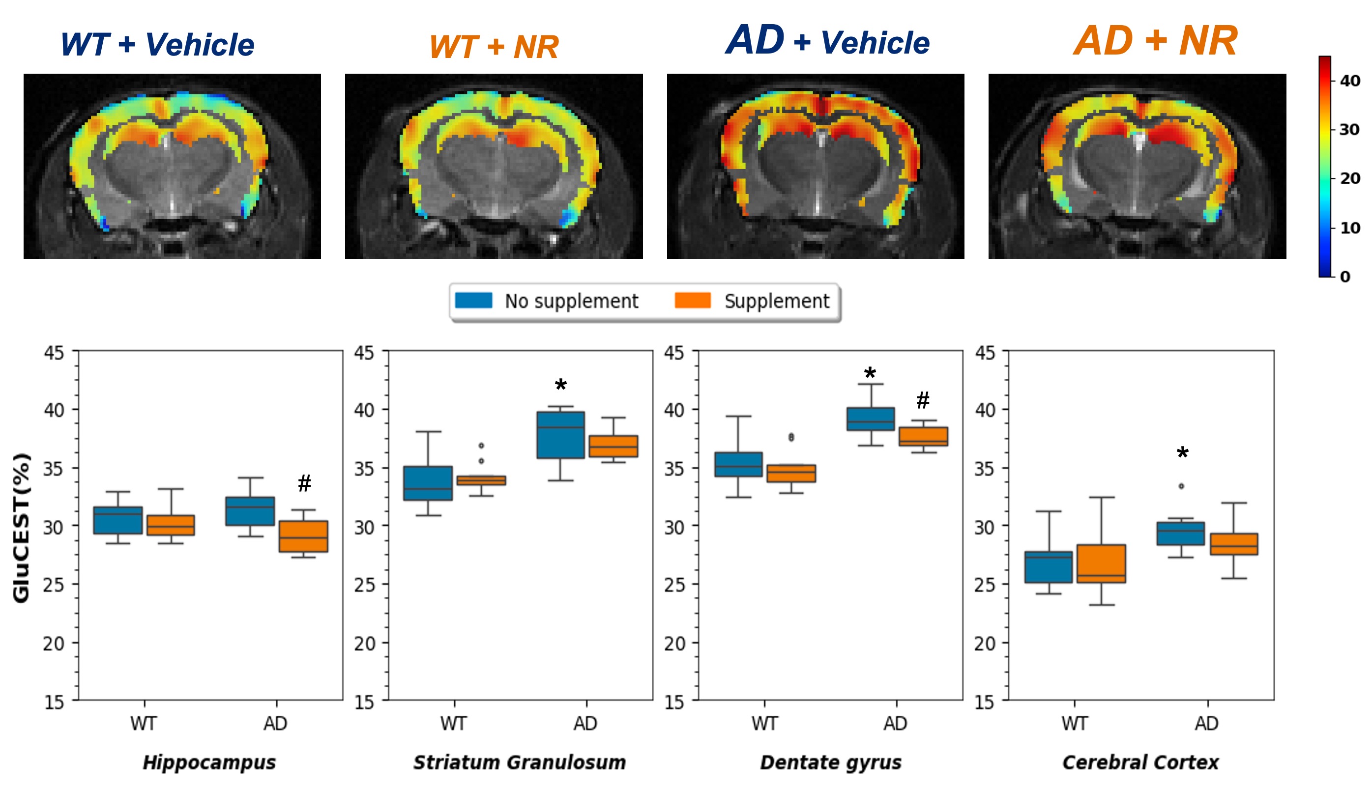
Top row shows representative GluCEST maps from WT and AD mice in cerebral GM regions. Visually, there is a noticeable decrease in GluCEST for AD mice in the hippocmpus following treatment compared to WT. There is a substantial increase in glutamate between WT and AD groups, suggesting excitotoxicity. Bottom row shows boxplots of GluCEST values for different disease types and treatment groups for the regions represented in the top row.
* - significant difference with respect to wild-type mouse genotype, p < 0.05
# - significant difference with respect to vehicle (un-treated) mice, p < 0.05
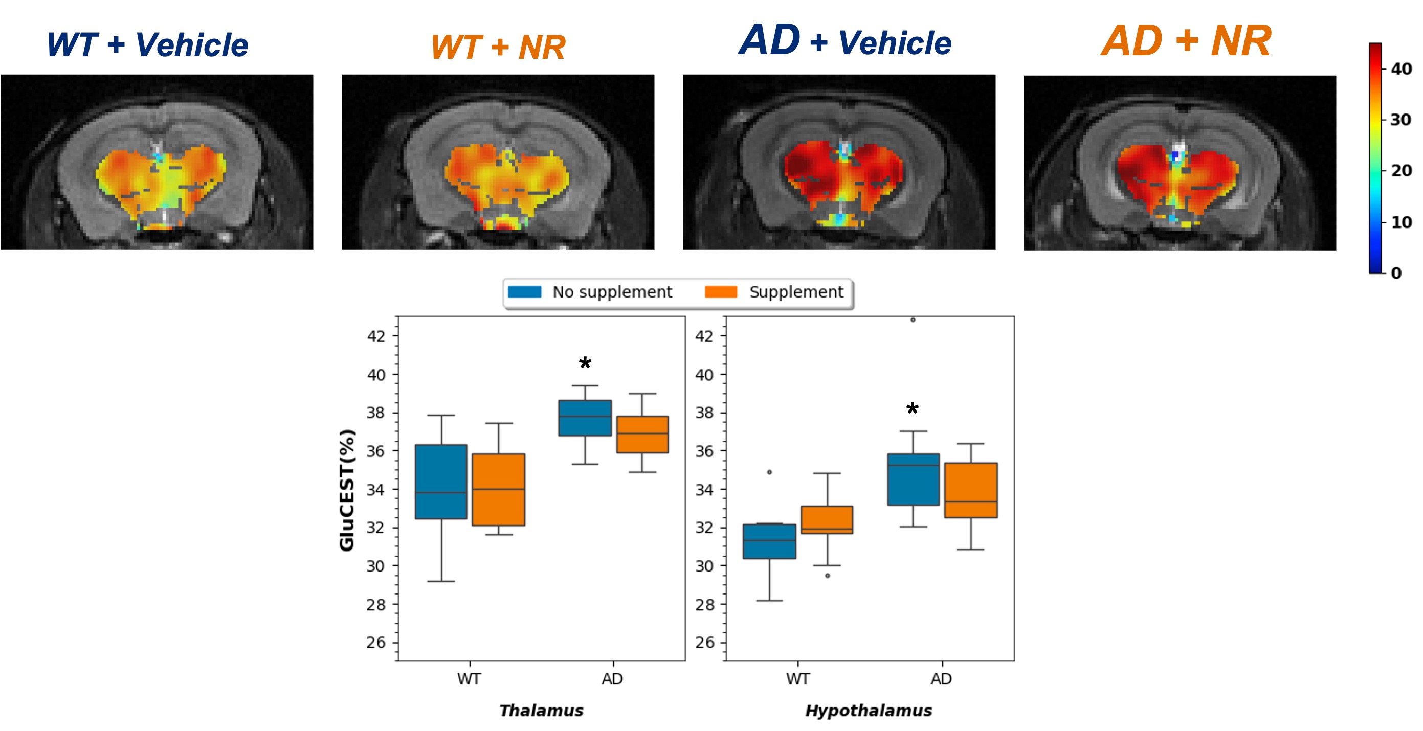
Top row shows representative GluCEST maps from WT and AD mice in midbrain GM regions. Visually, there is a noticeable decrease in GluCEST for AD mice in the thalamus following treatment compared to WT. There is a substantial increase in glutamate between WT and AD groups, suggesting excitotoxicity. Bottom row shows boxplots of GluCEST values for different disease types and treatment groups for the regions represented in the top row.
* - significant difference with respect to wild-type mouse genotype, p < 0.05
# - significant difference with respect to vehicle (un-treated) mice, p < 0.05
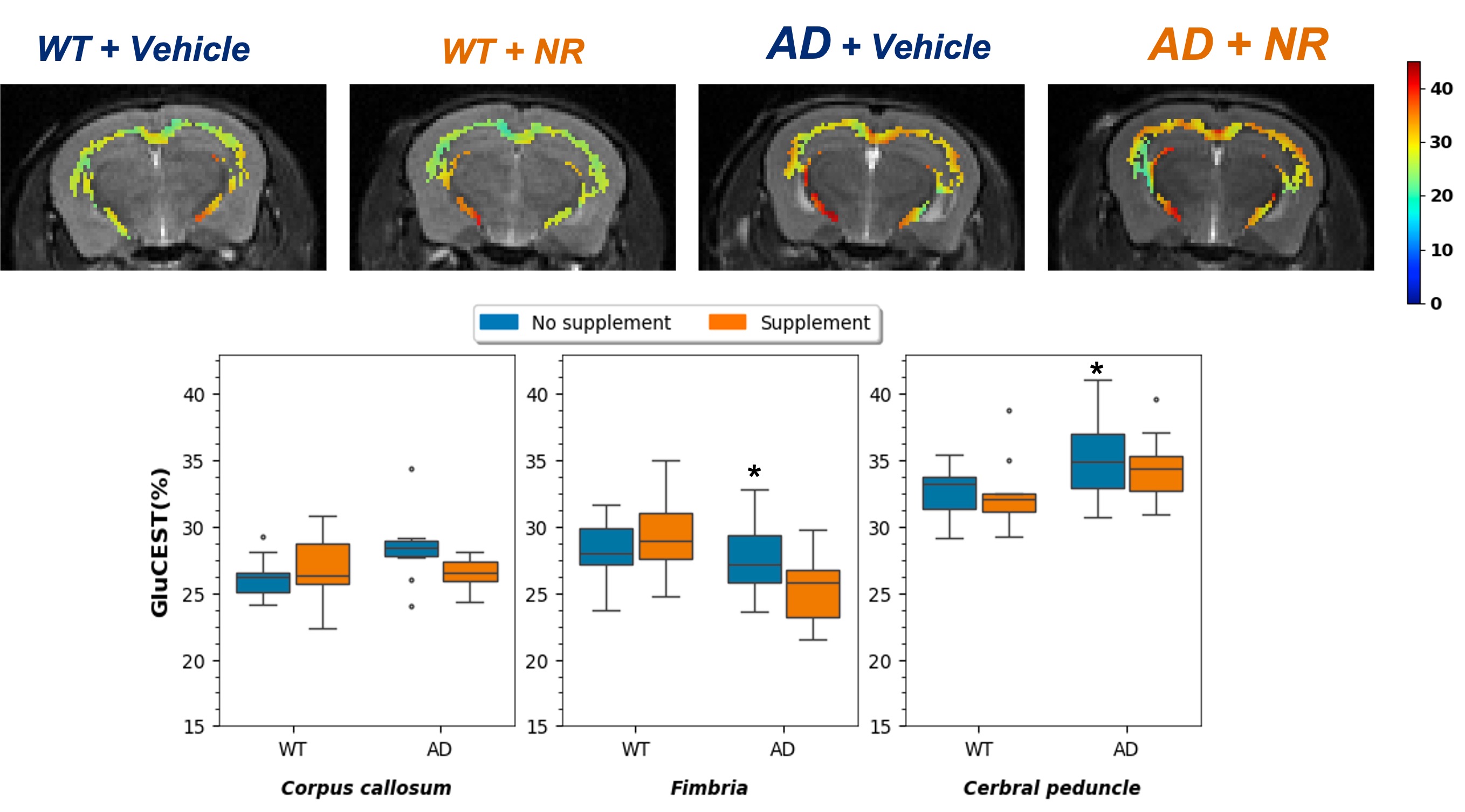
Top row shows representative GluCEST maps from WT and AD mice in WM regions. Visually, there is a noticeable decrease in GluCEST in the corpus callosum for AD mice following treatment compared to WT. There is a substantial increase in glutamate between WT and AD groups, suggesting excitotoxicity. Bottom row shows boxplots of GluCEST values for different disease types and treatment groups for the regions represented in the top row.
* - significant difference with respect to wild-type mouse genotype, p < 0.05
# - significant difference with respect to vehicle (un-treated) mice, p < 0.05