0675
Low-frequency magnetic signal detection using stimulus-induced rotary saturation sequence in ultra-low field MRI1Hamamatsu Photonics K.K., Hamamatsu, Japan, 2Kyoto University, Kyoto, Japan
Synopsis
Keywords: Low-Field MRI, Low-Field MRI
Motivation: Realization of biomagnetic measurement using MRI with high spatial resolution.
Goal(s): Detection of low-frequency magnetic signals below 50 Hz in ultra-low field MRI (ULF-MRI).
Approach: Magnetic signal detections were performed by stimulus-induced rotary saturation (SIRS) sequence in ULF-MRI. As magnetic signals, reference magnetic fields with 225 nTpp in amplitude and 10, 15, ..., 70 Hz in frequency were applied to the bottle phantom.
Results: The signal reduction of approximately 20% were observed when the reference magnetic field between 30 Hz and 45 Hz were applied. This indicates that low-frequency magnetic signals can be detected by the SIRS sequence in ULF-MRI.
Impact: We demonstrate the feasibility of biomagnetic measurement below 50 Hz such as brain activity first time by realizing low-frequency magnetic signal detection using stimulus-induced rotary saturation sequence in ULF-MRI with 7 mT in B0.
Introduction
Ultra-low field MRI (ULF-MRI) has advantages such as small inhomogeneity of B0 field and a margin for saturation absorption rate (SAR) due to low Larmor frequency. By these advantages, ULF-MRI is suitable for pulse sequences where long excitation pulses are utilized. On the other hand, a spin-lock sequence is a pulse sequence that enables the detection of oscillating magnetic fields, and is expected to be applied to biomagnetic measurements1,2. Since magnetic signal measurement using MRI has high spatial resolution, it is an effective measurement method for estimating the signal source in biomagnetic measurement. On the combination of spin-lock sequences and ULF-MRI scanners, there is additional advantages such as measurement of low-frequency magnetic field. The spin-lock frequency is proportional to the amplitude of the spin-lock pulse, and when the inhomogeneity of static magnetic field is larger compared to the spin-lock pulse, it is difficult to measure magnetic field. Previous reports investigated the rotary-echo spin-lock sequences3,4 employing the refocusing pulse in high-field MRI scanners. However, no one reported how much the advantage of ULF-MRI scanners in terms of homogeneity of the static magnetic field could contribute to solving this limitation. In this research, we demonstrate the detection of low-frequency magnetic signals of 50 Hz or less using a stimulus-induced rotary saturation (SIRS) sequence5, which is one of the spin-lock sequence, and investigate the feasibility for biomagnetic measurement such as brain activity measurement.Methods
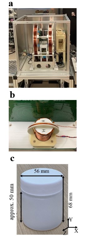
In this study, MR images were obtained by a homemade UFL-MRI system with a static magnetic field of approximately 7 mT (300 kHz in Larmor frequency), as shown in Figure 1a. An oscillating magnetic field of 225 nTpp was applied as a reference signal from a circular coil to the bottle phantom containing saline supplemented with 1 mM Magnevist® as shown in Figures 1b and 1c. Then, MR images were scanned using the SIRS-SE3D sequence with the imaging parameters as shown below: TR/TE: 500/25 ms, FoV: 96×60×60 mm3, Matrix: 32×20×12, Flip angle: 90 deg., Bandwidth: 100 Hz/pixel, NEX: 4, Spin-lock frequency: 50 Hz, Spin-lock duration: 50 ms.Results and Discussion
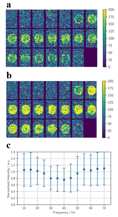
Images with and without 40 Hz magnetic field are shown in Figure 2a and 2b. In addition, images obtained at signal frequency of 10, 15, ..., 70 Hz were obtained and normalized by the image without signal. The region of interest was 3×3×5 voxels in the center of the bottle, we calculated the mean and standard deviation of them. As shown in Figure 2c, we plotted them as a function of the frequency of the signal magnetic field.From the results, the signal reduction of approximately 20% were observed when the reference magnetic field between 30 Hz and 45 Hz were applied. This indicates that low-frequency magnetic signals can be detected by the SIRS sequence in ULF-MRI. However, since the signal-to-noise ratios (SNRs) were insufficient, the standard deviations of the signal intensities were about 50% of the mean signal intensities. Therefore, it is essential to improve the SNR in the future. In addition, although the spin-lock frequency and spin-lock duration were fixed in these measurements, it is important to optimize such the imaging parameters towards more sensitive magnetic signal detection.
Conclusion
In this study, we revealed the feasibility of the low-frequency magnetic field detection with SIRS sequence. As a result, we were able to demonstrate the possibility of measuring magnetic signals of 225 nTpp between 30 Hz and 45 Hz without any refocusing pulses. Therefore, we confirmed that magnetic fields below 50 Hz such as brain activity were able to detect by SIRS sequence in the ULF-MRI scanner. In the future, we plan to improve the SNR and optimize the imaging parameters of SIRS towards the improvement of the detection sensitivity of low-frequency magnetic signals employing ULF-MRI scanners.Acknowledgements
This work was partially supported by Grants-in-Aid for Young Scientists (B) (JSPS KAKENHI Grant Number JP22K15621) from Japan Society for the Promotion of Science (JSPS), Japan.References
- Halpern-Manners NW, Bajaj VS, Teisseyre TZ, Pines A. Magnetic resonance imaging of oscillating electrical currents. Peoc Natl Acad Sci USA. 2010;107(19):8519-8524. doi:10.1073/pnas/1003146107
- Sveinsson B, Koonjoo N, Zhu B, Witzel T, Rosen MS. Detection of nanotesla AC magnetic fields using steady-state SIRS and ultra-low field MRI. J Neural Eng. 2020;17(3):34001. doi:10.1088/1741-2552/ab87fe
- Gram M, Seethaler M, Gensler D, Oberberger J, Jakob PM, Nordbeck P. Balanced spin-lock preparation for B1-insensitive and B0-insensitive quantification of the rotating frame relaxation time T1ρ. Magn Reson Med. 2021;85(5):2771-2780. doi:10.1002/mrm.28585
- Capiglioni M, Turco F, Wiest R, Kiefer C. Analysis of the robustness and dynamics of spin-locking preparations for the detection of oscillatory magnetic fields. Sci Rep. 2022;12(1):1-10. doi:10.1038/s41598-022-21232-1
- Witzel T, Lin F-H, Rosen BR, Wald LL. Stimulus-induced Rotary Saturation (SIRS): a potential method for the detection of neuronal currents with MRI. Neuroimage. 2008;42(4):1357-1365. doi:10.1016/j.neuroimage.2008.05.010
Figures